Depth probe for intracranial treatment

a depth probe and intracranial treatment technology, applied in the field of depth probes utilized for intracranial treatment, can solve the problems of inability to precisely electrically stimulate very small volumes of the brain, patients often experience diminishing returns of such treatment, delicate and often difficult to insert along specific trajectories or lines of insertion, etc., to achieve efficient and effective transmission of readings

Inactive Publication Date: 2006-03-16
TECH MEDICAL INSTR
View PDF57 Cites 16 Cited by
  • Summary
  • Abstract
  • Description
  • Claims
  • Application Information

AI Technical Summary

Benefits of technology

[0017] Another object of the invention is to provide a novel depth probe that is simple in structure and operation in order to facilitate intracranial procedures.
[0020] Another object of the invention is to provide a desirable depth probe having a lumen for receiving and guiding an inner catheter for the delivery of a drug to targeted brain tissue and that can remain in position when the inner catheter is removed, thereby permitting repeated insertions of different inner catheters without extended contact with brain tissue during insertion.
[0021] Another object of the invention is to provide an exceptional depth probe that provides an attached connector conductively connected to a plurality of monitoring and sensing elements for efficient and effective transmission of readings from the elements to external analysis and control devices.
[0022] Yet another object of the invention is to provide a novel depth probe having a distal portion provided with an inflatable balloon capable of sealing off the insertion tract formed by the probe to prevent a drug being introduced into the brain by the probe from migrating back through the tract and further allows for the monitoring of cellular function within the brain prior to and after introduction of the drug. SUMMARY OF THE INVENTION
[0030] Another highly desirable embodiment is where the proximal portion of the body has a first diameter and its distal portion has a second diameter such that the second diameter is less than the first diameter. Having this structure, the degree of contact with the tissue region by the body is reduced when the probe is inserted into the brain.

Problems solved by technology

However, the studies faced a major problem in that there was an inability to precisely electrically stimulate very small volumes of the brain.
While the electrical stimulation of brain tissue has been somewhat effective in the treatment of migraines, epilepsy and other neurological problems, patients often experience diminishing returns with such treatment.
While such thin, flexible probes are sufficiently bio-compatible, they are delicate and often difficult to insert along specific trajectories or lines of insertion.
Still, veering from the intended line of insertion is not altogether prevented by introduction of a stylet to the probe.

Method used


Image

  • Depth probe for intracranial treatment
  • Depth probe for intracranial treatment
  • Depth probe for intracranial treatment

Examples


Embodiment Construction

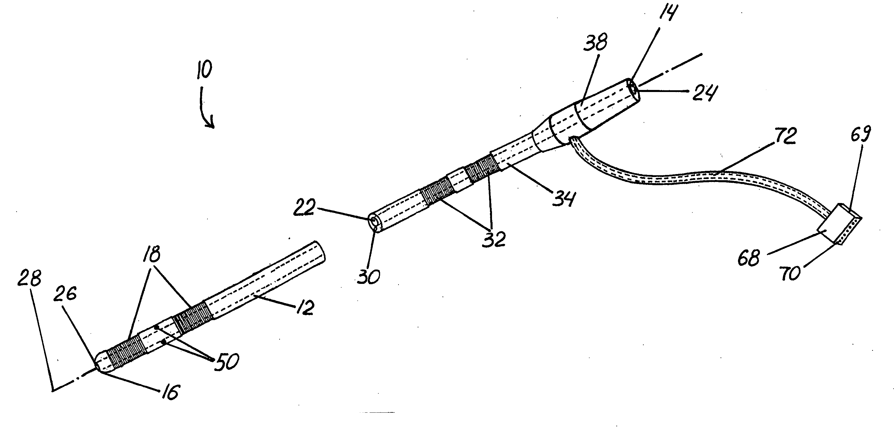
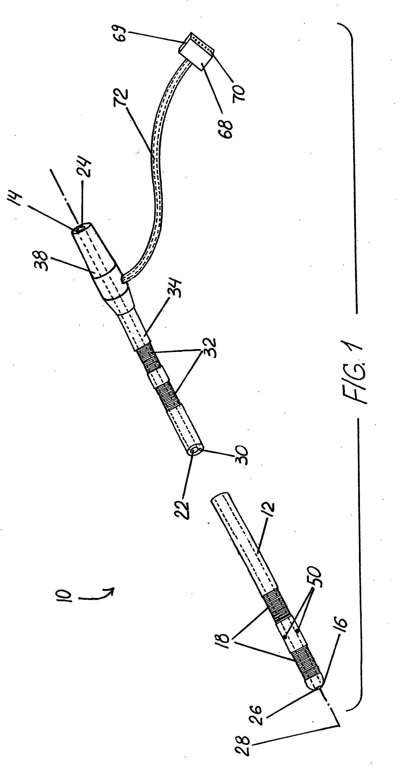
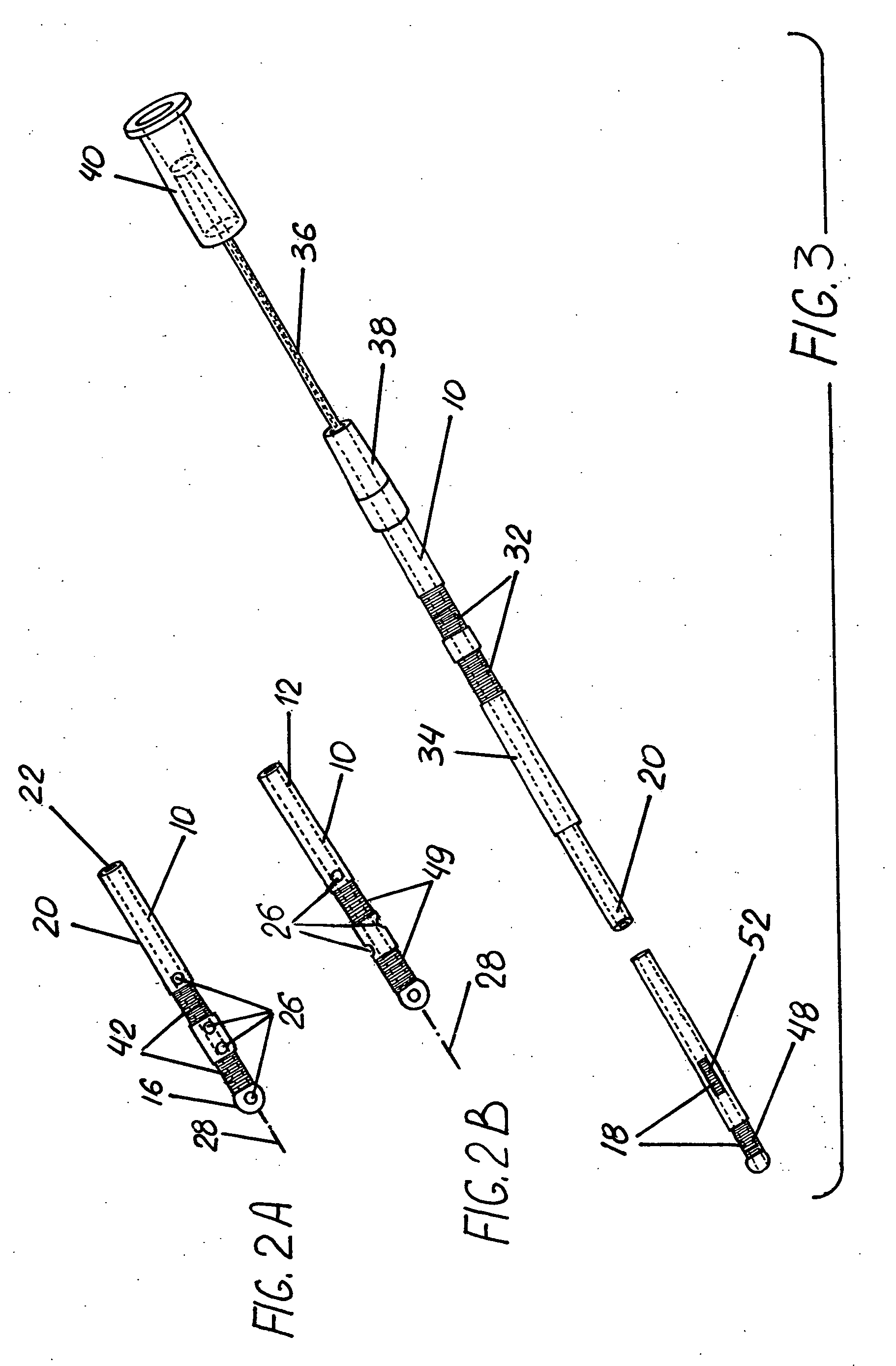
[0041] The figures illustrate preferred embodiments of an improved depth probe for intracranial treatment of a patient in accordance with this invention. FIG. 1 is a perspective view of depth probe 10 having an elongated, tubular body 12 extending from proximal end 14 to distal end 16. Body 12 preferably has a diameter between about 0.6 and 3.0 millimeters, most preferably about 1.0 millimeter.

[0042] As seen in FIG. 1, body 12 includes elements 18 secured to distal portion 20 at distal end 16. Body 12 is also provided with lumen 22 extending from opening 24 at proximal end 14 and in communication with aperture 26. Lumen 22 is a tubular channel extending for some length within body 12, preferably having a diameter of 0.5 millimeters or less. Body 12 is open at distal end 16 to form aperture 26. Opening 24 and aperture 26 are coaxial with lumen 22 along central axis 28 of body 12.

[0043] Elements 18 are conductively connected by leads 30 (seen in FIG. 1 running alongside lumen 22) to...


PUM


Abstract

A depth probe for intracranial treatment is provided having a body that includes a distal portion with at least one aperture and at least one element, a lumen defined by the body that communicates between an opening and the aperture, and a proximal portion with at least one proximal-contact. The proximal-contact is conductively connected with the element. The lumen is preferably sized to receive an inner catheter adapted to transfer a fluid such as a drug with a tissue region within the patient's brain. The depth probe can include a connector adapted to receive a plurality of proximal-contacts. A depth probe is disclosed that has a distal portion with an aperture and element, a lumen communicating between an opening and the aperture, and an inflatable balloon secured upon its distal portion. The balloon is adapted to seal upon inflation the tract created by the probe when inserted into the brain.

Description

RELATED APPLICATION [0001] This application claims the benefit of U.S. patent application Ser. No. 10 / 423,587, filed on Apr. 25, 2003.FIELD OF INVENTION [0002] The present invention relates to instrumentation utilized for intracranial treatment and, in particular, to depth probes utilized for intracranial treatment. BACKGROUND OF THE INVENTION [0003] Movement disorders such as epilepsy and Parkinson's disease have been estimated to affect some 1-2% of the developed world's population and up to 10% of people in underdeveloped countries. Currently, approximately 75% of those who suffer from movement disorders are responsive in some degree to drugs. [0004] Electrical stimulation has also been utilized to treat some movement disorders. In the treatment of epilepsy, studies have been performed in which awake patients undergoing temporal lobe surgery underwent cortical stimulation. Such stimulation of the visual and hearing areas of the brain reproducibly caused the patients to experience...

Claims


Application Information

Patent Timeline
Patent Type & Authority Applications(United States)
IPC IPC(8): A61M25/00A61B5/03A61B5/04A61F2/958A61MA61M25/06A61M31/00A61N1/05
CPCA61B5/031A61B5/04001A61B5/1473A61B5/6852A61M2210/0693A61M25/0662A61M25/10A61M2025/105A61M2025/1052A61M25/007A61B5/4094A61B5/24
Inventor PUTZ, DAVID A.
Owner TECH MEDICAL INSTR
Why Eureka
Try Eureka