Method and apparatus for automatic examination of cardiovascular functionality indexes by echographic imaging

an automatic examination and functional index technology, applied in the field of automatic determination of cardiovascular functional indexes, can solve the problems of normal unsteadiness, significant practical limits of typical ultrasonic examination by manual evaluation of echographic images, and phase shift between two events

Inactive Publication Date: 2006-03-23
CONSIGLIO NAT DELLE RICERCHE
View PDF20 Cites 21 Cited by
  • Summary
  • Abstract
  • Description
  • Claims
  • Application Information

AI Technical Summary

Benefits of technology

[0026] This way, the present invention has the advantage to provide a synchronized succession of functional c

Problems solved by technology

However, especially in cardiovascular analysis, a typical ultrasonic examination by means of manual evaluation of echographic images has relevant practical limits.
Furthermore, the phase shift of the two events depends on many factors and normally is unsteady with time.
Indeed, for example, the phase shift of the peak value of the R wave with respect to the maximum diameter of an artery is unsteady during a transient phase caused

Method used


Image

  • Method and apparatus for automatic examination of cardiovascular functionality indexes by echographic imaging
  • Method and apparatus for automatic examination of cardiovascular functionality indexes by echographic imaging
  • Method and apparatus for automatic examination of cardiovascular functionality indexes by echographic imaging

Examples


Embodiment Construction

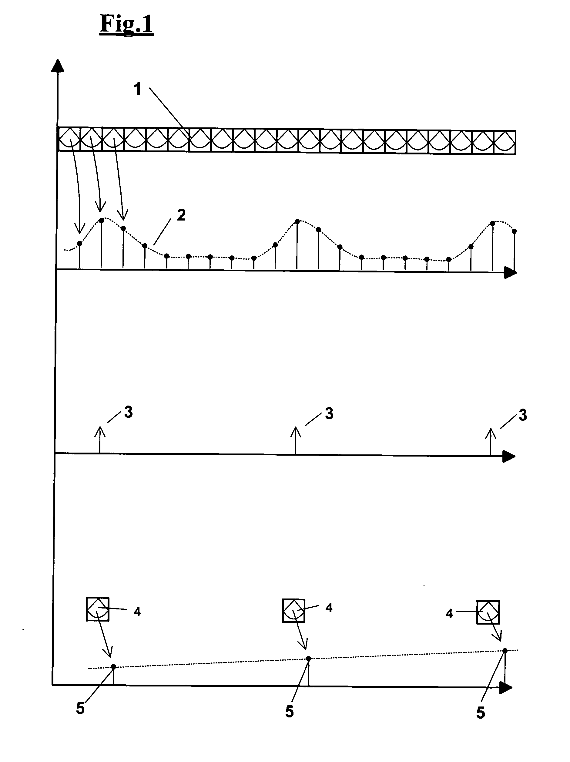
[0066] The present invention comprises an apparatus that computes a succession of starting images of an examined structure, obtaining a succession of images that are synchronized with respect to a cyclical movement of the structure and measures cardiovascular functionality indexes on this new synchronized succession of images.

[0067]FIG. 1 shows a possible embodiment of the various steps that carry out the present invention. The starting succession 1 is a set of B-mode images or color-Doppler images obtained with a high frame-rate, for example 25 or 30 frame / second. On each image of the succession at least one measure of a quantity is carried out obtaining a time-discrete signal 2, so-called “reference signal”, shown in the figure on a diagram that reports the time in abscissas and the measured quantity in ordinates. Said measure is chosen in order to determine a local kinetics induced by the heart beat. For example the reference signal 2 can be alternatively an area of the structur...


PUM


Abstract

A method for automatic examination of echographic images of the cardiovascular system, in particular, for computing functionality indexes of a segment of the cardiovascular system. Said method computes a succession of starting images (1), extracts a succession of images (4) synchronized with a cyclical movement (2) of the examined segment and measures cardiovascular functionality indexes (5) in the succession of synchronized images (4). The method, furthermore, describes the process for defining the edges of the examined structure starting from a starting ultrasonographic image in which the geometric features of the cardiovascular structure are known.

Description

FIELD OF THE INVENTION [0001] The present invention relates to an automatic method for determining cardiovascular functionality indexes by analyzing sequences of echographic images. In particular, but not exclusively, such images can be obtained from a patient after a treatment of mechanical or pharmacological type, as well as after a treatment that can enhance the capacity of reaction of an object under analysis and then its functionality. BACKGROUND OF THE INVENTION [0002] Ultrasonic examination of an organ's function is clinical practice that has quickly spread for its advantages of low invasivity, simplicity, and feasibility. However, especially in cardiovascular analysis, a typical ultrasonic examination by means of manual evaluation of echographic images has relevant practical limits. A typical ultrasonic examination, in fact, for being acceptable in the clinical routine, provides normally a few insulated measurements and introduces a considerable variability. For a more robus...

Claims


Application Information

Patent Timeline
IPC IPC(8): A61B8/02
CPCA61B8/08A61B8/0858A61B8/0891A61B8/14G06T2207/30101G06T7/0012G06T7/2033G06T2207/10132G06T2207/30048A61B8/488G06T7/246
Inventor DEMI, MARCELLOFAITA, FRANCESCOGEMIGNANI, VINCENZOGIANNONI, MASSIMOBENASSI, ANTONIO
Owner CONSIGLIO NAT DELLE RICERCHE
Why Eureka
Try Eureka