
Method of detecting or diagnosing of a neurodegenerative disease or condition

a neurodegenerative disease and diagnosis technology, applied in the field of neurodegenerative diseases or diagnosis methods, can solve the problems of difficult to know what biological factors to look for, altering the binding specificity of antibodies, and mystery of neurodegenerative diseases

Inactive Publication Date: 2006-06-29
REDOX REACTIVE REAGENTS LLC
View PDF11 Cites 30 Cited by
  • Summary
  • Abstract
  • Description
  • Claims
  • Application Information

AI Technical Summary

Benefits of technology

[0023] The objectives are further achieved by a method of detecting or diagnosing a neurodegenerative disease or condition in a subject by assaying first sample of cerebral spinal fluid from the subject to determine a level of at least one autoantibody of a selected specificity, treating a second sample from the subject with an oxidizing agent and assaying the oxidized second sample to determine a level the at least autoantibody having the selected specifici

Problems solved by technology

A continuing problem in the diagnosis of neurodegenerative diseases or conditions such as Alzheimer's has been to devise a reliable biomarker that provides a definitive indication of a physical pathology.
Further, the cause of neurodegenerative diseases has been a mystery, making it
difficult to know what biological factors to look for in terms of early warning signs that a neurodegenerative disease may be present.
A change in nitration could produce conformational changes in an antibody binding site that result in alteration of the binding specificity of the antibody.
Here again, it can be presumed that autoantibodies that may be present in cerebral spinal fluid of a normal individual in their masked form do not cause harm to the individual, and may play a yet unknown beneficial role; however, it is apparent that the autoantibodies could cause damage if they were to become unmasked in the cerebral spinal fluid.
For example, if transition metal sites are exposed by protein misfolding, such exposed sites could promote oxidation-reduction reactions that lead to unmasking of autoantibodies.
Unmasked antibodies such as antiphospholipid autoantibodies can interact with phospholipids and phospholipid-binding proteins in brain cells and may therefore cause many of the lesions and shrinkage of the brain that are seen in MRI studies of the Alzheimer patient brains and can cause the physical damage seen in other types of neurodegenerative diseases.
Moreover, as further discussed herein, it has been discovered that cerebral spinal fluid of confirmed Alzheimer patients that is treated with an oxidizing agent such as hemin does not show a dramatic increase in the amount of detectable autoantibodies, as compared with untreated cerebral spinal fluids, which indicate that an unmasking process has occurred in a diseased subject, such that the level of masked autoantibodies becomes depleted.

Method used


Image

  • Method of detecting or diagnosing of a neurodegenerative disease or condition
  • Method of detecting or diagnosing of a neurodegenerative disease or condition
  • Method of detecting or diagnosing of a neurodegenerative disease or condition

Examples


example 1

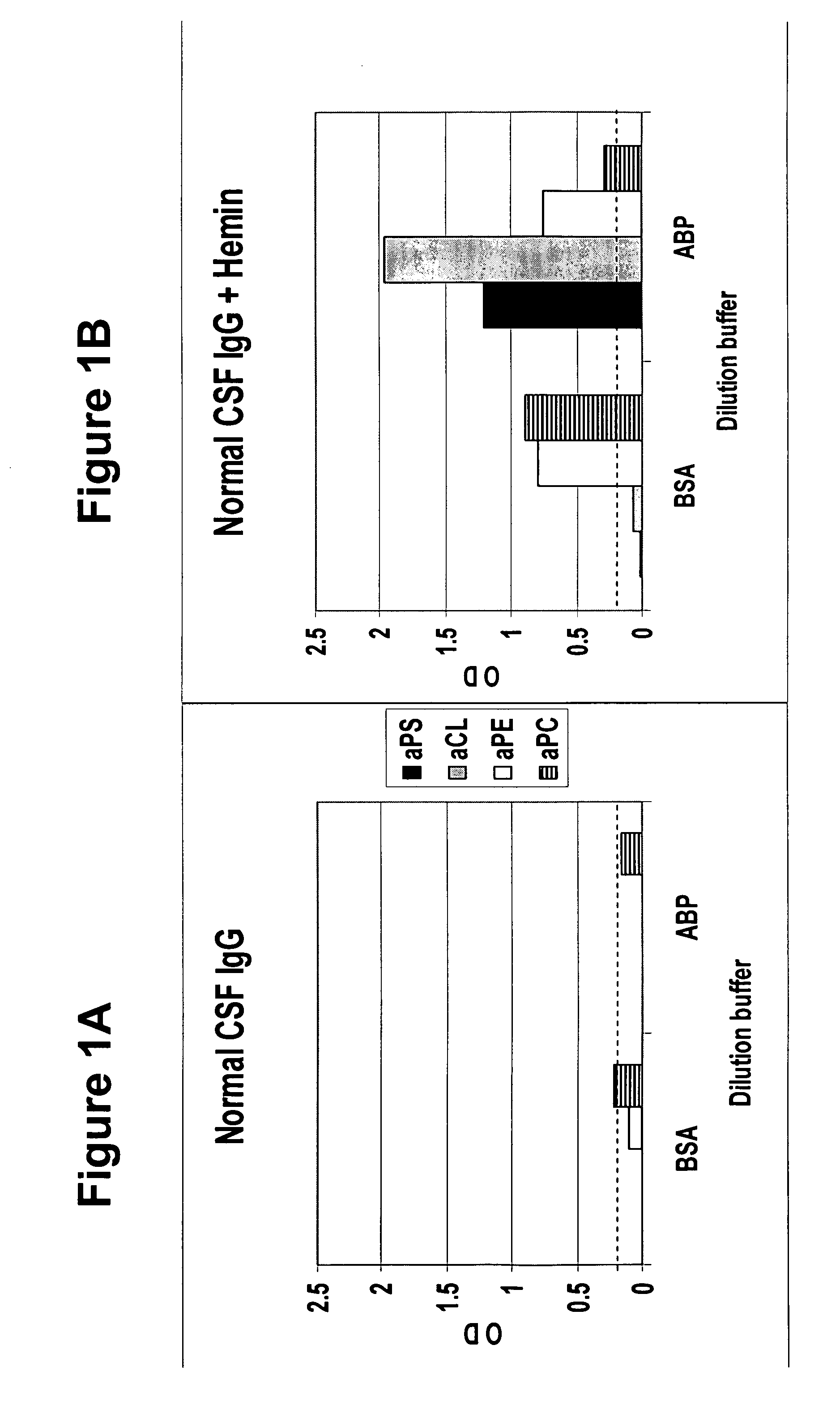
[0045] To determine whether human cerebral spinal fluid from a normal subject contains masked autoantibodies, spinal fluid was taken by spinal tap from a normal individual and samples in BSA and ABP dilution buffers were assayed by ELISA for aPS, aCL, aPE and aPC levels before and after oxidation treatment with hemin. As shown in FIGS. 1A and 1B, samples showed none or minimal levels of antiphospholipid antibodies before oxidation treatment (FIG. 1A), and substantial increases in the levels after oxidation treatment (FIG. 1B), with aPE and aPC showing the highest levels in a BSA buffer and aPS and aCL showing the highest levels in the ABP buffer.

example 2

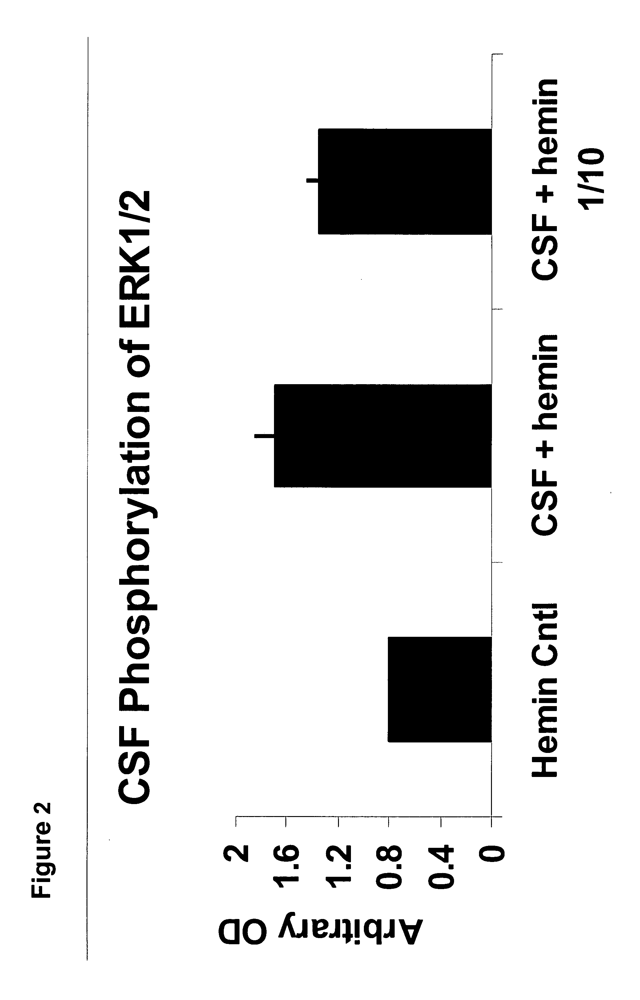
[0046] The presence of autoantibodies sensitive to oxidation-reduction reactions in normal cerebral spinal fluid samples and their absence in Alzheimer's disease patients' spinal fluid is remarkable, albeit it does not imply that these autoantibodies are functional and / or pathogenic. To test for functional activity, the cerebral spinal fluid samples containing redox-reactive autoantibodies were tested in a mouse model synaptosome assay. Cerebral spinal fluid samples were incubated for 10 minutes on the synaptosomes followed by lysing of the cells and probing for phosphorylation activity by Western blots. FIG. 2 is a histogram showing redox-reactive autoantibody ERK1 / 2 phosphorylation activity of a hemin buffer control, a hemin treated cerebral spinal fluid and a 1 / 10 dilution of the same hemin-treated cerebral spinal fluid.

[0047] The redox-reactive autoantibodies significantly increased the phosphorylation of the members of the MAPK cascade of signal transduction molecules, thus gi...

example 3

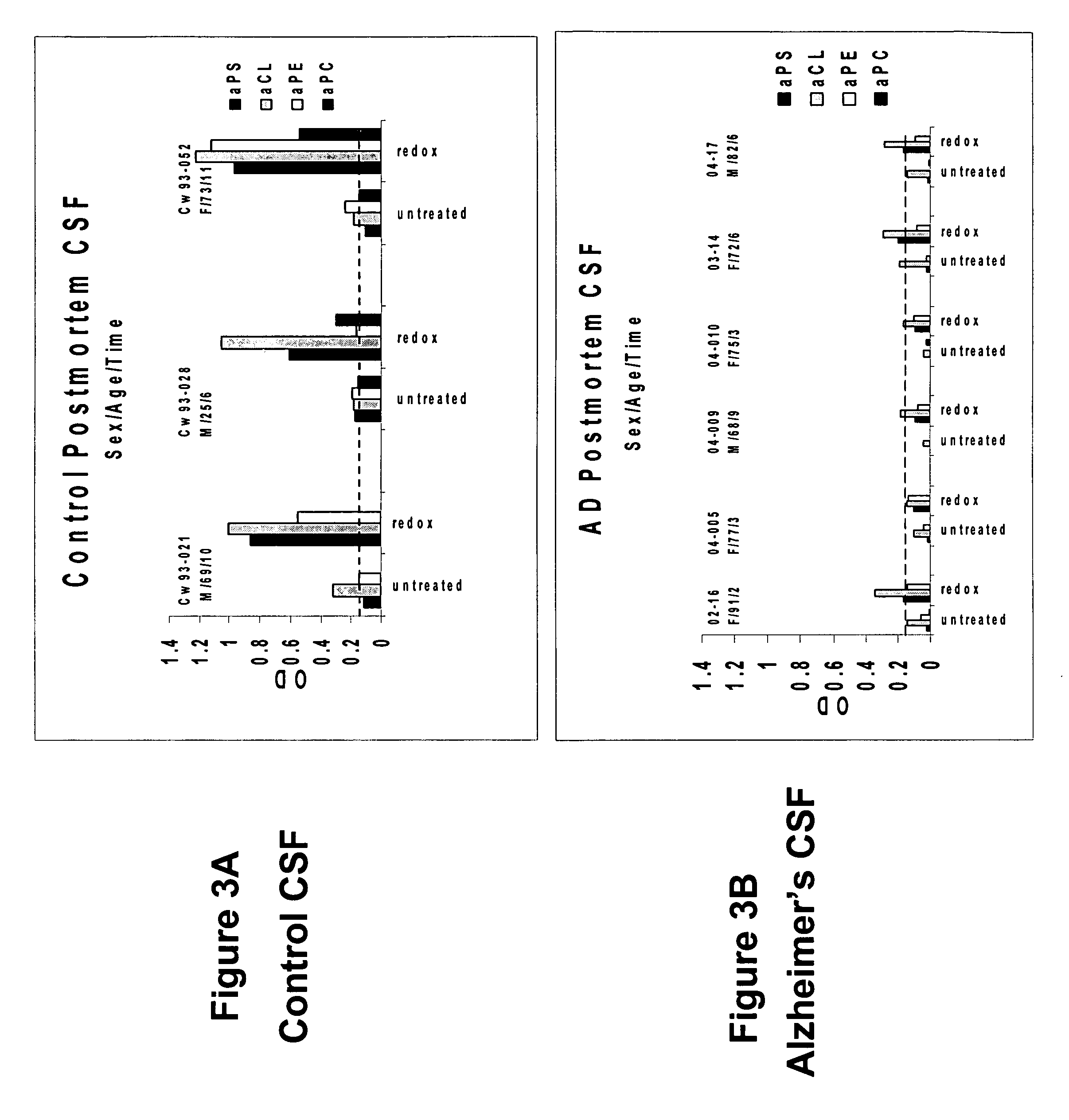
[0048] To determine whether human cerebral spinal fluid from a subject having Alzheimer's disease contains an elevated level of unmasked or active phospholipid autoantibodies, spinal fluid was taken post mortem from six patients diagnosed with Alzheimer's disease based on biopsy confirmation. Samples of cerebral spinal fluid in BSA and ABP dilution buffers were assayed for aPL levels. As shown in FIGS. 3A and 3B, samples from Alzheimer's patients (FIG. 3B) showed a decreased level or absence of aPL in comparison with the post mortem controls level (FIG. 3A) subsequent to oxidation with hemin. This indicates that oxidation-related autoantibodies have been unmasked and bound to their neuronal targets, a process shown to cause modifications in the normal synaptic mechanisms brain cells that can lead to dementia.


PUM

PropertyMeasurementUnit
Timeaaaaaaaaaa

Abstract

A neurodegenerative disease or condition is diagnosed in a subject by obtaining a sample of cerebral spinal fluid from the subject and assaying the sample by an assay method that detects the presence of at least one antiphospholipid autoantibody in the sample, wherein an elevated level of at least one antiphospholipid autoantibody in the sample of cerebral spinal fluid correlates with a neurodegenerative disease or condition in the subject. The neurodegenerative disease or condition may also be diagnosed by assaying a sample of cerebral spinal fluid to detect nitrosylated antibodies, wherein an elevated level of nitrosylated antibodies correlates with a neurodegenerative disease or condition in said subject. A neurodegenerative disease or condition is also detected or diagnosed by assaying a first sample of cerebral spinal fluid from the subject to determine a level of at least one autoantibody having a selected specificity, treating a second sample of cerebral spinal fluid with an oxidizing agent and assaying the oxidized second sample to determine a level the at least autoantibody having the selected specificity, and comparing the level of the at least one autoantibody in the first sample with the level of the at least one autoantibody in the oxidized second sample, wherein a lack of increase in the level of the at least one autoantibody in the oxidized second sample as compared to the level of the at least one autoantibody in the first sample correlates with a neurodegenerative disease or condition in said subject.

Description

[0001] The present application is a continuation-in-part of U.S. patent application Ser. No. 11 / 108,826, filed on Apr. 19, 2005, which application is a continuation-in-part of Ser. No. 10 / 863,365, filed Jun. 9, 2004, which application claims the benefit of U.S. Provisional Application Ser. No. 60 / 476,607, filed Jun. 9, 2003, the entire contents of which applications are incorporated by reference herein.FIELD OF INVENTION [0002] The present invention relates to methods of detecting or diagnosing a neurodegenerative disease or condition. In particular, the aspects of the present invention relate to methods of assaying cerebral spinal fluid of a subject to determine the presence or absence of autoantibodies. BACKGROUND OF THE INVENTION [0003] A continuing problem in the diagnosis of neurodegenerative diseases or conditions such as Alzheimer's has been to devise a reliable biomarker that provides a definitive indication of a physical pathology. Typically, a neurodegenerative disease is ...

Claims


Application Information

Patent Timeline
IPC IPC(8): G01N33/567G01N33/53
CPCG01N33/564G01N33/6896G01N33/92G01N2800/28G01N33/6854G01N2800/2821G01N2800/2835G01N2800/285G01N2800/2814
Inventor MCINTYRE, JOHN A.
Owner REDOX REACTIVE REAGENTS LLC
Learn More