Ultrasound diagnostic system and method of forming arbitrary M-mode images

a diagnostic system and ultrasonic technology, applied in diagnostics, medical science, applications, etc., can solve the problems of prolonging the diagnostic time and obtaining the m-mode imag

Inactive Publication Date: 2006-08-03
MEDISON CO LTD
View PDF5 Cites 46 Cited by
  • Summary
  • Abstract
  • Description
  • Claims
  • Application Information

AI Technical Summary

Benefits of technology

[0007] It is, therefore, an objective of the present invention to provide an ultrasound diagnostic system and a method of forming arbitrary M-mode images, which can

Problems solved by technology

Thus, it has drawbacks in that the M-mode image is obtained only in a proceeding direction of the ultrasound signals.
Accordingly, a user must artfully c

Method used


Image

  • Ultrasound diagnostic system and method of forming arbitrary M-mode images
  • Ultrasound diagnostic system and method of forming arbitrary M-mode images
  • Ultrasound diagnostic system and method of forming arbitrary M-mode images

Examples


Embodiment Construction

[0021] Hereinafter, a preferred embodiment of the present invention will be described below with reference to accompanying drawings.

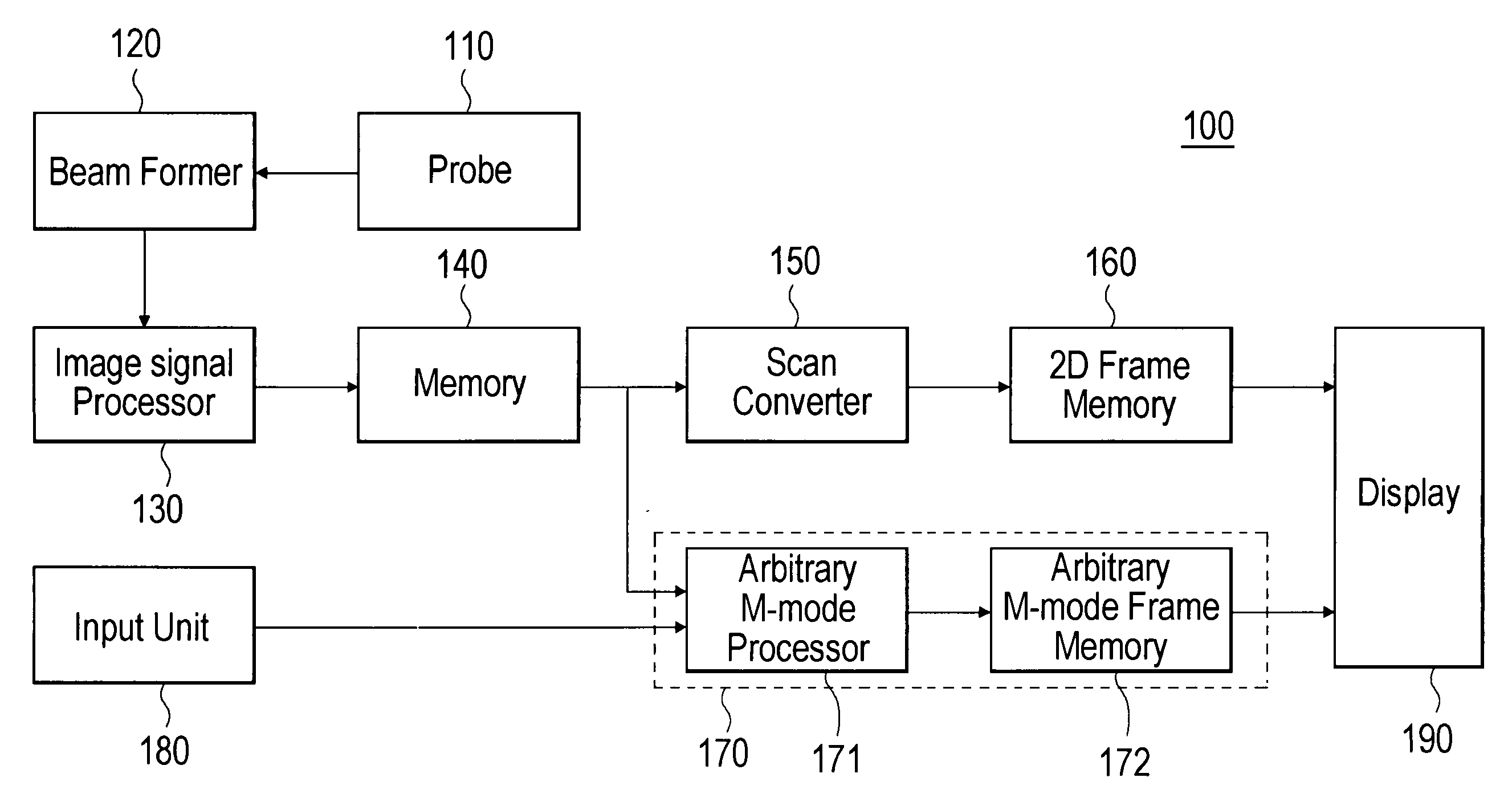
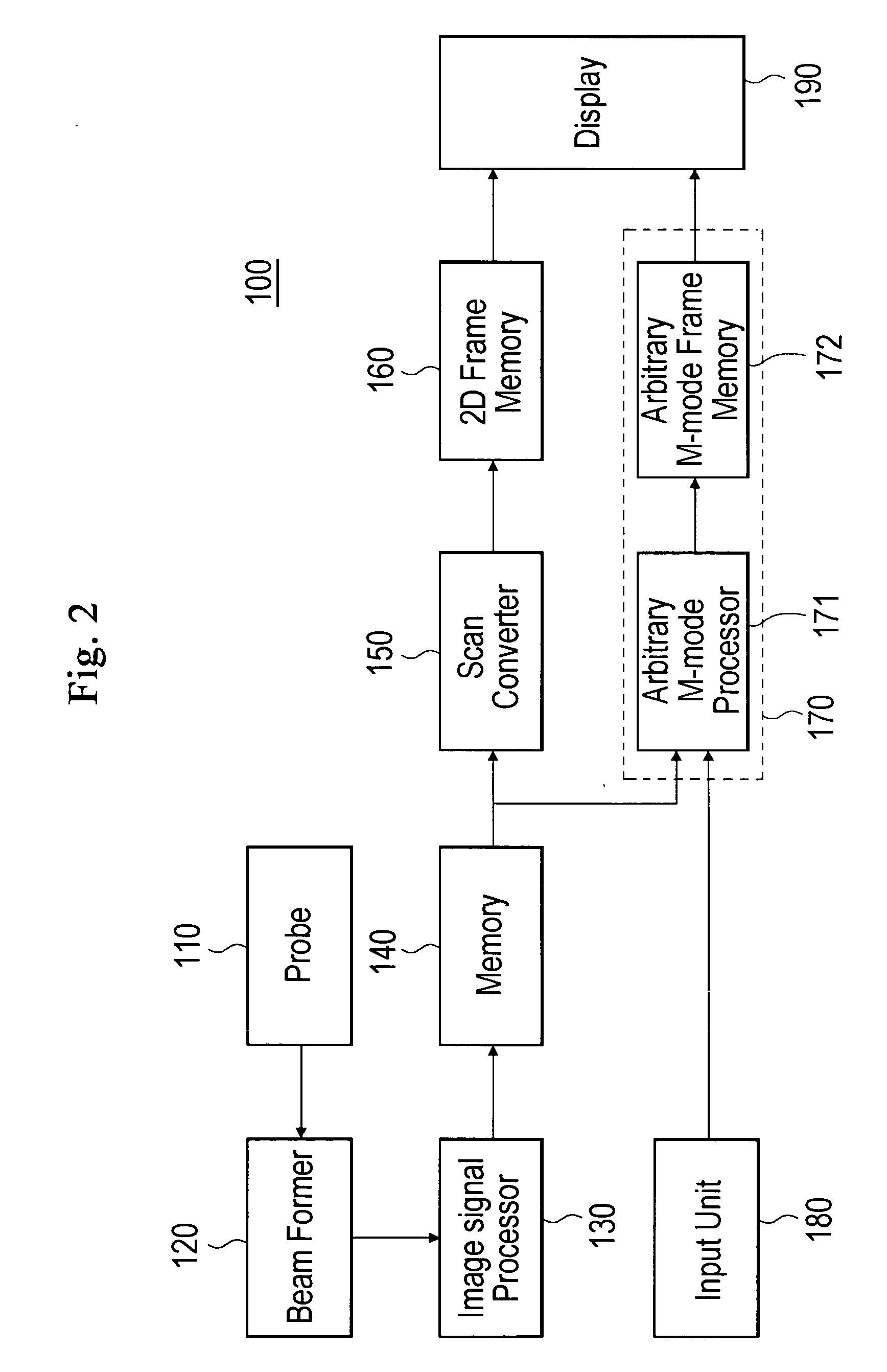
[0022]FIG. 2 is a block diagram schematically showing an ultrasound diagnostic system, which is constructed in accordance with one embodiment of the present invention.

[0023] Referring to FIG. 2, the ultrasound diagnostic system 100 includes a probe 110, a beam former 120, an image signal processor 130, a memory 140, a scan converter 150, a 2D frame memory 160, an arbitrary M-mode unit 170, a displaying device 190 and an input unit 180, wherein the arbitrary M-mode unit 170 has an arbitrary M-mode processor 171 and an M-mode frame memory 172.

[0024] A probe 110 includes a plurality of 1D or 2D transducers (not shown). The probe 110 transmits ultrasound signals to organs and receives the ultrasound signals reflected from the organs.

[0025] A beam former 120 adjusts a driving time of each transducer of a probe 110 when the probe 110 transmits the ultraso...


PUM


Abstract

The present invention relates to an ultrasound diagnostic system and a method of forming arbitrary M-mode ultrasound images. The ultrasound diagnostic system includes: a probe for transmitting ultrasound signals toward a desired part of a target object and receiving the ultrasound signals reflected from the desired part; a scan converter for converting an ultrasound image data into a B-mode image data, the ultrasound image data being obtained based on the ultrasound signal received from the probe; an input unit for receiving an arbitrary M-mode scan line set by a user; an arbitrary M-mode processor for generating an arbitrary M-mode image data corresponding to the arbitrary M-mode scan line; and a display unit for displaying at least one of the B-mode image data, the M-mode image data and the arbitrary M-mode scan line. In accordance with the present invention, the biological information of the target object can be observed and diagnosed from an arbitrary direction and path, regardless of the direction of the ultrasound signals. Additionally, the arbitrary M-mode image, which is more similar to the real image, can be provided for the user by generating the arbitrary M-mode images corresponding to the arbitrary M-mode scan line by using ultrasound image data before scan-conversion.

Description

FIELD OF THE INVENTION [0001] The present invention generally relates to an ultrasound diagnostic system and a method of displaying arbitrary M-mode images, and more particularly to an ultrasound diagnostic system and a method of forming arbitrary M-mode images for observing a transition trend of an ultrasound image information in a specific line of the ultrasound diagnostic image. BACKGROUND OF THE INVENTION [0002] An ultrasound diagnostic system is widely used for non-invasively obtaining tomographic images of soft tissue and blood flow within a human body. In such a system, the tomographic images are formed through the process of (1) transmitting an ultrasound signal from a surface of the human body toward a desired part within the human body, (2) receiving the ultrasound signal reflected from within the human body, and (3) performing signal processing upon the received ultrasound signal (echo signal). Compared to other medical imaging systems (e.g., X-ray diagnostic system, X-ra...

Claims


Application Information

Patent Timeline
IPC IPC(8): A61B8/14
CPCA61B8/14A61B8/463A61B8/486F16L3/137
Inventor KIM, JEONG HWAN
Owner MEDISON CO LTD
Why Eureka
Try Eureka