Method for expanding the display of a volume image of an object region

a technology of object region and volume image, applied in image enhancement, tomography, instruments, etc., can solve the problem of only offering a limited field of view for visualization

Inactive Publication Date: 2006-11-30
SIEMENS HEALTHCARE GMBH
View PDF8 Cites 25 Cited by
  • Summary
  • Abstract
  • Description
  • Claims
  • Application Information

AI Technical Summary

Benefits of technology

[0006] An object of the present invention is to provide a method for expanding the viewport of a volume image of ...

Problems solved by technology

These modalities, however, offer only...

Method used


Image

  • Method for expanding the display of a volume image of an object region
  • Method for expanding the display of a volume image of an object region
  • Method for expanding the display of a volume image of an object region

Examples


Embodiment Construction

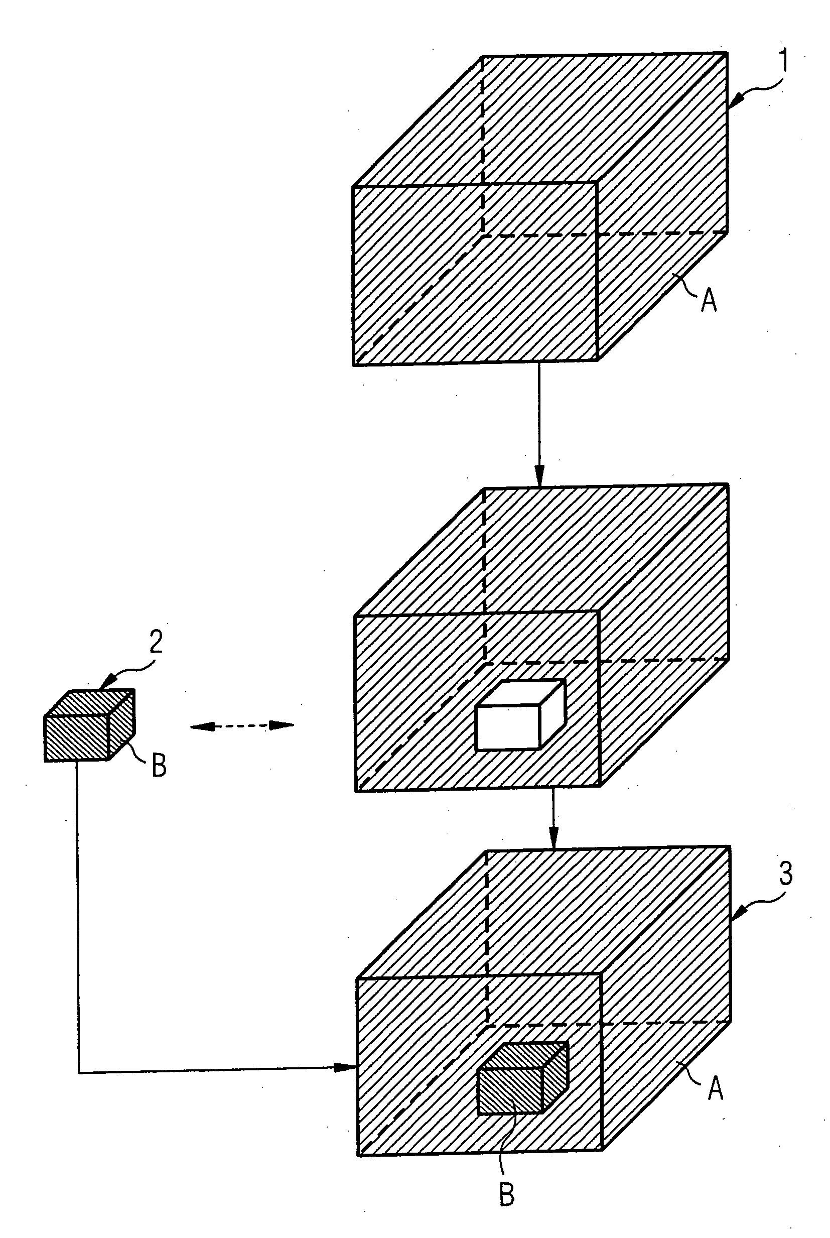
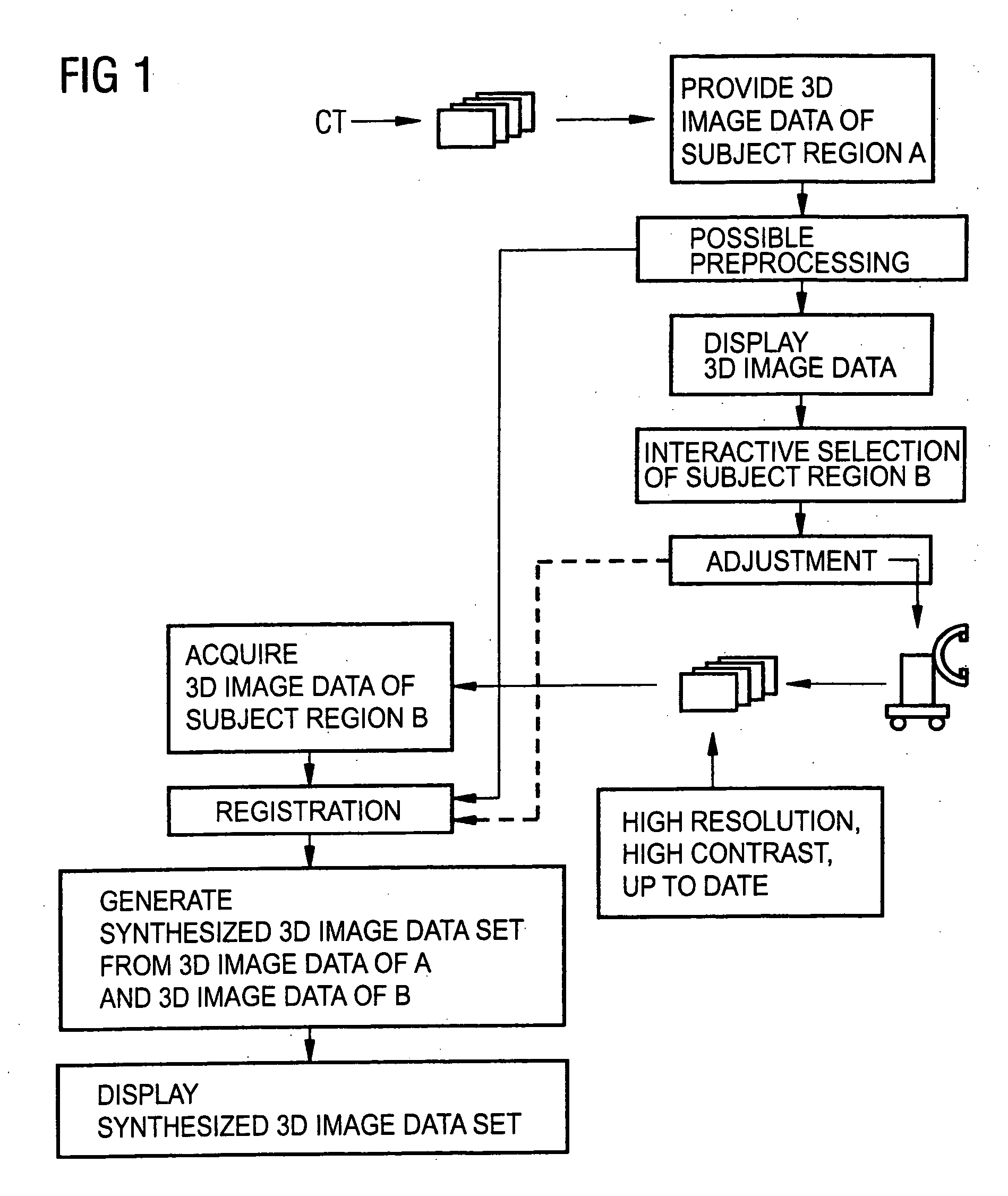
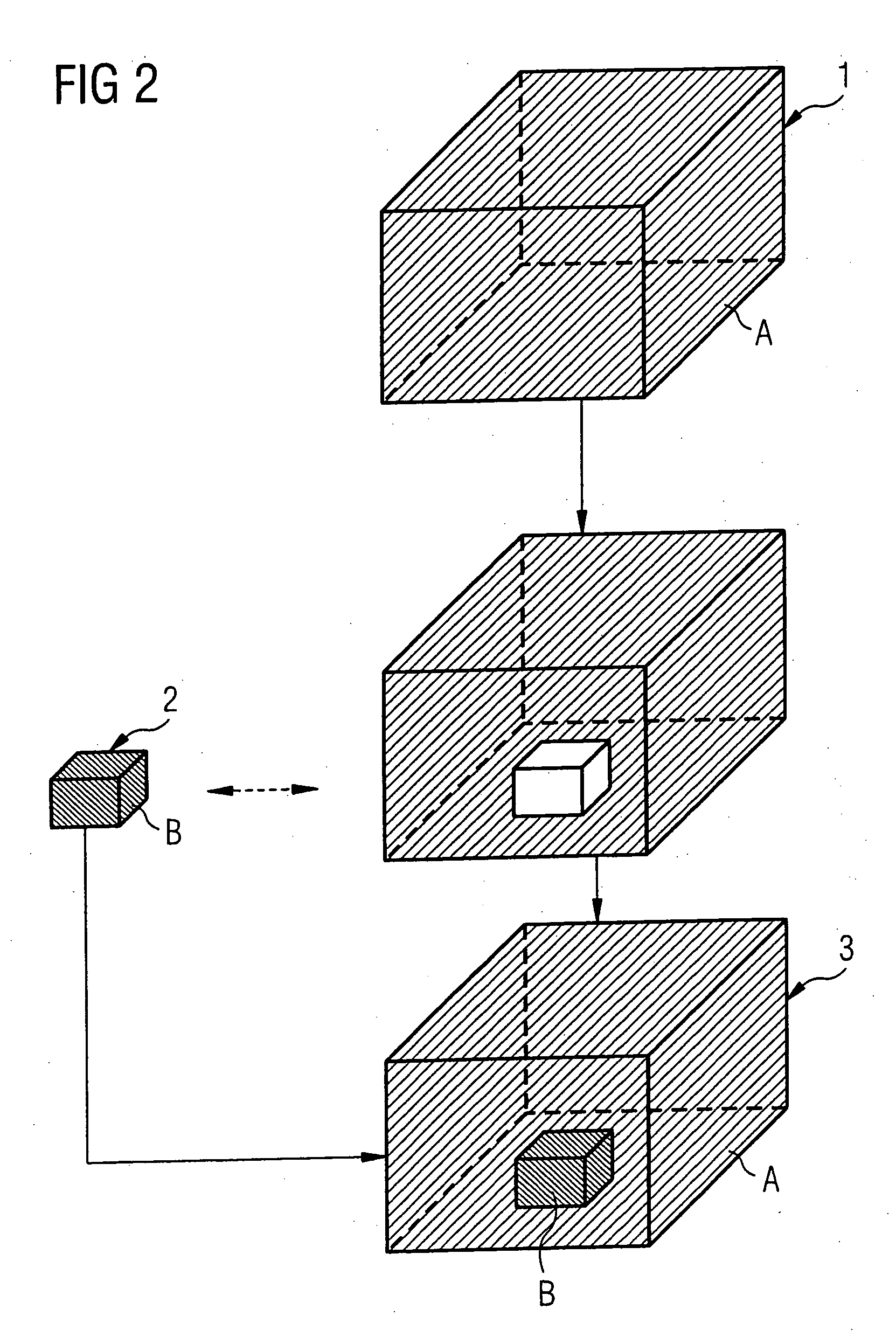
[0026] The present method will be described again in detail using the example of an operation, e.g., after a pelvic fracture, during which a 3D image of the treatment region is acquired and displayed with a mobile 3D C-arm device.

[0027] Currently many 3D image data sets are often used both for planning as well as for implementing a method procedure, the 3D image data sets being generated, e.g., from CT volume images prior to the operation. By means of the visualization of these 3D image data sets a complete, large-scale overview results of the total relevant body environment. In the case of surgery, however, the physician must for various reasons proceed based on a current imaging, for example, is obtained by means of imaging endoscopy, ultrasound or mobile 3D x-ray imaging. In the case of repeated imaging during the operation, the physician can also immediately track changes in this way. The current imaging is already necessary for safety, since changes in the anatomy of the patie...


PUM


Abstract

In a method for expanding the display of a volume image of an object region, in particular in medical applications, in which at least one first image data set of a larger object region is made available, at least one second image data set of a smaller object region is acquired, the smaller object region lying within the larger object region, and the first 3D image data set is brought into registration with the second 3D image data set. A synthesized 3D image data set is generated from the first and second 3D image data sets and visualized in a display. The first 3D image data in the first 3D image data set represent the smaller object region, if necessary after a suitable adaptation of the first and/or of the second 3D image data set, are replaced by second 3D image data of the second 3D image data set with identical scaling and alignment of the image data sets. An overview of the larger object region thus is possible, and the smaller object region of interest the image can be represented with up-to-date data as well as a higher resolution and/or a higher contrast.

Description

BACKGROUND OF THE INVENTION [0001] 1. Field of the Invention [0002] The present invention relates to a method for expansion of the display of a volume image of an object region, of the type wherein a first image data set of a larger object region is made available, a second image data set of a smaller object region is acquired, the smaller object region lying within the larger object region, and the image data sets are displayed in combination. [0003] 2. Description of the Prior Art [0004] In surgery, compact imaging systems such as mobile x-ray devices, ultrasound devices or endoscope / laparoscope are often used. These modalities, however, offer only a limited field of view for visualization. For surgeons, it is desirable to be able to see the local environment of the treatment region in a larger field of view. [0005] Various options are known in whereby at a time interval prior to surgery, 3-D images of the body, e.g., by means of computed tomography or magnetic resonance tomograph...

Claims


Application Information

Patent Timeline
IPC IPC(8): G06T15/00A61B6/03A61B8/00G06T19/20
CPCA61B6/12A61B6/4405A61B6/4441A61B6/466A61B6/5235G06T2207/30004G06T7/0028G06T7/0038G06T19/00G06T2200/04A61B6/5247G06T7/33G06T7/38
Inventor BARFUSS, HELMUTBARTH, KARLWESSELS, GERD
Owner SIEMENS HEALTHCARE GMBH
Why Eureka
Try Eureka