5-Androstenediol As An Inhibitor of Gliomas
a technology of androstenediol and inhibitors, which is applied in the field of methods of treating gliomas, can solve the problems of difficult surgery of tumors, and high risk of cancer types
- Summary
- Abstract
- Description
- Claims
- Application Information
AI Technical Summary
Problems solved by technology
Method used
Image
Examples
example 1
Inhibition of Proliferation of Human and Rodent Glioma Cells
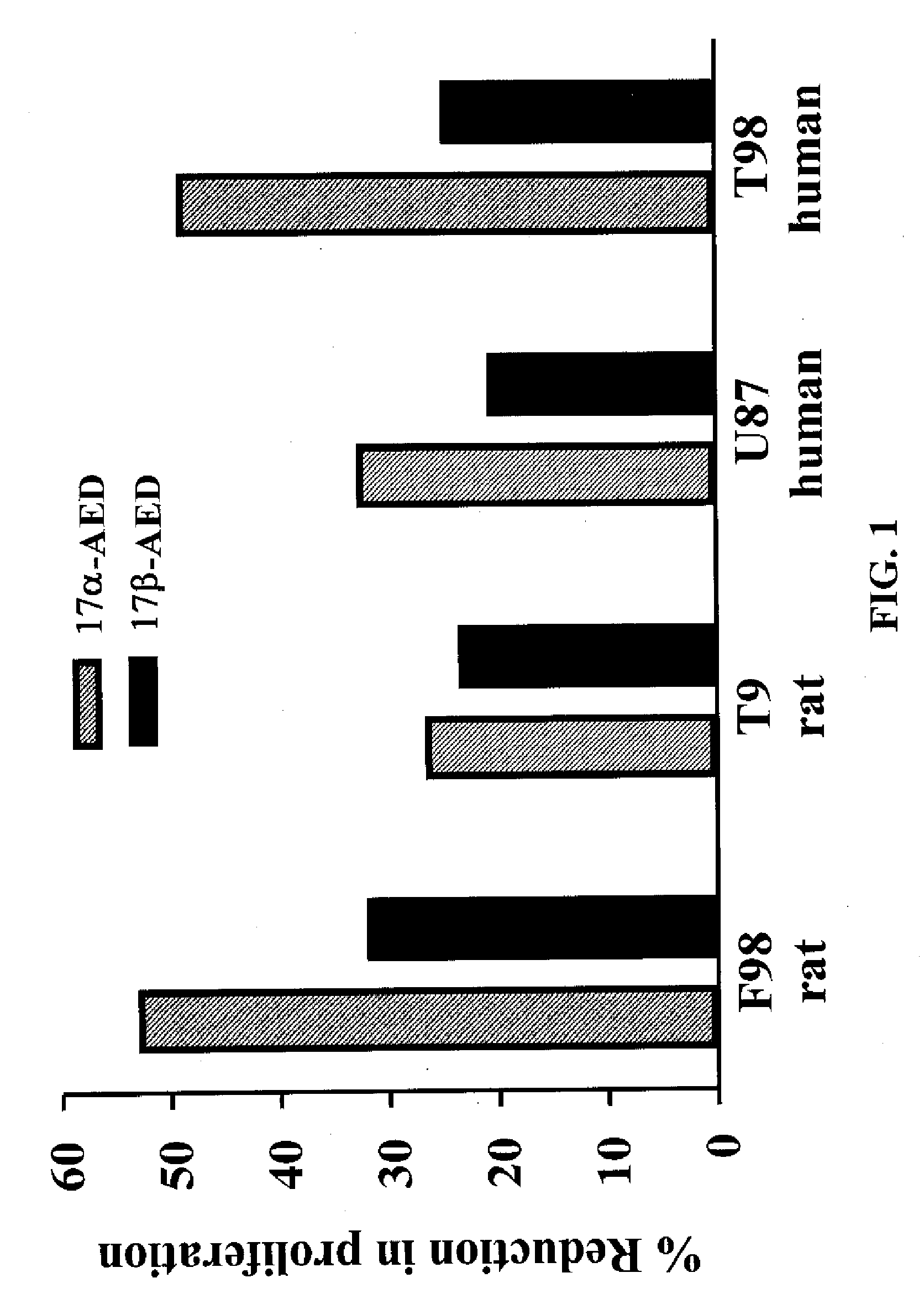
[0059] Rat F98 and T9 glioma cells and Human F98 and U87 glioma cells were cultured in 96 well plates at a density of approximately 1×10⁴ cells per well. Cells were treated with 500 μm of αAED or βAED or vehicle and cultured for 3 days. The cultures were pulsed with 3H-TdR for the last 15 hours. The incorporation of 3H-TdR was used to measure cellular proliferation. At the end of 3 days, the proliferation of the cells were measured as described above.
[0060] As seen in FIG. 1, AED had a significant negative effect on the proliferation of the glioma cell lines. More particularly, αAED inhibited cell growth of rat F98 glioma cells, rat T9 glioma cells, human U87 glioma cells, and human T98 glioma cells when compared to controls. Similarly, βAED inhibited cell growth of the rat F98 glioma cells, rat T9 cells, human U87 glioma cells, and human T98 glioma cells. This data demonstrates that both epimers of AED have a significant...
example 2
Suppression of Glioma Growth is Dose Dependent
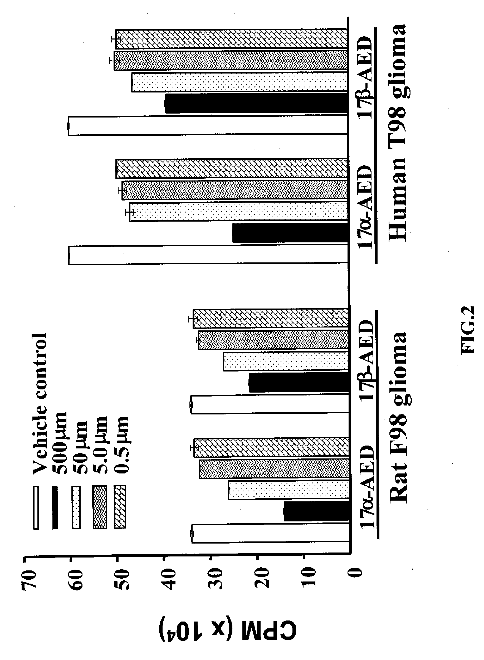
[0061] Rat F98 glioma cells and human F98 glioma cells were grown and cultured as in example 1 above. The cells were cultured in the presence of αAED or βAED in titrated concentrations of 500 μm, 50 μm, 5.0 μm, and 0.5 μm. After 3 days, glioma cell proliferation was measured as described above.
[0062] As seen if FIG. 2, αAED and βAED show significant antiproliferative effects on the rat F98 glioma cells and human T98 glioma cells. Furthermore, as seen in FIG. 2, the antiproliferative effect is dose dependent. At 500 μm concentrations, αAED and βAED inhibited the proliferation of rat F98 glioma cells and human T98 glioma cells. As can be seen in FIG. 2, as the concentrations of αAED and βAED decreased, so does the corresponding antiproliferative effect. These results show that growth inhibition by AED is dose dependent.
example 3
Morphological Data Showing Effects of Androsterene on Rat F98 Gliomas
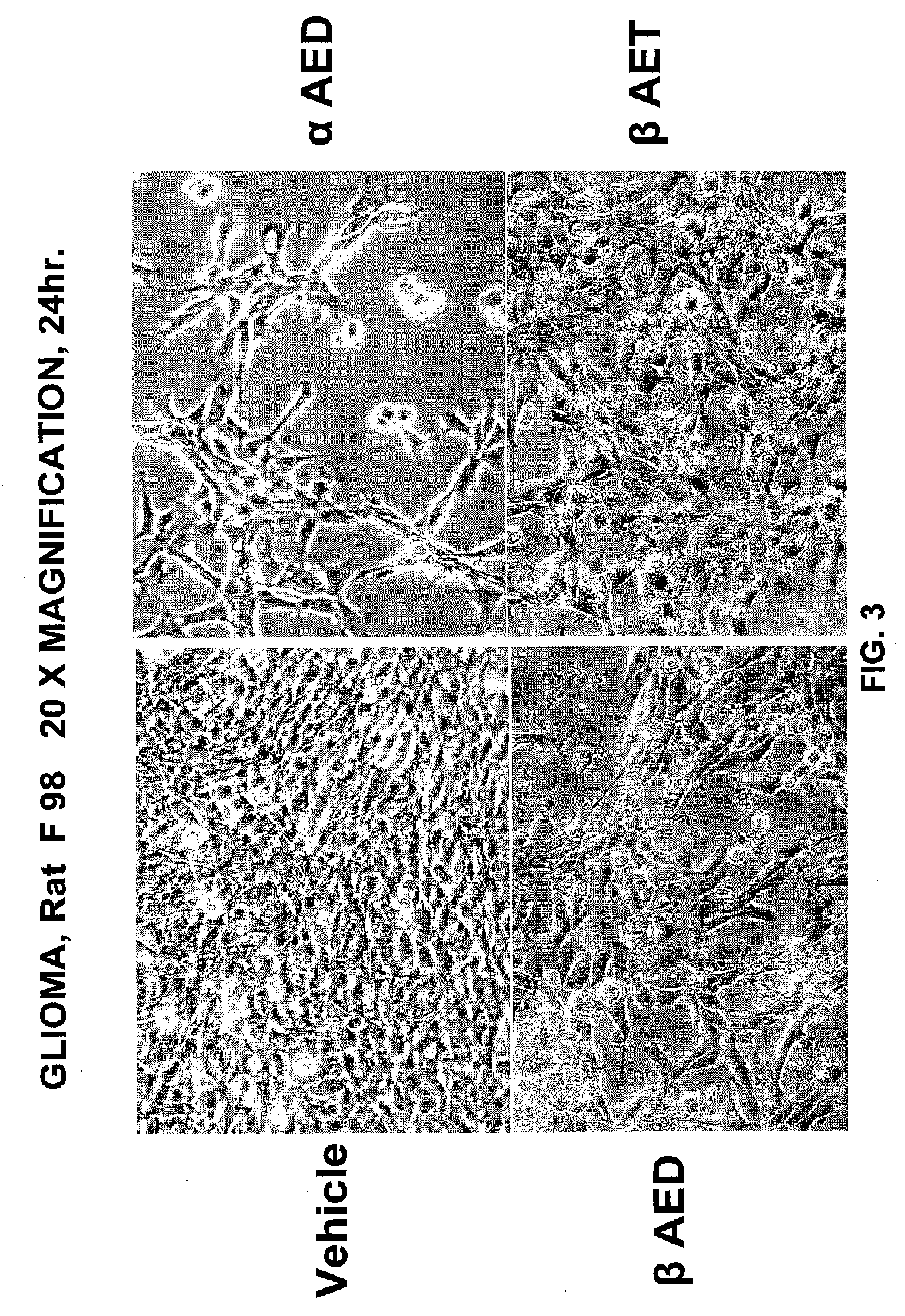
[0063] Cells were cultured as described in example 1 for 24 hours. Cells were cultured with 500 mm of αAED, βAED, βAET, and vehicle. After culture, the cells were viewed in situ under 20× magnification using a reverse image microscope.
[0064] As can be seen in FIG. 3, significant morphological changes occurred in the rat F98 glioma cells cultured with αAED, βAED, or βAET as compared to vehicle treated cells. The results indicate that αAED exhibits the most drastic morphological changes as compared to the cells treated with vehicle. As demonstrated in FIG. 3, the vehicle treated cells appear normal, whereas the αAED, βAED, and βAET treated cells display morphology of apoptasis including cell shrinkage, rounding, extensive vascuolization, partial detachment and further demonstrate the lobulated appearance of apoptotic cells. FIG. 3 shows that βAED and βAET produce significant morphological changes indicative of anti...
PUM
 Abstract
Description
Claims
Application Information
 - R&D
- Intellectual Property
- Life Sciences
- Materials
- Tech Scout
- Unparalleled Data Quality
- Higher Quality Content
- 60% Fewer Hallucinations
Browse by: Latest US Patents, China's latest patents, Technical Efficacy Thesaurus, Application Domain, Technology Topic, Popular Technical Reports.
© 2025 PatSnap. All rights reserved.Legal|Privacy policy|Modern Slavery Act Transparency Statement|Sitemap|About US| Contact US: help@patsnap.com


