Method and apparatus for minimally invasive surgery using endoscopes

a technology of endoscopes and endoscopes, which is applied in the field of image guidance for minimally invasive surgery, can solve the problems of difficult to judge the depth of penetration from only the image of the endoscope, the limited field of view of the thoracoscope, and the difficulty of guiding instruments such as the thoracoscope to a specific vertebra

Active Publication Date: 2006-12-14
SIEMENS MEDICAL SOLUTIONS USA INC
View PDF48 Cites 161 Cited by
  • Summary
  • Abstract
  • Description
  • Claims
  • Application Information

AI Technical Summary

Problems solved by technology

Performing such spinal surgery without opening a patient's chest and, instead, using minimally invasive surgical techniques, poses several challenges.
First, guiding instruments such as a thoracoscope to a specific vertebrae is difficult since a physician cannot simply count vertebrae along the spine from inside the thorax.
Second, the thoracoscope's field of view is somewhat limited and, therefore, it is difficult to judge depth of penetration from only the endoscope's image.
However, while this method provides added benefit over the use of an endoscope alone, it does not significantly add to the ability of navigating an endoscope in more difficult and delicate procedures.
However, this method only uses the virtual images and the location of a pointer with no endoscopic or other local images.
Therefore, such a method is not sufficient for the aforementioned delicate or difficult procedures.

Method used


Image

  • Method and apparatus for minimally invasive surgery using endoscopes
  • Method and apparatus for minimally invasive surgery using endoscopes
  • Method and apparatus for minimally invasive surgery using endoscopes

Examples


Embodiment Construction

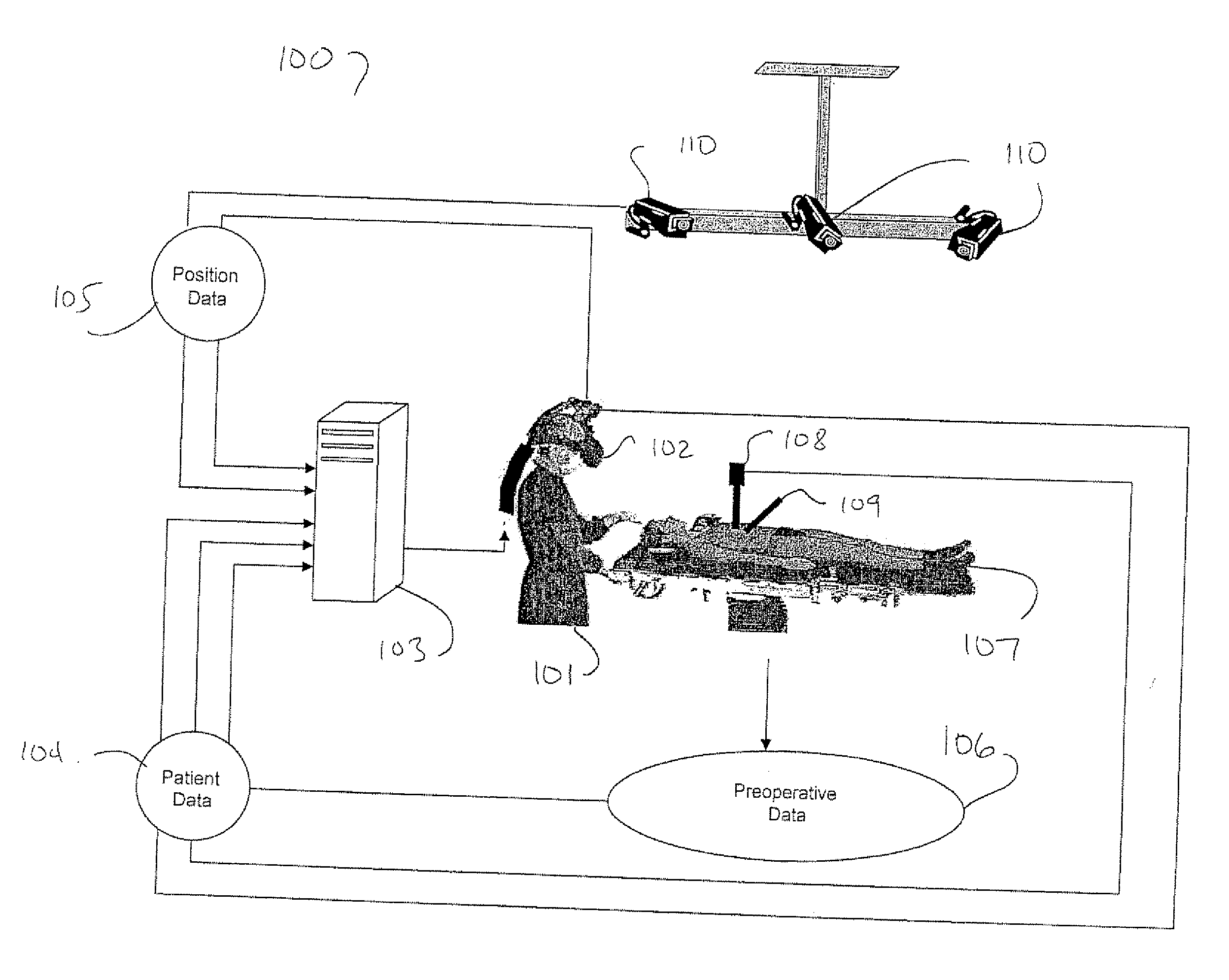
[0015] As discussed above, in order to perform difficult surgical tasks such as those difficult minimally-invasive tasks discussed above, it is desirable that a physician 101 has as much information as possible in regards to the positioning of the surgical instruments and endoscope with respect to anatomical structures. Therefore, in accordance with the principles of the present invention, a flexible navigation and visualization system is provided. FIG. 1 shows an illustrative endoscopic imaging, navigation and visualization system 100 in accordance with an embodiment of the present invention. Referring to that figure, physician 101 is illustratively a surgeon performing thoracoscopic surgery on patient 107 tying on surgical table 111. Surgeon 101 manipulates surgical instrument 109 to accomplish predetermined surgical tasks, such as removing material from between vertebrae in the spinal column of patient 107 or, alternatively, fusing multiple vertebrae together, as discussed above....


PUM


Abstract

A method and apparatus for presenting three-dimensional data to a physician is provided to facilitate the flexible navigation of an endoscope and surgical instruments with respect to anatomical structures. In accordance with a first embodiment a first set of data corresponding to a three dimensional model of a patient's anatomy is received. This three-dimensional model may be rendered from images taken in CT or MRI scanning, as discussed above In accordance with this embodiment, this model is then combined with a second set of data corresponding to a view obtained from an endoscope. In another embodiment, the view from the illustrative endoscope is displayed as an inset image on the display of the three-dimensional image. In yet another embodiment, the three-dimensional image comprises a graphical representation of at least a first surgical instrument, such as said endoscope. The surgeon may select among various combinations of views and may zoom in or out from any particular view.

Description

[0001] This patent application claims the benefit of U.S. Provisional Application No. 60 / 690,339, filed Jun. 14, 2005, which is hereby incorporated by reference herein in its entirety.BACKGROUND OF THE INVENTION [0002] The present invention relates generally to image guidance for minimally invasive surgery and, more particularly, to augmented reality visualization using endoscopes. [0003] Minimally invasive surgery is becoming increasingly widespread due to the benefits to both the patient (i.e., decreased morbidity and faster recovery) as well as the benefits to the insurance industry (i.e., less hospitalization time and lower cost). Typically, minimally invasive surgery involves creating one or more small incisions and performing any surgical tasks through those incisions using specialized, frequently elongated, surgical tools. [0004] As one skilled in the art will recognize, visual feedback is essential to such minimally invasive surgery. Typically, visual feedback in such surger...

Claims


Application Information

Patent Timeline
Patent Type & Authority Applications(United States)
IPC IPC(8): A61B1/04
CPCA61B1/04A61B19/52A61B1/0005G06F19/3437A61B2019/5289A61B90/36A61B2090/364G16H50/50A61B34/20A61B2090/376
Inventor SAUER, FRANKKHAMENE, ALIVOGT, SEBASTIAN
Owner SIEMENS MEDICAL SOLUTIONS USA INC
Why Eureka
Try Eureka