

Physiology workstation with real-time fluoroscopy and ultrasound imaging
a workstation and ultrasound imaging technology, applied in the field of physiology workstations with real-time fluoroscopy and ultrasound imaging workstations, can solve problems such as complex and sensitiv
- Summary
- Abstract
- Description
- Claims
- Application Information
AI Technical Summary
Benefits of technology
Problems solved by technology
Method used
Image

Examples
Embodiment Construction
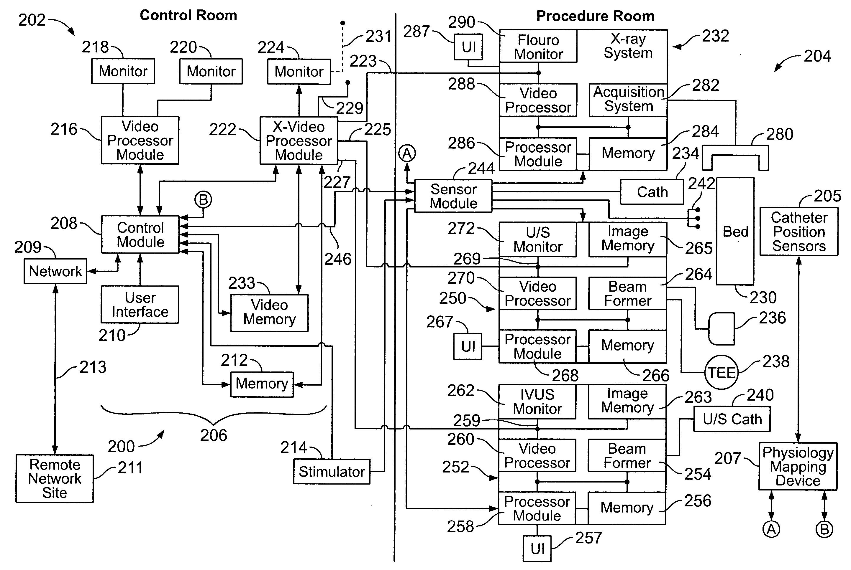
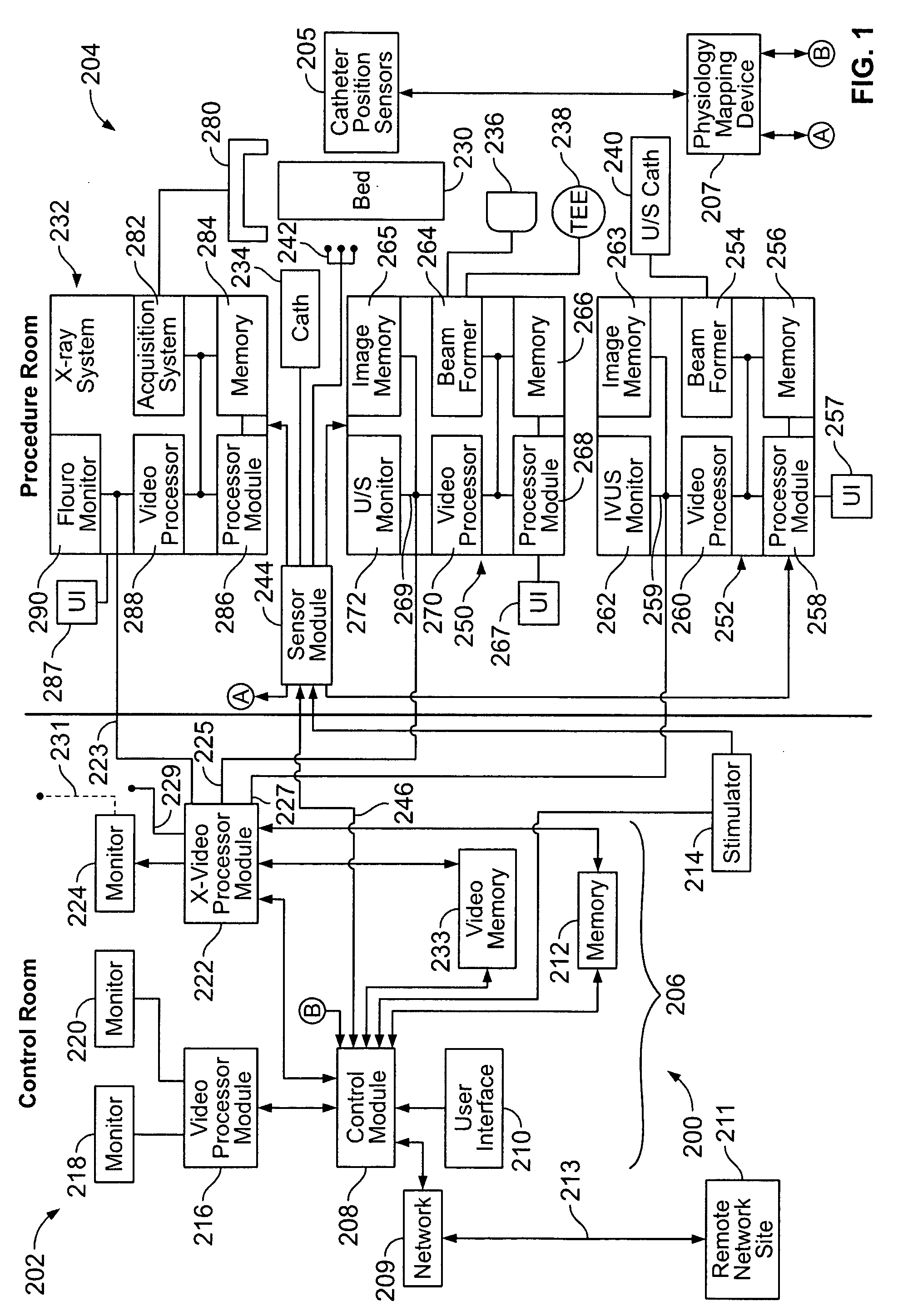
[0021]FIG. 1 illustrates an image management system 200 formed in accordance with an embodiment of the present invention. The image management system 200 may be distributed between a control room 202 and procedure room 204 or, alternatively, may be all located in the procedure room 204. Thus the image management system 200 may be located entirely in the procedure room 204. A physiology workstation 206 (e.g., EP or HD workstation) is provided to control and coordinate EP or HD procedures, ablation procedures and the like. The physiology workstation 206 includes a control module 208 that is controlled by an operator through user interface 210. Memory 212 stores various information as will be explained below in more detail. A stimulator 214 is provided to generate stimulus signals delivered to the patient in the procedure room 204. A physiology video processor module 216 communicates with the control module 208 and controls monitors 218 and 220. An external video processor module 222 i...
PUM
 Abstract
Description
Claims
Application Information