Capsulated medical equipment
a medical equipment and capsulated technology, applied in the field of capsulated medical equipment, can solve the problems of countermeasures that permit early detection of the clogged state of the capsulated body
- Summary
- Abstract
- Description
- Claims
- Application Information
AI Technical Summary
Benefits of technology
Problems solved by technology
Method used
Image
Examples
first embodiment
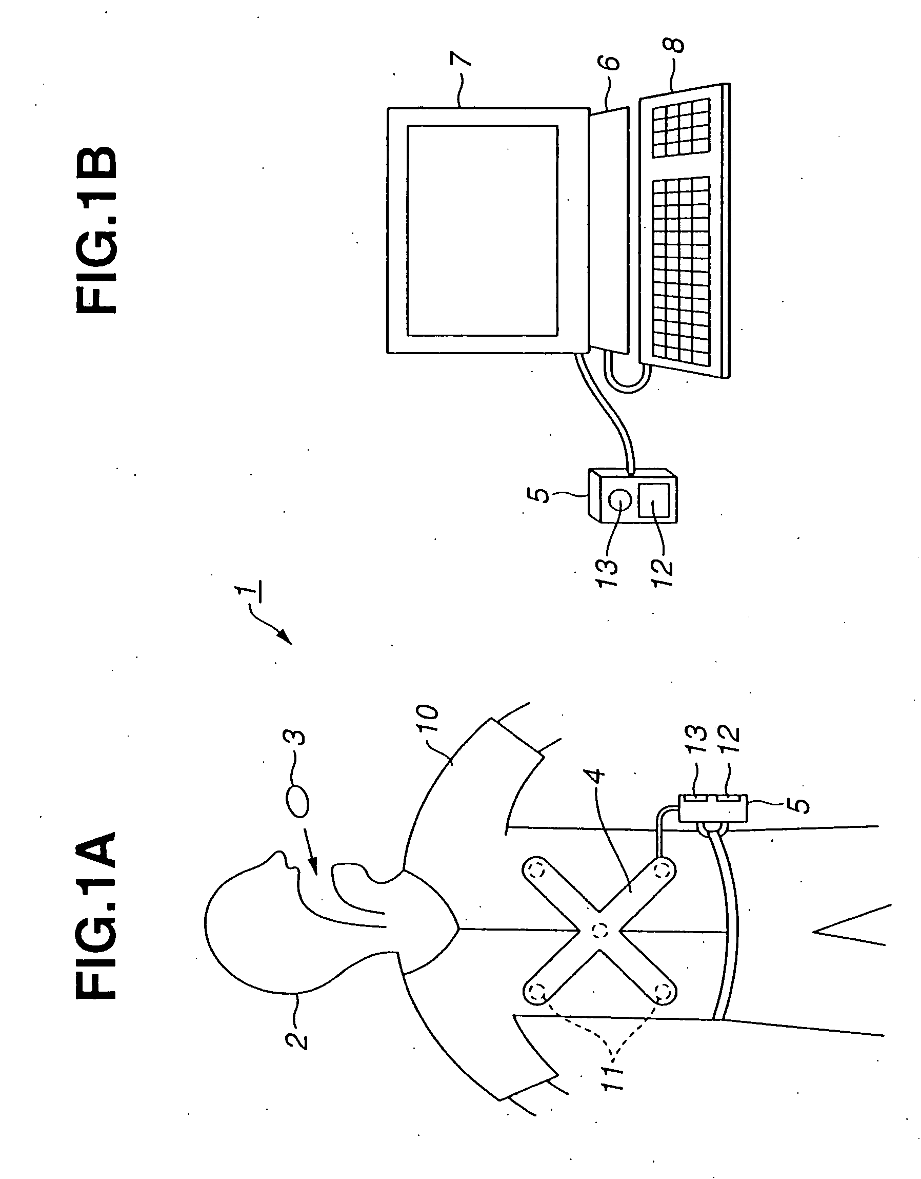
[0036] Referring to FIG. 1A to FIG. 9, the first embodiment of the present invention will be described below.
[0037] As shown in FIG. 1A, a capsulated endoscope system 1 in accordance with the first embodiment of the present invention consists mainly of a capsulated endoscope 3 (that is a capsulated body) and an extracorporeal unit 5 (placed away from the patient 2). The capsulated endoscope 3 is gulped down through the mouth of a patient 2, and transmits by radio an image signal that represents an optical image of the inner wall of an intracavitary duct while passing through the intracavitary duct. The extracorporeal unit 5 receives the signal sent from the capsulated endoscope 3 through an antenna unit 4 mounted on the extracorporeal region of the patient 2, and has a facility that preserves images.
[0038] As shown in FIG. 1B, the extracorporeal unit 5 is connected to a personal computer 6 so that it can be disconnected freely. The personal computer 6 fetches an image preserved in...
second embodiment
[0080] Next, the second embodiment of the present invention will be described with reference to FIG. 10 to FIG. 12. An object of the present embodiment is to provide capsulated medical equipment having a feature of detecting clogging of a capsulated body (a capsulated endoscope 3C in the present embodiment) in an early stage, and of automatically unclogging the capsulated body. FIG. 10 shows the capsulated endoscope 3C employed in the second embodiment.
[0081] The capsulated endoscope 3C is different from the capsulated endoscope 3 employed in the first embodiment in a point that the capsulated endoscope 3C has a plurality of pressure sensors 43 located on the periphery of a portion thereof having the largest diameter. Moreover, (a stator) of a pager motor (vibrating motor) 44 that is compact and vibrates is locked in the capsulated endoscope at the opposite end of the capsulated endoscope relative to the objective 15. The motor 44 is driven by a motor driver 45 as shown in FIG. 11....
third embodiment
[0099] Next, the third embodiment of the present invention will be described with reference to FIG. 13 and FIG. 14. FIG. 13 shows a capsulated endoscope system 51 in accordance with the third embodiment. The system 51 consists mainly of a capsulated endoscope 3D, an extracorporeal unit 52, a personal computer 53, an actuator control circuit 54, actuators 55a and 55b, and electromagnets 56a and 56b. The extracorporeal unit 52 preserves image data produced by the capsulated endoscope 3D and calculates the position of the capsulated endoscope 3D. The personal computer 53 is connected to the extracorporeal unit 52. The actuator control circuit 54 is connected to the personal computer 53. The actuators 55a and 55b are driven with a driving signal sent from the actuator control circuit 54. The electromagnets 56a and 56b are three-dimensionally moved with thrusts produced by the actuators 55a and 55b respectively.
[0100] The capsulated endoscope 3D has the components thereof arranged as sh...
PUM
 Abstract
Description
Claims
Application Information
 - R&D
- Intellectual Property
- Life Sciences
- Materials
- Tech Scout
- Unparalleled Data Quality
- Higher Quality Content
- 60% Fewer Hallucinations
Browse by: Latest US Patents, China's latest patents, Technical Efficacy Thesaurus, Application Domain, Technology Topic, Popular Technical Reports.
© 2025 PatSnap. All rights reserved.Legal|Privacy policy|Modern Slavery Act Transparency Statement|Sitemap|About US| Contact US: help@patsnap.com


