Enhanced ultrasound image display

a technology of ultrasound image and display, applied in the field of medical imaging, can solve the problems of difficult interpretation of ultrasound images, and may not be able to match the bright and dark areas of the fan with the features of the fan, and achieve the effect of improving the diagnostic usefulness of the ultrasound catheter

Active Publication Date: 2007-11-29
BIOSENSE WEBSTER INC
View PDF15 Cites 18 Cited by
  • Summary
  • Abstract
  • Description
  • Claims
  • Application Information

AI Technical Summary

Benefits of technology

[0007] According to disclosed embodiments of the invention, using specialized cardiac catheters for image acquisition, features of the heart are readily identifiable on an ultrasound image, based on a previously generated electrical activation map of the heart. The electrical activation map is automatically registered with the ultrasound image using information obtained from position sensors in the catheters. Features identifiable on the electrical activation map, presented

Problems solved by technology

However, many physicians find it difficult to interpret ultrasound images, which typically appear as a two-dimensional fan-shaped pattern.
Although the physician knows wha

Method used


Image

  • Enhanced ultrasound image display
  • Enhanced ultrasound image display
  • Enhanced ultrasound image display

Examples


Embodiment Construction

[0024] In the following description, numerous specific details are set forth in order to provide a thorough understanding of the present invention. It will be apparent to one skilled in the art, however, that the present invention may be practiced without these specific details. In other instances, well-known circuits, control logic, and the details of computer program instructions for conventional algorithms and processes have not been shown in detail in order not to obscure the present invention unnecessarily.

System Overview

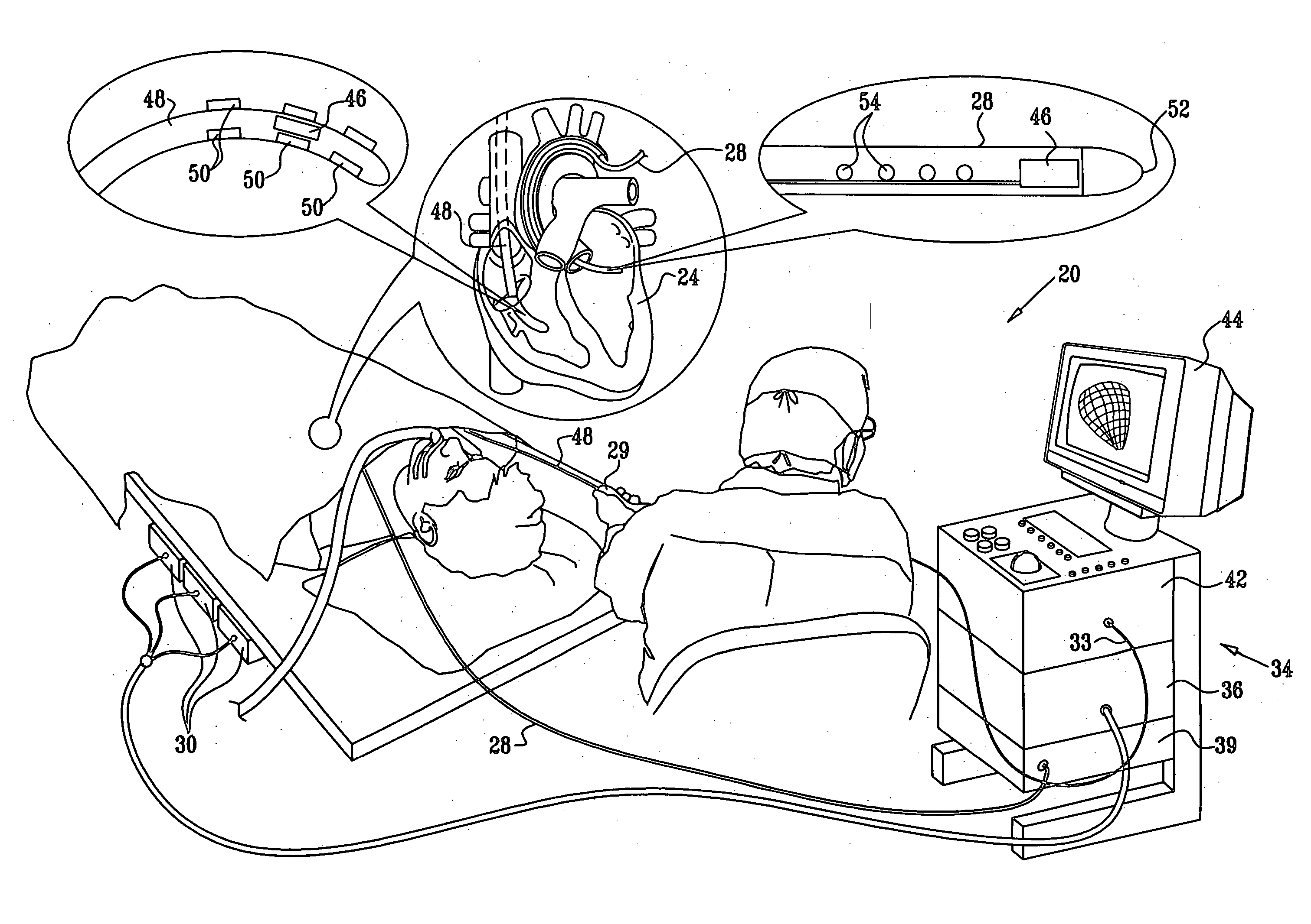
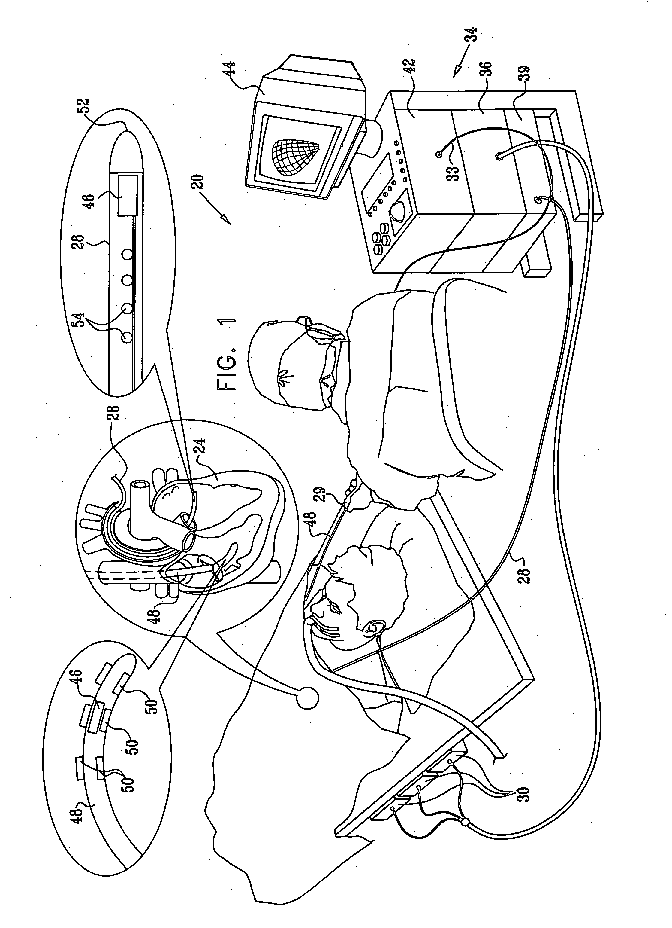
[0025] Turning now to the drawings, reference is initially made to FIG. 1, which is an illustration of a system 20 for imaging and generating electrical activation maps of a heart 24 of a patient, and which is suitable for performing diagnostic or therapeutic procedures involving the heart 24, in accordance with an embodiment of the present invention. The system comprises a catheter 28, which is percutaneously inserted by a physician into a chamber or vascul...


PUM


Abstract

Using specialized cardiac catheters for image acquisition, features of the heart are readily identifiable on an ultrasound image, based on a previously generated electrical activation map of the heart. The electrical activation map is automatically registered with the ultrasound image using information obtained from position sensors in the catheters. Features identifiable on the electrical activation map, presented as points, tags, design lines, and textual identification, are projected into the plane of the ultrasound fan and overlaid on the ultrasound image, thereby clarifying the features that are visible on the latter.

Description

BACKGROUND OF THE INVENTION [0001] 1. Field of the Invention [0002] This invention relates to medical imaging. More particularly, this invention relates to improvements in the identification of topologic features in cardiac ultrasound images. [0003] 2. Description of the Related Art [0004] Ultrasound imaging is now well established as a modality for imaging the heart. For example, U.S. Pat. No. 6,066,096, whose disclosure is incorporated herein by reference, describes an imaging probe for volumetric intraluminal ultrasound imaging. The probe, configured to be placed inside a patient's body, includes an elongated body having proximal and distal ends. An ultrasonic transducer phased array is connected to and positioned on the distal end of the elongated body. The ultrasonic transducer phased array is positioned to emit and receive ultrasonic energy for volumetric forward scanning from the distal end of the elongated body. The ultrasonic transducer phased array includes a plurality of ...

Claims


Application Information

Patent Timeline
IPC IPC(8): A61B5/05
CPCG06T7/0026G06T2207/30048G06T7/0042G06T7/32G06T7/73A61B8/00G01S15/00
Inventor TAL, ROY
Owner BIOSENSE WEBSTER INC
Why Eureka
Try Eureka