
Methods of screening agents for cytotoxic and antimicrobial activity

Inactive Publication Date: 2008-01-24
BOARD OF RGT THE UNIV OF TEXAS SYST
View PDF0 Cites 1 Cited by
  • Summary
  • Abstract
  • Description
  • Claims
  • Application Information

AI Technical Summary

Benefits of technology

[0008] Thus, in accordance with the present invention, there is provided a method of screening a test substance for cytotoxic and antimicrobial activity comprising (a) providing at least one eukaryotic cell and at least one pathogenic organism, each of the cells expressing a fluorescent marker protein or stained with a fluorescent dye; (b) contacting each of the cells with the test substance; and (c) assessing a fluorescent signal from each of the cell or organism, wherein a decrease in signal from only the eukaryotic cell indicates that the test substance is a cytotoxic agent, and wherein a decre

Problems solved by technology

Unfortunately, the HeLa-GFP assay the inventors previously reported could not be applied to high-throughput analyses as it relied on the visual detection of cell death through fluorescence microscopy.

Method used


Image

  • Methods of screening agents for cytotoxic and antimicrobial activity
  • Methods of screening agents for cytotoxic and antimicrobial activity
  • Methods of screening agents for cytotoxic and antimicrobial activity

Examples


example 1

Materials and Methods

[0141] Cell culture. HeLa-GFP cells were cultured in DMEM media supplemented with 10% heat-inactivated newborn calf serum as previously described (Montoya et al., 2004). Stock cultures were prewashed twice with PBS before the treatment of trypsin for cell resuspension and subsequently re-plated at the concentration of 4,000 cells / well. E. coli-GFP cells were grown in LB broth media while the MAC104 M. avium strain was grown in 7H9-ADC media.

[0142] HeLa-GFP fluorometric cytotoxicity assay. All cell lines were seeded in clear-bottomed 96-well assay plates (3603, Costar, Corning Inc., Corning, N.Y.) in order to minimized background fluorescence. HeLa cells were grown in a total volume of 0.2 ml of medium and incubated overnight for proper cell attachment. The quinone compounds were then added to each well (in triplicate) and then incubated for a period of 18 hr. For the determination of fluorescence, the assay plates were then read using the Fluoroskan Ascent F1 ...

example 2

Results and Discussion

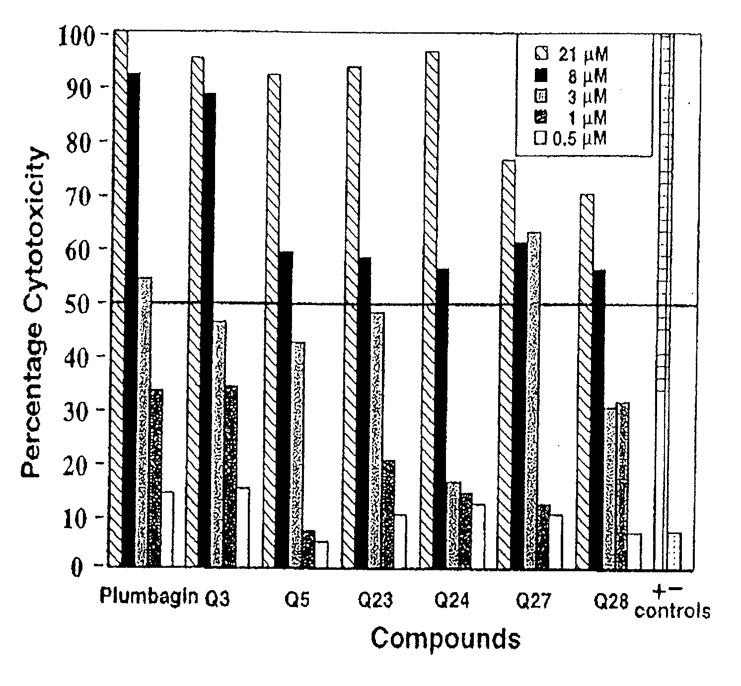
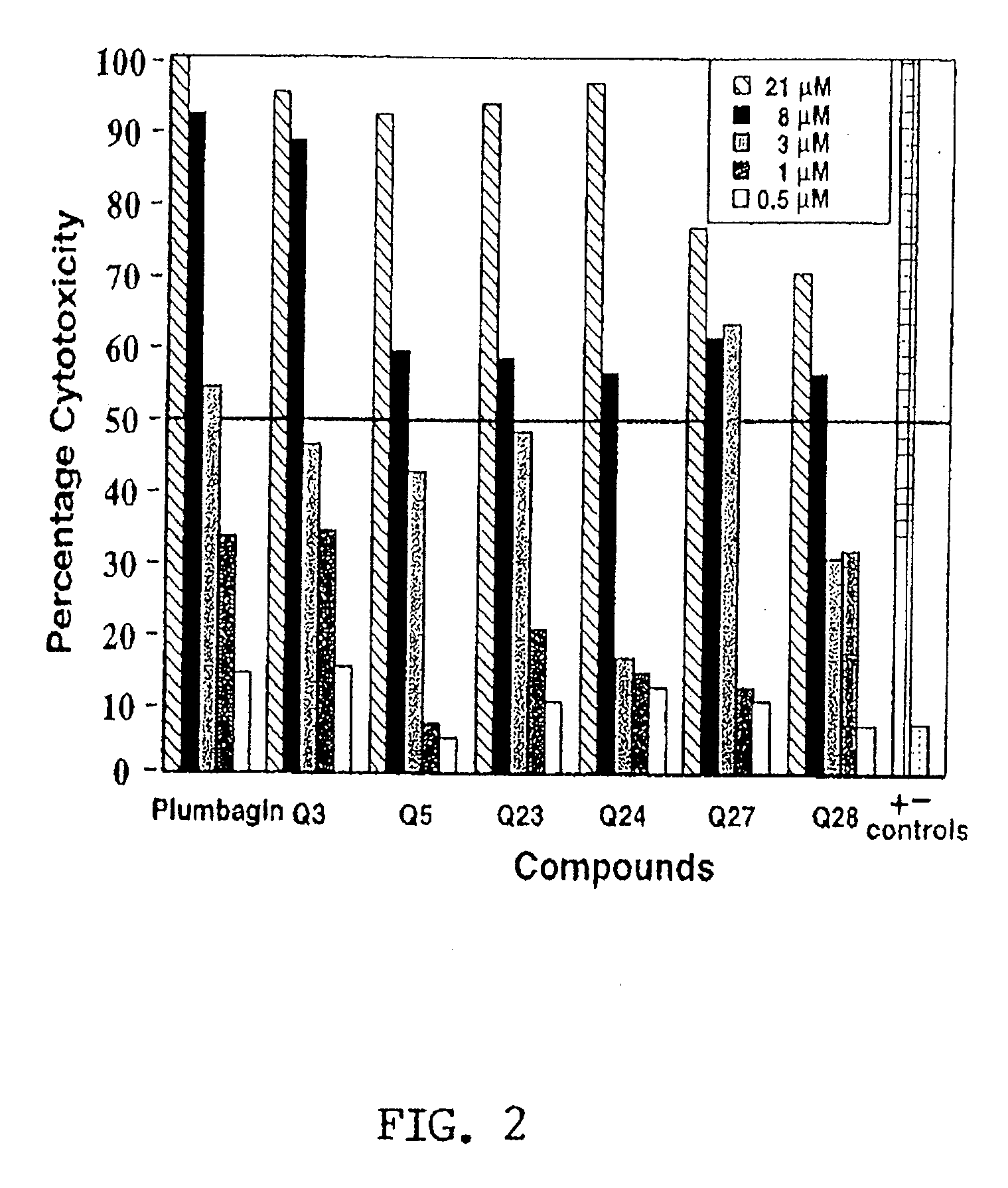
[0147] In order to rapidly screen novel compounds for their toxic properties, a relatively simple GFP-based assay was recently utilized to simultaneously screen several compounds without having to perform other elaborate assays (Montoya et al., 2004). Although easy to implement, this assay could not be applied to high-throughput analysis as it relied on the visualization of cell death (Montoya et al., 2004). Given this obvious limitation, the assay was modified to facilitate the simultaneous screening of multiple compounds in a 96-well format using an automated fluorescence plate reader. Since small quantities of compounds are required, this assay is particularly well suited for the screening of combinatorial chemical libraries. Another advantage of these microplate assays is the ability to perform all assays in duplicate or triplicate to derive more consistent results, and for this reason, all experiments were performed in triplicate.

[0148] As proof of conce...


PUM


Abstract

The present invention provides for methods of screening agents for cytotoxic and / or anti-microbial activity. In particular embodiments, the loss of a fluorescent signal from the treated cells, due to cell death or inhibition of growth, is measured for several different cell types in parallel.

Description

[0001] This application claims benefit of priority to U.S. Provisional Application Ser. No. 60 / 807,963, filed Jul. 21, 2006, the entire contents of which are hereby incorporated by reference.[0002] This invention was made with government support under grant numbers 2G12RR08124, S06 GM8012-34 and AI01812-02 awarded by the National Institutes of Health. The government has certain rights in the invention.BACKGROUND OF THE INVENTION [0003] 1. Field of the Invention [0004] The present invention relates generally to the fields of cell biology, molecular biology, oncology and microbiology. More particularly, it concerns the use of novel screening assays for cytotoxic and anti-microbial agents. [0005] 2. Description of Related Art [0006] As large quantities of novel chemical compounds are generated, companies and researchers are faced with the growing challenge of screening these compounds for their potential therapeutic and / or toxic effects; thus leading to a growing demand for novel cell-...

Claims


Application Information

Patent Timeline
IPC IPC(8): C12Q1/02
CPCG01N33/5014C12Q1/18
Inventor AGUILERA, RENATO J.
Owner BOARD OF RGT THE UNIV OF TEXAS SYST
Learn More