

Method for hardening correction in medical imaging
a correction method and technology for medical imaging, applied in the field of medical imaging, can solve the problems of complex correction methods, adversely affecting diagnosis, and difficult detection of smaller lesions in cases of density caused by hardening, and achieve the effect of removing the influence of noise in the projection image profil
- Summary
- Abstract
- Description
- Claims
- Application Information
AI Technical Summary
Benefits of technology
Problems solved by technology
Method used
Image

Examples
Embodiment Construction
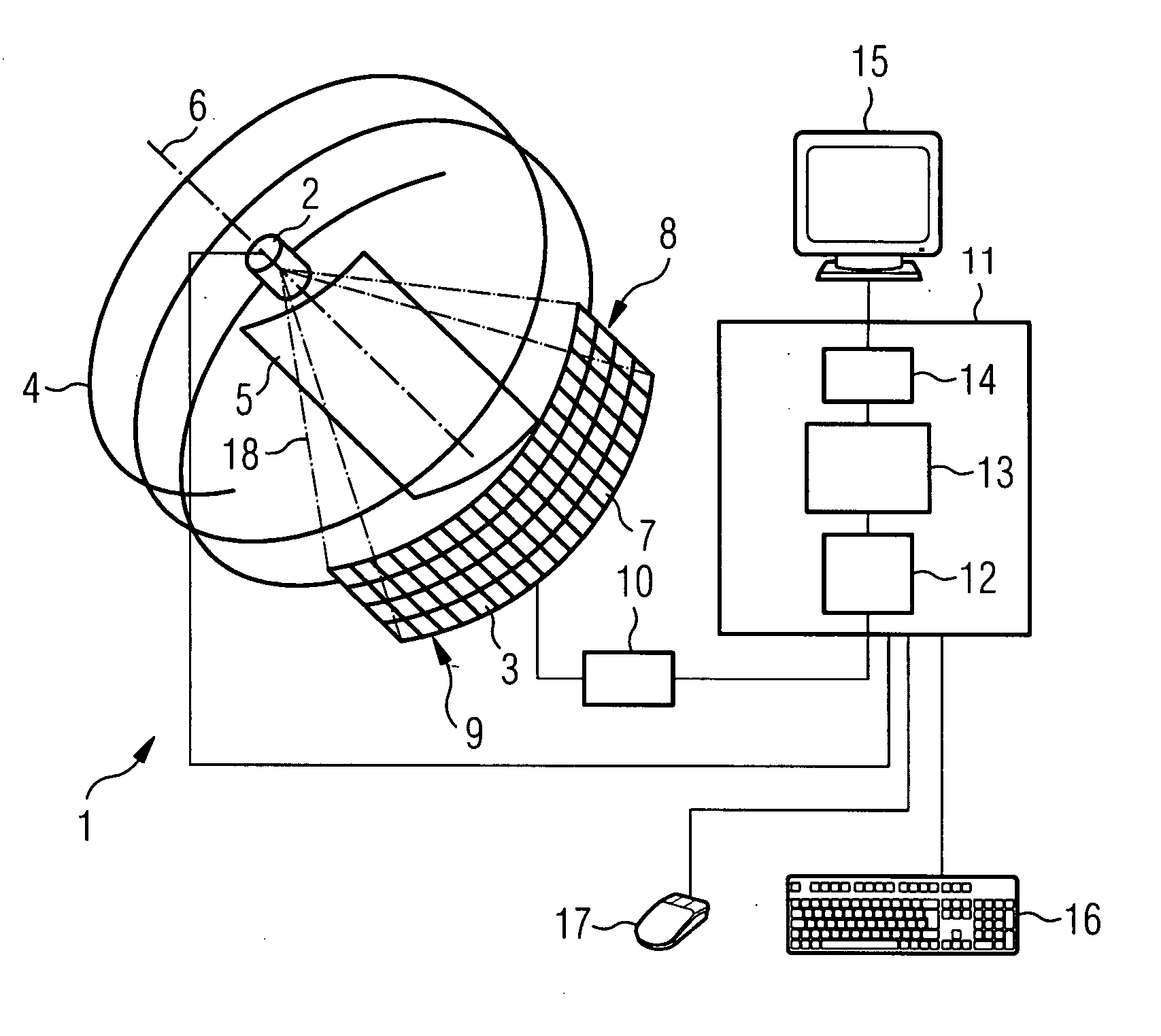
[0049]FIG. 1 shows a computer tomography device 1 which features an x-ray tube 2. The x-ray tubes 2 and the x-ray detectors 3 move on a track 4 around a patient support table 5, on which a patient not shown in FIG. 1 is located during the examination. Usually the x-ray detector 3 and the x-ray tubes 2 are guided by a yoke and run in the yoke on a circular path around the patient support table 5. The yoke and the patient support table 5 can move relative to one another along a longitudinal axis 6. The track 4 is in this case spiral-shaped in relation to the patient support table 5.
[0050] The x-ray detector 3 is preferably a digital flat-panel detector which is made up of a plurality of detector elements 7, the so-called pixels. The detector elements 7 are preferably arranged in rows 8 and columns 9. Furthermore a readout circuit 10 and also an evaluation circuit 10 are connected downstream from the x-ray detector 3. The evaluation unit 11 can for example be a commercially-available ...
PUM
 Abstract
Description
Claims
Application Information