Method and Arrangement Relating to X-Ray Imaging

a technology of x-ray imaging and method, applied in the field of method and arrangement of x-ray imaging, can solve the problems of low dose, noisy, unfavorable use of two projection images, etc., and achieve the effect of low dose and better image quality

Active Publication Date: 2008-08-21
PHILIPS DIGITAL MAMMOGRAPHY SWEDEN
View PDF5 Cites 54 Cited by
  • Summary
  • Abstract
  • Description
  • Claims
  • Application Information

AI Technical Summary

Benefits of technology

[0006]According to the present invention, tomosynthesis has advantages relative to stereo imaging. Tomosynthesis provide better image quality, and future radiologists will be used to tomosynthesis images. Tomosynthesis also avoids trouble when finding corresponding points in two 2-dimensional images, which would otherwise be difficult for clusters of micro-calc or diffuse contours. In the future, many clinics will have only a tomosynthesis apparatus, and that apparatus must be extended to perform biopsy. A typical tomosynthesis apparatus requires less complicated extensions than transforming the ap

Problems solved by technology

Each projection image is acquired with a very low dose and thus noisy.
Therefore it i

Method used


Image

  • Method and Arrangement Relating to X-Ray Imaging
  • Method and Arrangement Relating to X-Ray Imaging
  • Method and Arrangement Relating to X-Ray Imaging

Examples


Embodiment Construction

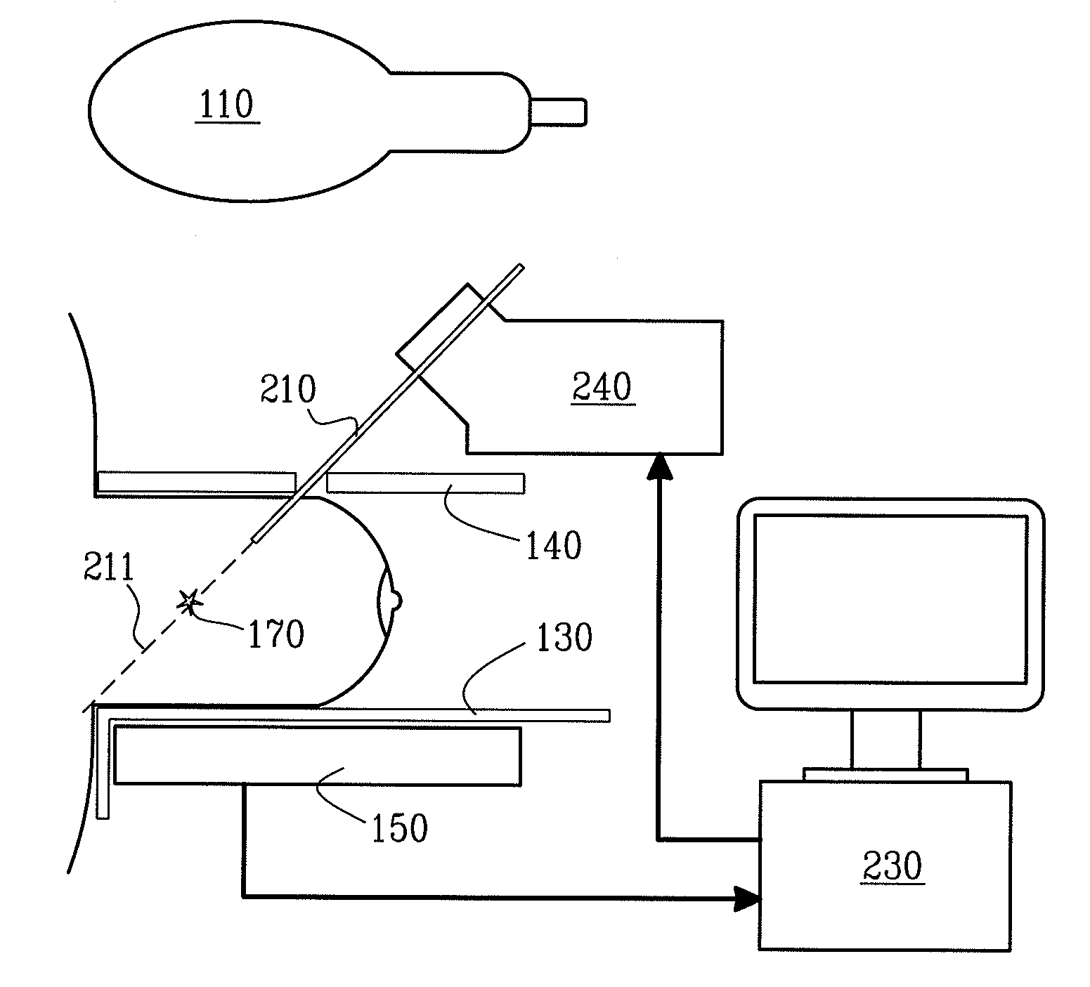
[0022]The preferred embodiment of the present invention comprises a patient support 130, and a compression paddle 140 for compression of a breast 170 containing a location with some tissue to extract 171. The compression paddle 140 contains a hole for inserting a needle 210 towards said tissue to extract.

[0023]Furthermore, the preferred embodiment comprises an acquisition system for obtaining tomosynthesis image data, including a set of projection images (preferably 10-30) and reconstructing a three-dimensional image volume. The acquisition system comprises a detector unit 150, an x-ray source 110, and a computer 230 for reconstruction of a three-dimensional image volume from the set of projection images. The projection images are views of the breast from slightly different angles. The computer 230 reconstructs a three-dimensional image volume from said projection images, and displays the image volume to the operator.

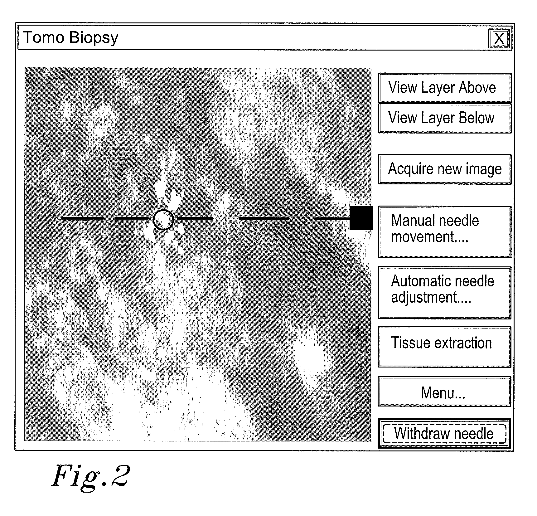
[0024]The computer also comprises an algorithm for measuring the n...


PUM


Abstract

Three-dimensional imaging of a breast tissue is achieved by acquiring a series of X-ray projection images from various angles. A three-dimensional image constructed from the projection images by a computer provides information to the operator to select a coordinate point within the breast from which to obtain a biopsy sample. Acquisition of projection images and construction of three-dimensional images is continued during the insertion of the needle into the breast, during positioning the needle towards the coordinate point, during the sampling and after the sampling. The advantages of utilizing tomosynthesis for breast tissue imaging over stereo imaging include better image quality, easier coordinate measurements within a tissue, ease of use of the equipment by the medical personnel, and fewer image artifact problems.

Description

TECHNICAL FIELD OF THE INVENTION[0001]The present invention relates to a method and arrangement in X-ray imaging, in particular three-dimensional imaging, and more especially tomosynthesis and biopsy.BACKGROUND OF THE INVENTION[0002]Tomosynthesis is used to create a three-dimensional image volume of a person's body part, e.g. her breast, or an object, using X-rays. Currently, tomosynthesis breast imaging is available only for research purposes, but an increasing number of market analysts believe it will become more widely used than conventional two-dimensional mammography.[0003]Tomosynthesis is essentially a limited form of Computed Tomography or CT. Normally, several projection images, e.g. 5 or 30, are acquired in a range of different angles, e.g. −10 to +10 degrees. Each projection image is essentially a conventional 2-dimensional digital X-ray image of the examined object. The projection images are then combined using special purpose software for producing a 3-dimensional image ...

Claims


Application Information

Patent Timeline
IPC IPC(8): A61B6/04
CPCA61B6/466A61B6/502A61B19/50A61B19/201A61B10/0233A61B6/025A61B34/10A61B90/11
Inventor HJARN, TORBJORNHEMMENDORFF, MAGNUS
Owner PHILIPS DIGITAL MAMMOGRAPHY SWEDEN
Why Eureka
Try Eureka