

Ultrasound image displaying method and ultrasound diagnostic apparatus
a technology which is applied in the field of ultrasound diagnostic apparatus and ultrasound image display method, can solve the problems of difficult to distinguish a 3d structure such as a blood vessel, and achieve the effect of facilitating the distinction of the blood vessel
- Summary
- Abstract
- Description
- Claims
- Application Information
AI Technical Summary
Benefits of technology
Problems solved by technology
Method used
Image

Examples
Embodiment Construction
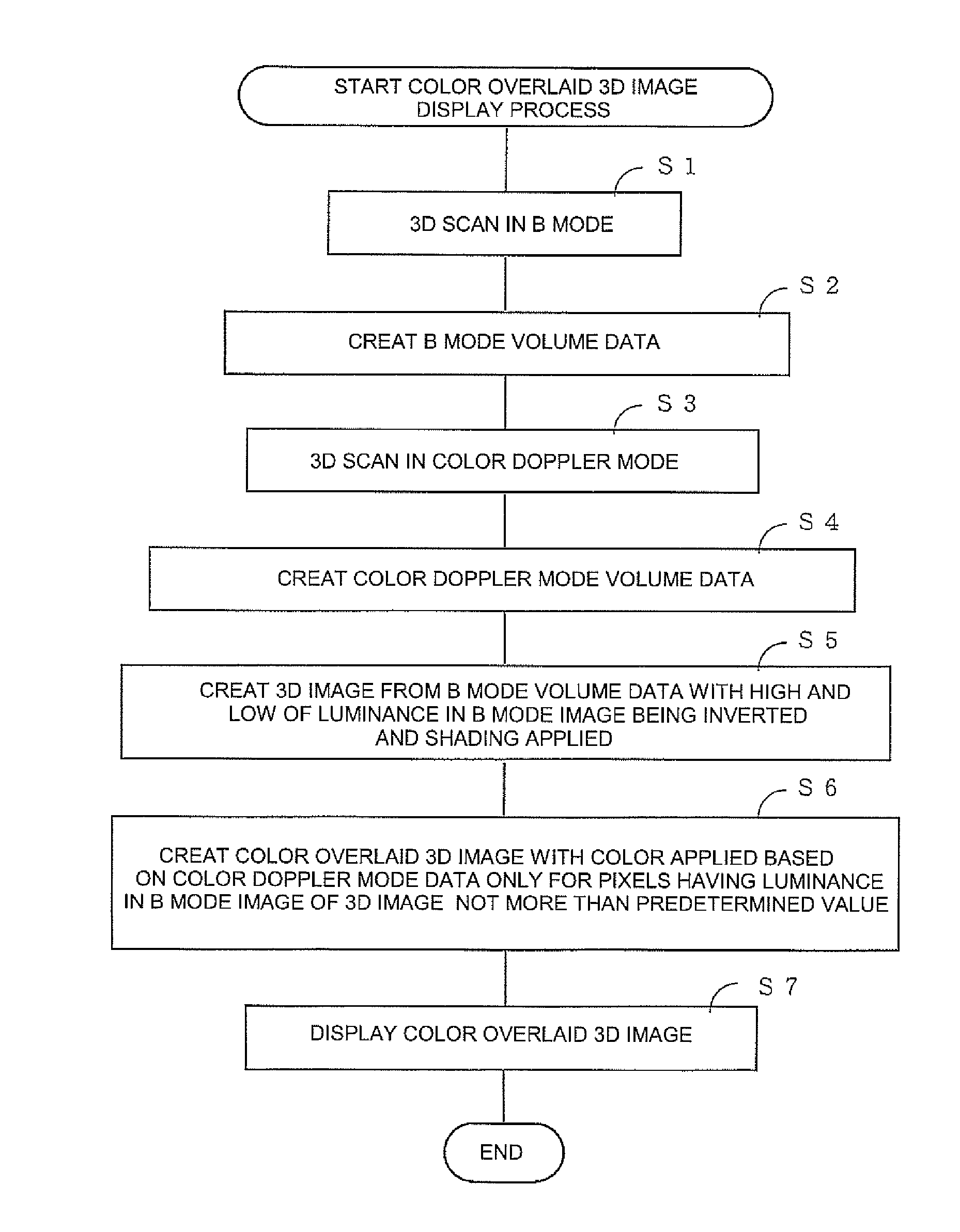
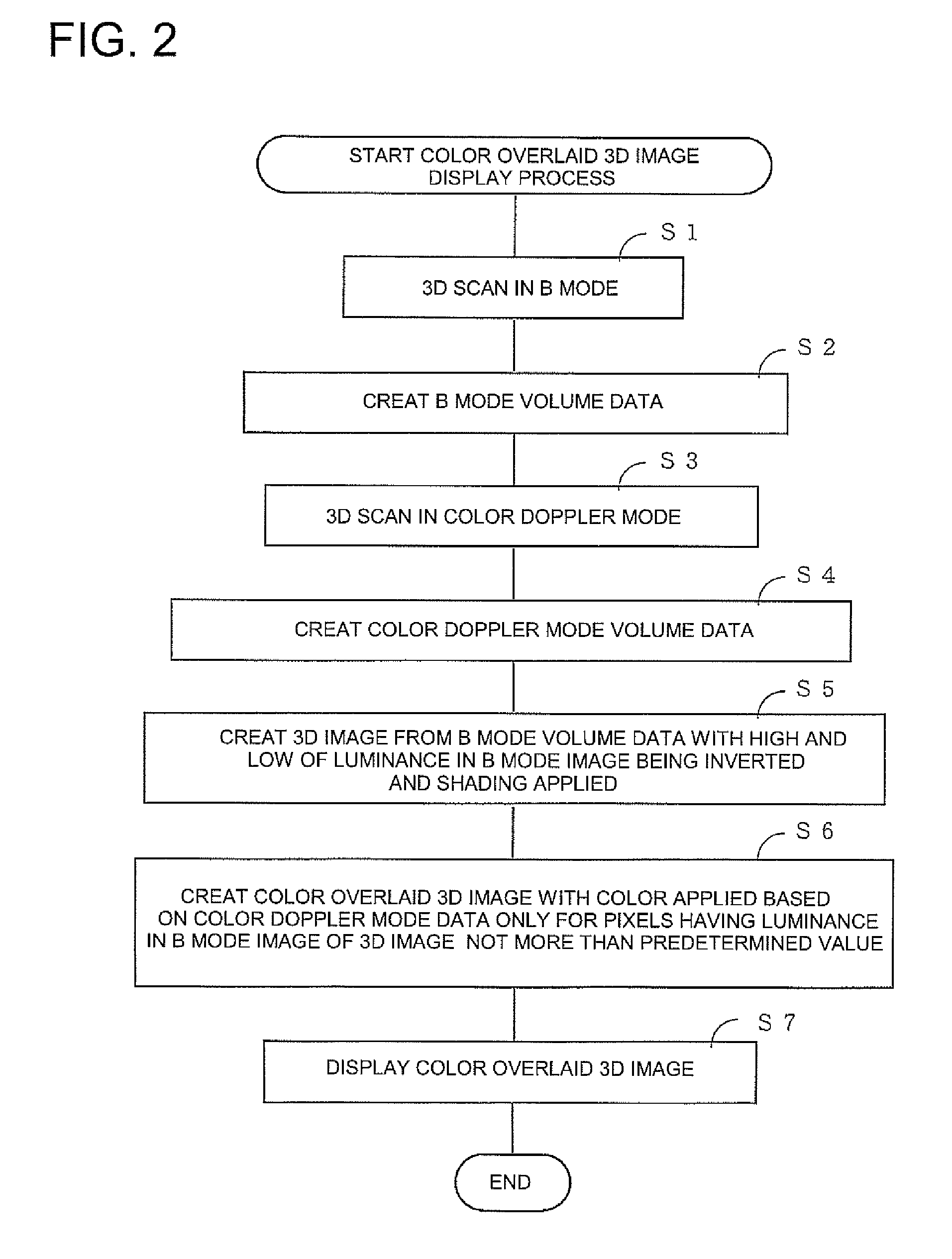
[0047]The invention will be described in greater details herein below with reference to some embodiments shown in the accompanying drawings. It should be noted here that the disclosed embodiments may not be considered to limit the invention.
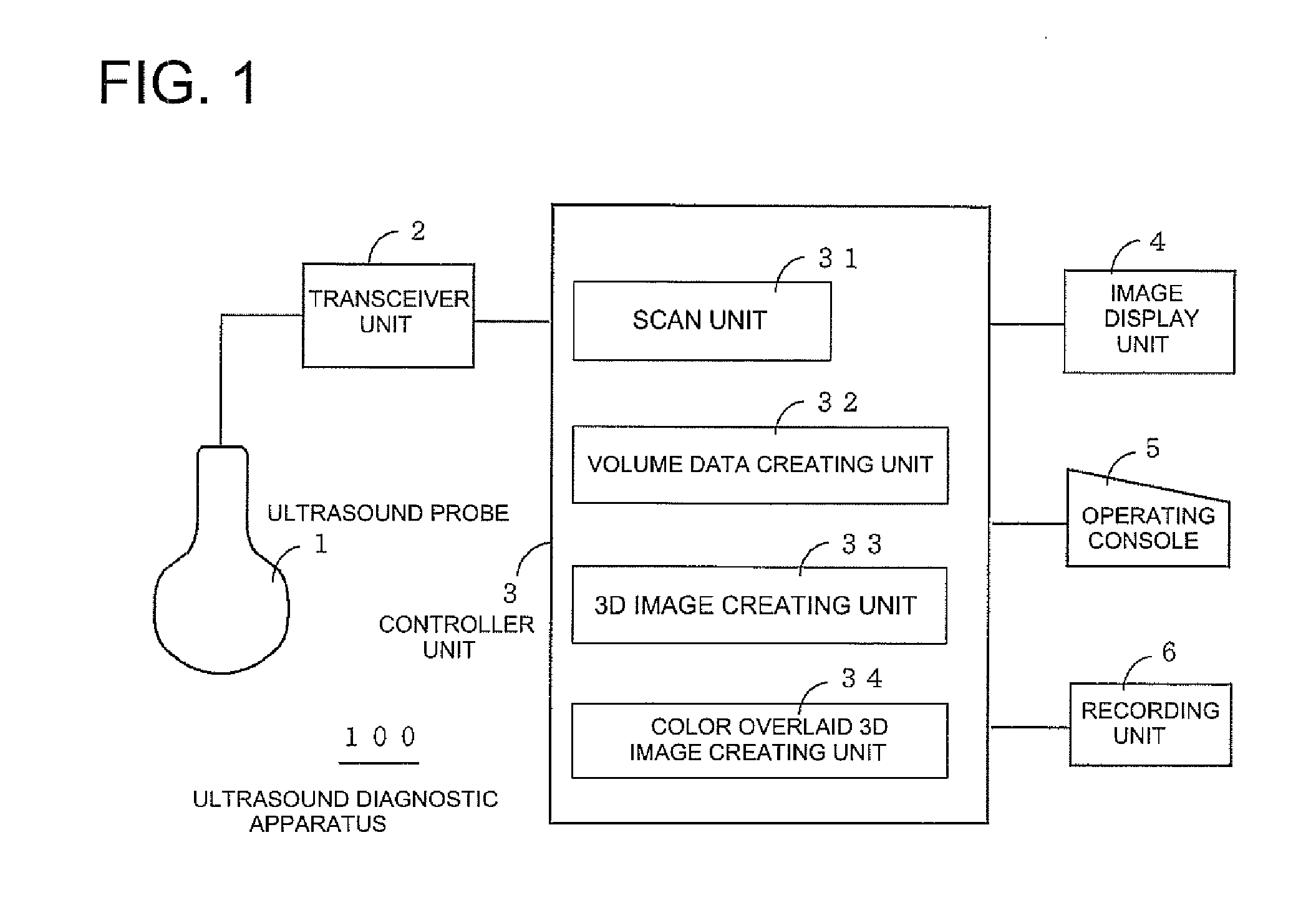
[0048]FIG. 1 shows a schematic block diagram of an ultrasound diagnostic apparatus 100 in accordance with the first embodiment.
[0049]The ultrasound diagnostic apparatus 100 includes an ultrasound probe 1 capable of 2D scan by electronic scan as well as of 3D scan by electric scan (scan by means of a motor) or electronic scan; a transceiver unit 2 for driving the ultrasound probe 1 to perform 2D scan and 3D scan within the body of an examinee by means of ultrasound beam; a controller unit 3; an image display unit 4 for displaying an ultrasound image; an operating console 5 for inputting an instruction or data by an operator; and a recording unit 6 for recording the ultrasound images.
[0050]The controller unit 3 includes a scan unit 31 for controlli...
PUM
 Abstract
Description
Claims
Application Information