System and Method for Segmenting a Region in a Medical Image

a medical image and segmentation technology, applied in the field of image segmentation, can solve the problems of increasing the risk of further complications, difficult segmentation of ultrasound images of structures with low contrast such as the prostate, and insufficient local image processing techniques such as edge detectors,

Active Publication Date: 2008-10-30
MERATIVE US LP
View PDF15 Cites 77 Cited by
  • Summary
  • Abstract
  • Description
  • Claims
  • Application Information

AI Technical Summary

Problems solved by technology

Symptoms due to carcinoma of the prostate are generally absent until extensive growth occurs and once the tumour extends beyond the prostate the risk of further complications increases.
However, an ultrasound image of structures having low contrast such as the prostate can be difficult to segment.
Typical local image processing techniques such as edge detectors have been found to be inadequate in and of themselves for finding the boundary, due to speckle, shadowing and other image artefacts.

Method used


Image

  • System and Method for Segmenting a Region in a Medical Image
  • System and Method for Segmenting a Region in a Medical Image
  • System and Method for Segmenting a Region in a Medical Image

Examples


Embodiment Construction

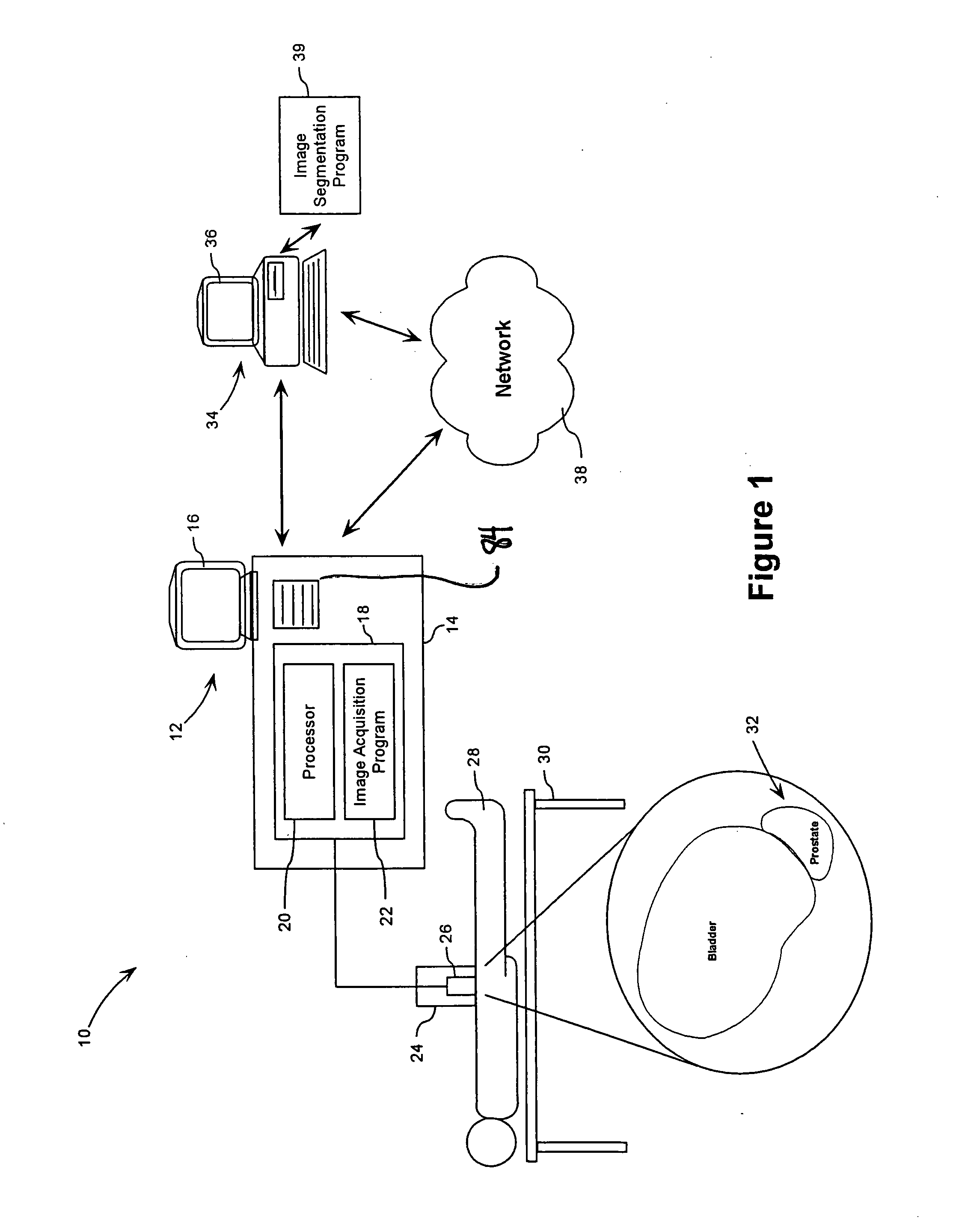
[0024]Referring therefore to FIG. 1, an imaging environment, in this example an ultrasound imaging environment is generally denoted by numeral 10. The environment 10 comprises an ultrasound imaging apparatus 12, an examination table 30 and a patient 28 positioned on the table 30. The apparatus 12 comprises an ultrasound machine 14, a display and control interface 16, and an ultrasound probe 24. The probe 24 includes one or more transducers 26 that emit sound waves and receive echoes of such sound waves as is well known in medical imaging.

[0025]The machine 14 comprises an imaging module 18 for controlling the probe 24. The imaging module 18 comprises a processor 20 and a computer implemented image acquisition program 22 for obtaining one or more images of a region of interest in the patient 28. Preferably, the ultrasound apparatus 12 is capable of obtaining 3-D ultrasound images wherein an array of transducers 26 or a moveable transducer 26 is used in order to obtain an array or stac...


PUM


Abstract

A method and computer program are provided for segmenting a prostate from a medical image such as an ultrasound image. Each slice in a study is analysed and uses a manual initialization to obtain an initial contour, in which a user selects initial points that are automatically rendered on the image. An automatic refinement stage then snaps the contour to the prostate boundary in the image based on a pre-stored anatomical atlas and edge information obtained from the image. A manual adjustment stage may then be performed and if selected, the contour is automatically propagated to the next image slice to avoid the manual initialization. An auxiliary image slice may be referred to, which indicates how the prostate shape changes from slice to slices e.g. by providing a perpendicular profile.

Description

[0001]This application claims priority from U.S. Application No. 60 / 828,894 flied on Oct. 10, 2006, the contents of which are incorporated herein by reference.FIELD OF THE INVENTION[0002]The present invention relates to image segmentation and has particular utility in segmenting a region in a medical image.DESCRIPTION OF THE PRIOR ART[0003]As the frequency of prostate cancer diagnoses in men increases, so to does the need for early detection. Symptoms due to carcinoma of the prostate are generally absent until extensive growth occurs and once the tumour extends beyond the prostate the risk of further complications increases. However, when diagnosed at an early stage, prostate cancer is curable and even at later stages, treatment can be effective. Treatment options can vary depending on the extent of the cancer and the prognosis generally worsens when the diagnosis is at an advanced stage.[0004]The challenges that face physicians for handling prostate cancer diagnoses include being a...

Claims


Application Information

Patent Timeline
Patent Type & Authority Applications(United States)
IPC IPC(8): G06K9/46
CPCA61B8/13A61B8/469G01S7/52036G01S7/52074G01S7/52084G01S15/8993G06K9/4638G06K2009/366G06K2209/051G06T7/0083G06T7/0089G06T2207/10132G06T2207/20092G06T7/12G06T7/149G06V10/248G06V10/457G06V2201/031
Inventor GEIGER, PAULACCOMAZZI, VITTORIO
Owner MERATIVE US LP
Why Eureka
Try Eureka