Ultra Low Radiation Dose X-Ray CT Scanner

a ct scanner and ultra-low radiation technology, applied in tomography, instruments, nuclear engineering, etc., can solve the problems of difficult perception and characterization of small lesions, unable to detect low contrast, and 30 percent of breast cancers missed in conventional x-ray screening mammography, etc., to achieve excellent low contrast detectability and high spatial resolution in planes

Active Publication Date: 2008-10-30
WISCONSIN ALUMNI RES FOUND
View PDF1 Cites 36 Cited by
  • Summary
  • Abstract
  • Description
  • Claims
  • Application Information

AI Technical Summary

Benefits of technology

[0010]The present invention provides a line scan cone beam CT imaging system that provides both high in-plane spatial resolution and excellent low contrast detectability, thereby combining the advantages of tomosynthesis and conventional CT. Such a system is extremely advantageous for imaging breasts. In one embodiment, an x-ray cone beam irradiates an object and a two dimensional x-ray detector array is used to acquire multiple views of projection data at predefined view angles. Between views, the cone beam is moved along a translational axis in a plane at a constant perpendicular distance (z) form an imaging plane (xy) in the object, and the detector array is likewise moved in the opposite direction along a parallel translational axis located on the opposite side of the object.
[0011]Another aspect of the present invention is an image reconstruction method that enables an image to be produced from a limited range of view angles acquired using the oppositely directed translational motion of the x-ray source and detector. In one embodiment, a projection data set is acquired form a limited range of view angles, and within this limited range Fourier space is vastly undersampled, thereby limiting the radiation exposure of the object being imaged. For example, projection data from 10-30 views is obtained within a limited view angle range of −60 degrees to +60 degrees. An image is then generated by converting a cone beam projection data set of a real object into a parallel-beam projection data set corresponding to a virtual object and using a novel image reconstruction method. The image reconstruction method generates an initial image of the virtual object (initial reconstructed virtual image) and calculates a metric which measures image quality, such as total variation, wherein the values of pixels in the reconstructed virtual image are updated using that calculated metric. The updated virtual image is then transformed into Fourier space, and this Fourier space data is constrained by replacing certain values with the measured Fourier coefficients in the set of converted parallel-beam projections. The constrained Fourier space data is transformed into a constrained reconstructed virtual image, and the image quality metric is again calculated. The steps are repeated until the constrained virtual image meets a preset quality as determined by the calculated metric. The constructed virtual image can then be transformed into an image of the real object.

Problems solved by technology

At this time, there is no certain way to prevent breast cancer.
However, about 30 percent of breast cancers are missed in conventional x-ray screening mammography.
The structure noise makes it difficult to perceive and characterize small lesions.
Despite the fact that conventional x-ray CT has been found useful in detecting lesions and implants in the breast, it is not a suitable technique for breast cancer screening.
This is primarily due to the fact that x-ray photons have to penetrate through non-breast tissues in a conventional CT scan, which leads to substantial dose inefficiency.
However, due to the limitations of the state of the art image reconstruction methods in limited view angle tomosynthesis, significant image blurring between different image planes still remains.
Thus, a significant amount of structure noise remains in the reconstructed images.
This results in inferior contrast resolution in tomosynthesis.

Method used


Image

  • Ultra Low Radiation Dose X-Ray CT Scanner
  • Ultra Low Radiation Dose X-Ray CT Scanner
  • Ultra Low Radiation Dose X-Ray CT Scanner

Examples


Embodiment Construction

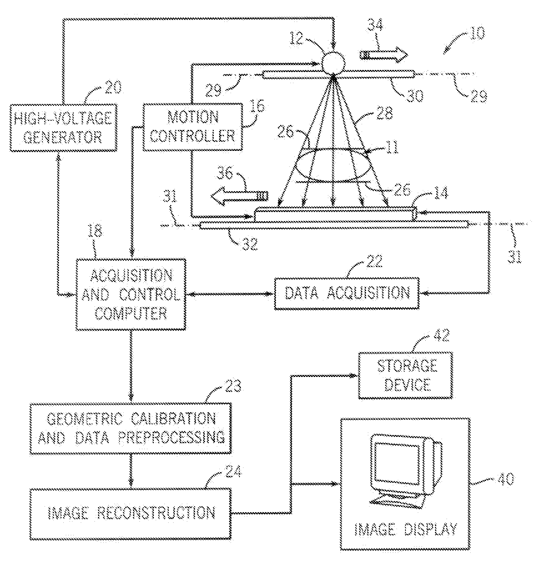
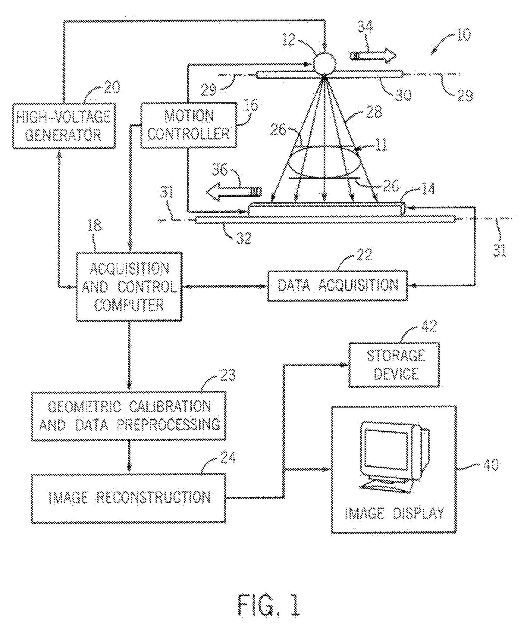
[0020]FIG. 1 is a schematic illustration of the line scan cone beam CT imaging system 10 for generating an image of an object 11 such as a breast. CT imaging system 10 includes an x-ray source 12, a detector array 14, and a motion controller 16 to control the motion of the x-ray source 12 and the detector array 14, with the motion controller 16 under the direction of an acquisition and control computer 18. the acquisition and control computer 18 also controls a high voltage generator 20 for activating the x-ray source 12 as desired and further controls a data acquisition module 22. A geometric calibration and data preprocessing module 23 and an image reconstruction module 24 are also included to provide processing capabilities for the CT imaging system 10.

[0021]When used to image breasts, system 10 can be constructed as a table top system so that during an imaging procedure, similar to conventional x-ray mammography, a patient is standing and the breast is held by two plastic plates...


PUM


Abstract

A line scan cone beam CT imaging system irradiates an object with an x-ray cone beam for multiple views. A projection data set of the object is acquired at each view. Between views, the cone beam and detector array are translated along parallel lines in opposite directions. An image is generated by converting the cone beam projection data set of the real object into a parallel-beam projection data set corresponding to a virtual object and using a total variation minimization image reconstruction algorithm to reconstruct a virtual image of the virtual object. The reconstruction algorithm includes the constraint that the Fourier transform of the reconstructed virtual image matches the known Fourier coefficients in the set of converted parallel-beam projections of the virtual object. The constructed virtual image is then transformed into an image of the real object.

Description

STATEMENT REGARDING FEDERALLY SPONSORED RESEARCH OR DEVELOPMENT [0001]This invention was made with United States government support awarded by the following agencies: NIH Grants: R01EB005712. The United States has certain rights in this invention.CROSS-REFERENCE TO RELATED APPLICATIONS[0002]Not Applicable.BACKGROUND OF THE INVENTION[0003]Breast cancer is the most common cancer among women today. According to recent statistics, all women in the U.S. have a baseline risk of developing breast cancer of approximately 11-12 percent over the course of their lifetime and between 30-40 percent of women who develop breast cancer die from it. At this time, there is no certain way to prevent breast cancer. The best strategy is to detect breast cancer early and treat it before the cancer metastasizes.[0004]X-ray screening mammography has been the most effective method for early detection of breast cancer and clinical studies have shown that it reduces mortality by 30 to 50 percent. However, abo...

Claims


Application Information

Patent Timeline
Patent Type & Authority Applications(United States)
IPC IPC(8): G06K9/00
CPCA61B6/025A61B6/032A61B6/502G06T11/006G06T2211/424G06T2211/436
Inventor CHEN, GUANG-HONG
Owner WISCONSIN ALUMNI RES FOUND
Why Eureka
Try Eureka