Surgical procedures

a surgical and surgical technology, applied in the field of surgical procedures, can solve the problems of difficult analysis of each sample by the cytologist, limited number of limitations, and often cannot be removed, and achieve the effects of less reliance, efficient and reliable location and identification of target tissue, and more precise and reliable excision of tissu

Inactive Publication Date: 2008-11-13
HEALTH BEACONS
View PDF32 Cites 63 Cited by
  • Summary
  • Abstract
  • Description
  • Claims
  • Application Information

AI Technical Summary

Benefits of technology

[0032]At the conclusion of or following surgery, but before closing the surgical field or incision, the radio frequency probe or scanner is again employed to scan one or the other or both of the surgical field, to determine the absence therefrom of the PIT tag or tags, and the excised tissue, to determine the presence therein of the tag or tags, thereby to ensure that the target tissue, organ or object has been successfully excised. This step eliminates or at least minimizes the need for radiological re-imaging in the operating room and / or transporting excised tissue from the operating room to an imaging facility for re-imaging and x-ray analysis and / or for re-excision of tissue.
[0033]The invention thus assures more efficient and reliable location and identification of target tissue, more precise and reliable excision of the tissue, significantly less reliance on and / or need for re-imaging and re-excision, and less time consuming and more efficient and practicable surgical procedures then provided by prior practices.

Problems solved by technology

Despite the advances made in technologies such as medical imaging to assist the physician in early stage diagnosis and treatment of patients with possible atypical tissue such as cancer, it is often necessary to sample difficult to reach organs or tissue lesions by biopsy to confirm the presence or absence of abnormalities or disease.
Not only does this technique require multiple samples, but each sample is difficult for the cytologist to analyze as the specimen cells are isolated outside the context of healthy surrounding tissue.
Despite the advantages of wire localization techniques to locate the suspect tissue for the surgeon, they have a number of limitations.
If a wire is inaccurately placed, it frequently cannot be removed except by surgical excision.
Also, if the tip of the wire penetrates the lesion, the surgeon may sever the lesion in cutting through the tissue along the wire.
Even if a wire does not migrate after placement, the surgeon generally must follow the wire, which is rarely the most cosmetically desirable path to the lesion (such as a circumareolar approach).
Because the distal tip of the wire is often placed in the center of the suspect tissue, a problem known as “track seeding” can rarely occur in which cancerous or precancerous cells disturbed by the wire or surgical pathway are distributed to unaffected tissue during the procedure.
Additionally, the use of a localization wire marker presents logistical problems.

Method used


Image

  • Surgical procedures
  • Surgical procedures

Examples


Embodiment Construction

[0038]The following is a detailed description of an embodiment of the invention presently deemed by the inventors to be the best mode of carrying out their invention.

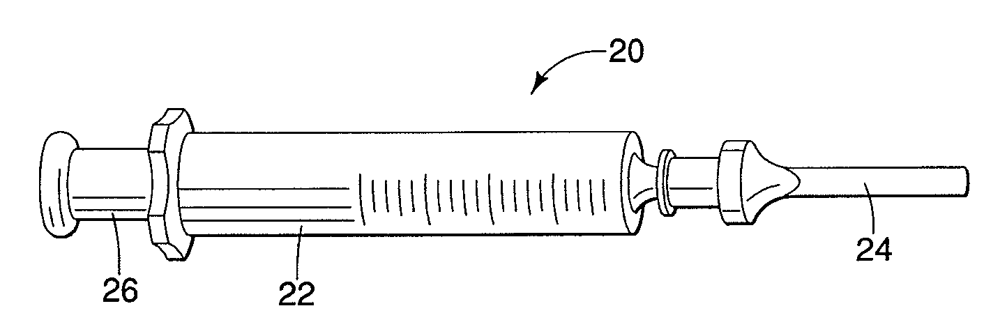
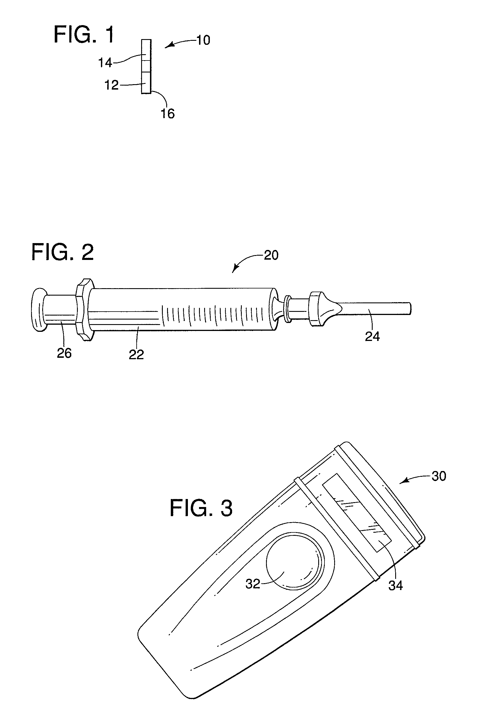
[0039]The invention described herein is appropriate for a range of applications for marking specific volumes of tissue or foreign objects for surgical excision or other purposes. Although the description is largely in the context of marking nonpalpaple lesions in breast tissue for subsequent excision, the invention is not so limited. For instance, the invention may be used to mark tissue in a variety of locations in the body of a human being or an animal, such as the liver, the lungs, muscle tissue, bones, or other tissue or organs where the advantages of the invention may be realized. It may also be used to mark foreign objects in tissue or body cavities, such as a bullet or the like. Accordingly, the invention as described and claimed below is not limited to the marking and removal of lesions in breast tissue.

[0040]FI...


PUM


Abstract

A method of locating and identifying, during surgery, target regions within a body intended for excision of suspect tissue and/or removal of diseased organs or foreign objects, resides in implantation within the body proximate the suspect tissue or object prior to surgery of one or more passive integrated transponder tags and, at the time of and/or during surgery, scanning of the body with a radio frequency scanner or reader that activates the tag or tags and provides the surgeon with one or more signals indicative of the approximate location and unique identification of each of the tags, thereby to aid the surgeon in performance of the surgery. Verification of the success of the surgical procedure is obtained following surgery by scanning the site for absence of the tag or tags and/or by scanning the excised tissue for presence of the tag or tags.

Description

CROSS-REFERENCE TO RELATED APPLICATIONS[0001]This application is a continuation-in-part application filed under 35 U.S.C. §111(a), of International Application PCT / US2007 / 002313, filed Jan. 25, 2007, and claims the benefit of U.S. Provisional Application No. 60 / 761,851, filed Jan. 25, 2006, entitled “Method of Locating Tissue or Foreign Objects Using Implantable Removable Transponders”.FIELD OF THE INVENTION[0002]This invention relates to surgery performed on the bodies of humans and animals for removal of suspect tissue, such as cancer, malignancies, tumors and diseased organs, and for removal of foreign objects, such as bullets. The invention is concerned with the identification and the location of suspect tissue for the benefit of an operating surgeon or veterinarian. More particularly, this invention relates to improved methodology to identify the region of surgical interest and the location of tissue that is to be removed by surgery, and subsequent verification of removal of su...

Claims


Application Information

Patent Timeline
Patent Type & Authority Applications(United States)
IPC IPC(8): A61B5/05A61B19/00
CPCA61B19/54A61B2019/448A61B2019/547A61B2019/5487A61B90/39A61B90/98A61B2090/397A61B2090/3987
Inventor PETCAVICH, ROBERT J.REICHER, MURRAY
Owner HEALTH BEACONS
Why Eureka
Try Eureka