Image processing apparatus, image processing method and recording medium

Active Publication Date: 2009-01-01
FUJIFILM CORP
View PDF1 Cites 6 Cited by
  • Summary
  • Abstract
  • Description
  • Claims
  • Application Information

AI Technical Summary

Benefits of technology

[0021]As described above, according to the aspects of the present invention, when a user only selects a

Problems solved by technology

However, the approaches described in the above two Japanese patents had a problem that a ma

Method used


Image

  • Image processing apparatus, image processing method and recording medium
  • Image processing apparatus, image processing method and recording medium
  • Image processing apparatus, image processing method and recording medium

Examples


Example

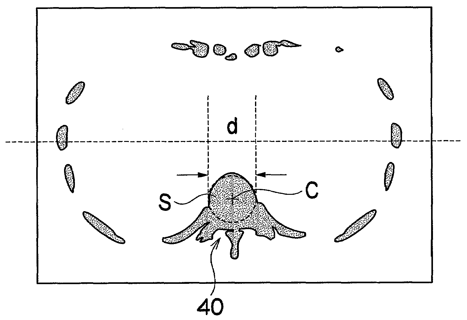
[0032]Now, an image processing apparatus and an image processing method according to the present invention will be explained in detail below with reference to the drawings. In the following embodiments, an X-ray CT image is used for explanation, but the present invention may be also applied to an image such as a synthesized image from an X-ray CT image and a PET image.

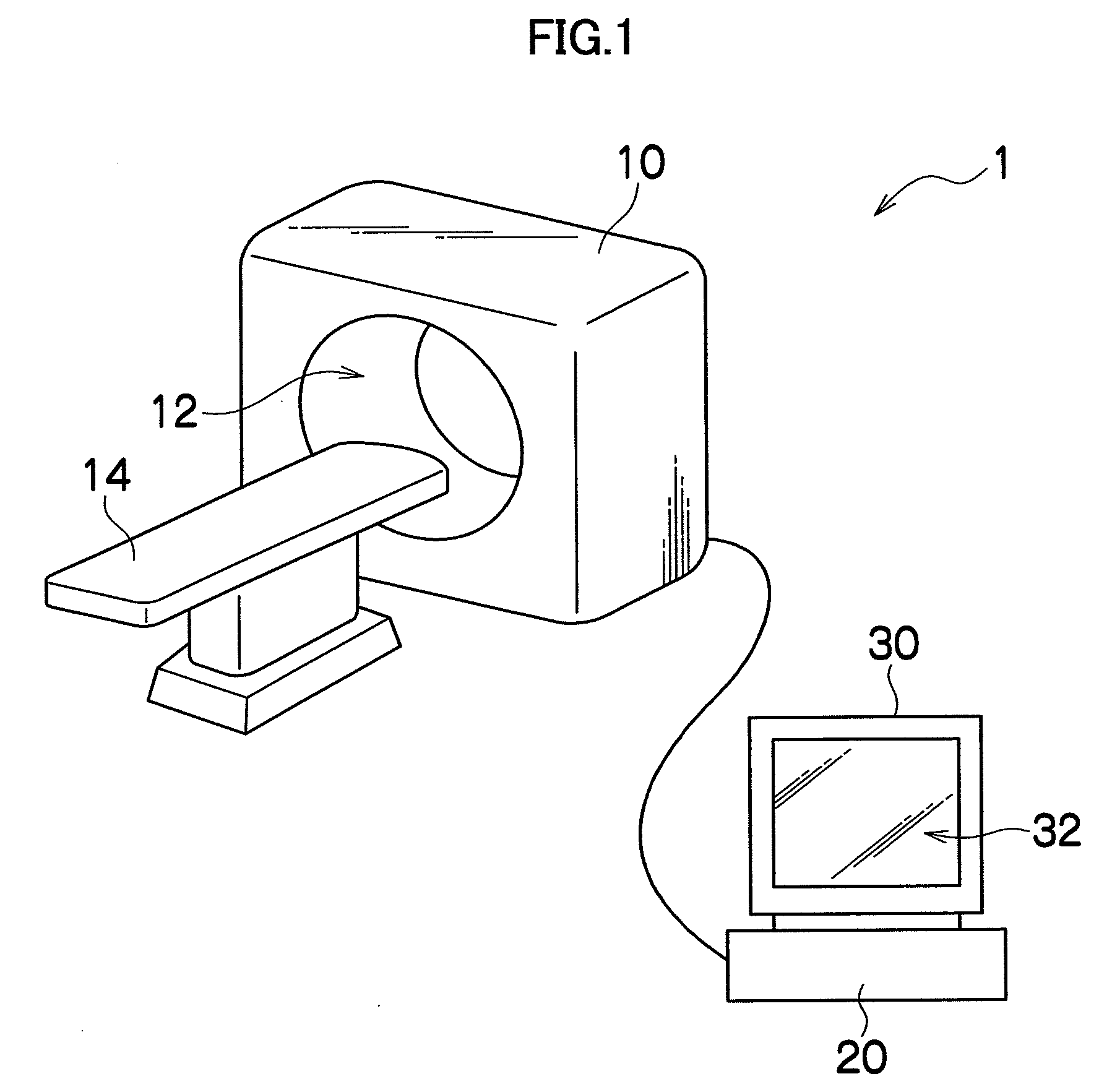
[0033]FIG. 1 is a structural diagram showing an outline of an X-ray CT apparatus according to the present invention which performs image processings for extracting a bone area from a CT image and displaying a bone in units of pieces.

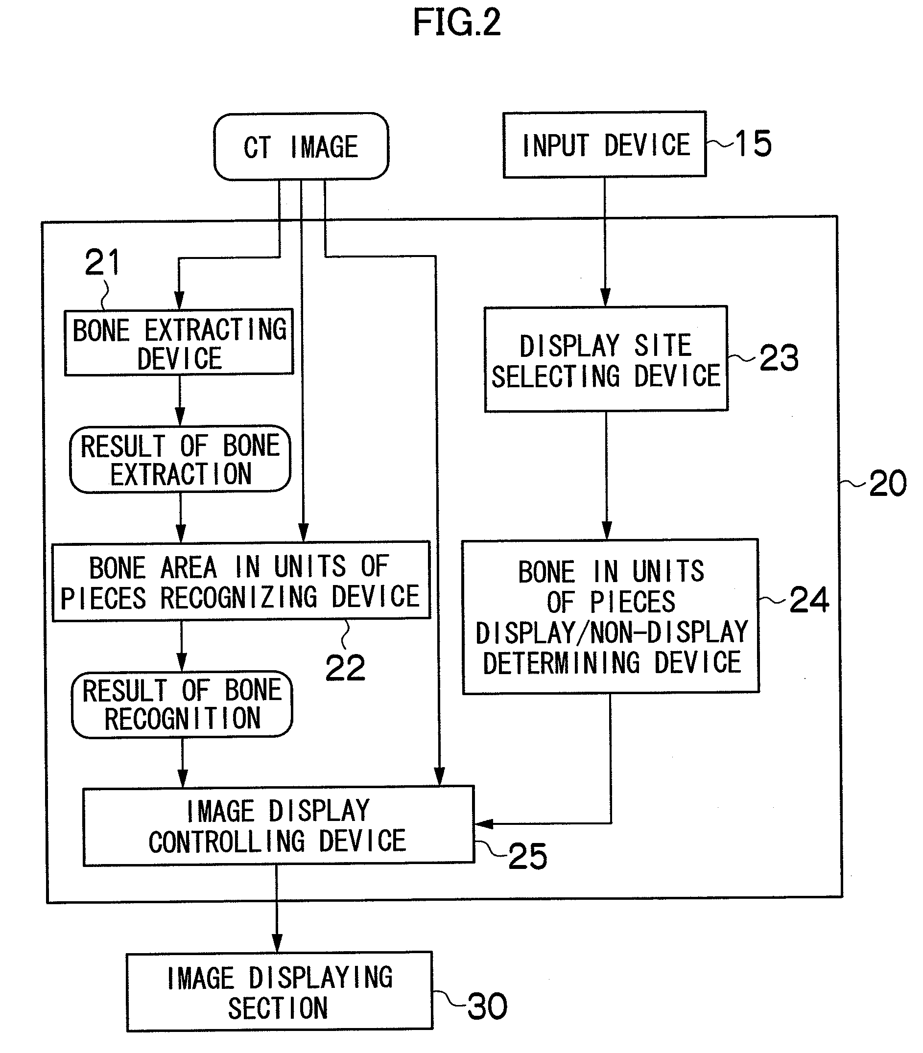
[0034]As shown in FIG. 1, an X-ray CT apparatus 1 generally includes: a scanner 5 gantry section 10 which takes a CT slice image; an image processing section 20 which performs various image processings to the obtained CT slice image; and an image displaying section 30 which displays the processed CT image.

[0035]The scanner gantry section 10 has an opening 12 formed therein, so that a subjec...


PUM


Abstract

An image processing apparatus includes: a recognizing device which recognizes a bone area extracted from a tomogram, the bone area including a bone site composed of several bone pieces, in units of bone pieces; a display site selecting device which selects a bone site to be displayed; a display/non-display determining device which determines a bone piece to be displayed with respect to the selected bone site; and a display controlling device which controls to display a bone piece corresponding to the selected bone site based on the determination by the display/non-display determining device. Thus, only a selection of a bone site to be displayed allows a display of the selected bone site in units of bone pieces.

Description

BACKGROUND OF THE INVENTION[0001]1. Field of the Invention[0002]The present invention relates to an image processing apparatus, an image processing method, and recording medium on which a program causing a computer to perform the steps of the method is recorded. More specifically, the present invention relates to an image processing apparatus, an image processing method and a recording medium for displaying a bone site specified from a bone area extracted from a tomogram image, in units of bone pieces.[0003]2. Description of the Related Art[0004]Conventionally, tomograms of human body such as CT (Computer Tomography), MRI (Magnetic Resonance Imaging), and PET (Positron Emission Tomography) have been widely used for medical diagnosis, and Computer Aided Diagnosis of Medical Images is known.[0005]In the diagnosis, a bone area is automatically extracted from an X-ray CT image as a preprocessing for displaying a MIP (Maximum Intensity Projection) image, recognizing a bone number, or dis...

Claims


Application Information

Patent Timeline
IPC IPC(8): G06K9/00G16H30/40
CPCG06F19/321G16H30/40
Inventor WANG, CAIHUA
Owner FUJIFILM CORP
Why Eureka
Try Eureka