Image processing apparatus, image diagnostic apparatus and image processing method

a diagnostic apparatus and image processing technology, applied in the field of image processing apparatus, image diagnostic apparatus and image processing method, can solve the problems of inability to evaluate subendocardial ischemia, bull's eye not providing precise information with respect to blood,

Active Publication Date: 2009-01-01
TOSHIBA MEDICAL SYST CORP
View PDF8 Cites 66 Cited by
  • Summary
  • Abstract
  • Description
  • Claims
  • Application Information

AI Technical Summary

Benefits of technology

[0022]The present invention as described above makes it possible to display information concerning a myocardial blood flow perfusion in a myocardial thickness direction.

Problems solved by technology

Accordingly, the conventional bull's-eye does not provide precise information with respect to blood flow perfusion that represents the range of ischemia along the myocardial thickness direction.
However, the conventional development image display lacks information with respect to the myocardium along the thickness direction, leading to a problem in that the subendocardial ischemia cannot be evaluated.
This is common problem not only for magnetic resonance imaging apparatuses but for any other diagnostic medical imaging apparatuses which are capable of acquiring image data with high spatial resolutions.

Method used


Image

  • Image processing apparatus, image diagnostic apparatus and image processing method
  • Image processing apparatus, image diagnostic apparatus and image processing method
  • Image processing apparatus, image diagnostic apparatus and image processing method

Examples


Embodiment Construction

[0037]An image processing apparatus, an image diagnostic apparatus and an image processing method according to an embodiment of the present invention will be described with reference to the accompanying drawings.

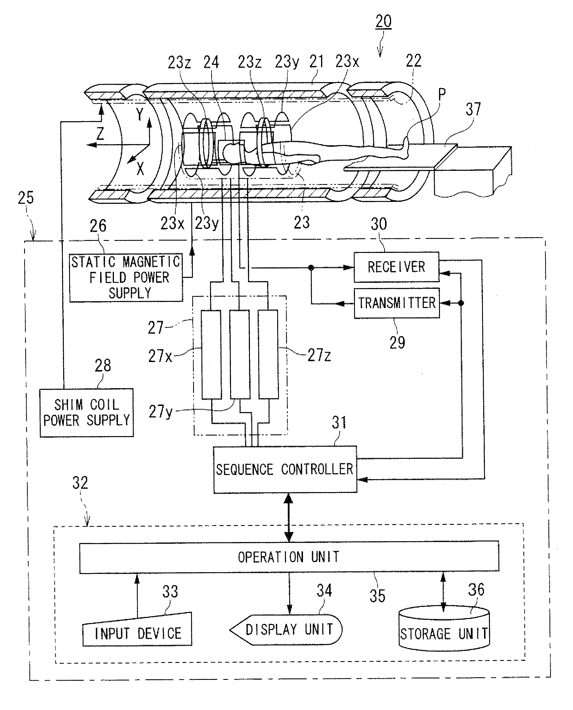
[0038]FIG. 5 is a block diagram showing an embodiment of magnetic resonance imaging apparatus as an example of image diagnostic apparatus having an image processing apparatus according to of the present invention.

[0039]A magnetic resonance imaging apparatus 20 includes a static field magnet 21 for generating a static magnetic field, a shim coil 22 arranged inside the static field magnet 21 which is cylinder-shaped, a gradient coil 23 and a RF coil 24. The static field magnet 21, the shim coil 22, the gradient coil 23 and the RF coil 24 are built in a gantry (not shown).

[0040]The magnetic resonance imaging apparatus 20 also includes a control system 25. The control system 25 includes a static magnetic field power supply 26, a gradient power supply 27, a shim coil power supply...


PUM


Abstract

An image processing apparatus includes an image data acquisition unit, a development generating unit and a display unit. The image data acquisition unit acquires slice image data with regard to a heart of an object. The development generating unit obtains blood flow perfusion information with regard to a myocardial thickness direction based on the slice image data and generates development data according to a desired development format for displaying the blood flow perfusion information. The display unit displays the development data.

Description

BACKGROUND OF THE INVENTION[0001]1. Field of the Invention[0002]The present invention relates to an image processing apparatus, an image diagnostic apparatus and an image processing method, and more particularly, to an image processing apparatus, an image diagnostic apparatus and an image processing method which display a myocardial perfusion image showing a myocardial blood flow perfusion.[0003]2. Description of the Related Art[0004]Magnetic Resonance Imaging (MRI) is an imaging method which excite nuclear spin of an object set in a static magnetic field with a radio frequency (RF) signal having the Larmor frequency magnetically and reconstruct an image based on a nuclear magnetic resonance (NMR) signal generated due to the excitation.[0005]In the diagnosis of ischemic heart disease, as a method for evaluating myocardial blood flow perfusion by means of an MRI, a method is known in which multi-slice dynamic longitudinal relaxation (T1) weighted imaging is performed for obtaining a ...

Claims


Application Information

Patent Timeline
Patent Type & Authority Applications(United States)
IPC IPC(8): A61B5/055
CPCG01R33/5601G06T17/00G06F19/3406G01R33/56366G16H40/63
Inventor SUGIURA, SATOSHI
Owner TOSHIBA MEDICAL SYST CORP
Why Eureka
Try Eureka