
Reconstruction Support Regions For Improving The Performance of Iterative SPECT Reconstruction Techniques

a reconstruction support and reconstruction technique technology, applied in the field of nuclear medicine, can solve the problems of inability to achieve the above-described effort, computationally prohibitive model the system matrix,

Active Publication Date: 2009-04-02
SIEMENS MEDICAL SOLUTIONS USA INC
View PDF2 Cites 10 Cited by
  • Summary
  • Abstract
  • Description
  • Claims
  • Application Information

AI Technical Summary

Benefits of technology

The present invention is a method for reconstructing tomographic images of an object using data collected from a detector. The method involves obtaining data, processing it to create a reconstruction support region, and then creating an image based on this support region. The method can use multiple projection images, detector locations, and attenuation images to create a more accurate and detailed image. The technical effect of this invention is to provide a reliable and accurate method for reconstructing tomographic images of an object.

Problems solved by technology

However, modeling the system matrix can be computationally prohibitive both in terms of the amount of memory required and / or the time that is required.
However, the above-described efforts are generally unsatisfactory.

Method used


Image

  • Reconstruction Support Regions For Improving The Performance of Iterative SPECT Reconstruction Techniques
  • Reconstruction Support Regions For Improving The Performance of Iterative SPECT Reconstruction Techniques
  • Reconstruction Support Regions For Improving The Performance of Iterative SPECT Reconstruction Techniques

Examples


Embodiment Construction

[0020]The present invention will now be described and disclosed in greater detail. It is to be understood, however, that the disclosed embodiments are merely exemplary of the invention and that the invention may be embodied in various and alternative forms. Therefore, specific structural and / or functional details disclosed herein are not to be interpreted as limiting the scope of the claims, but are merely provided as an example to teach one having ordinary skill in the art to make and use the invention.

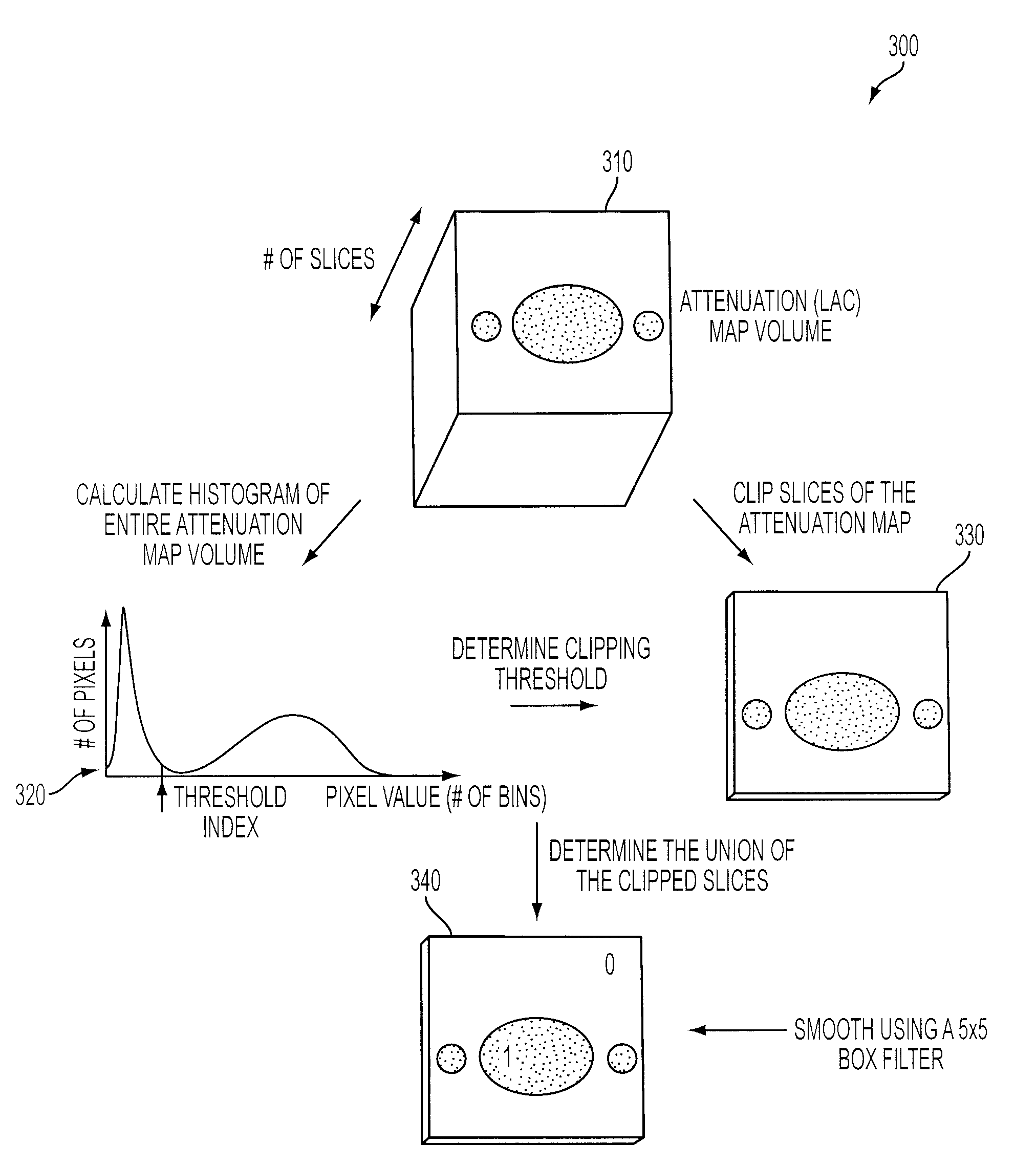
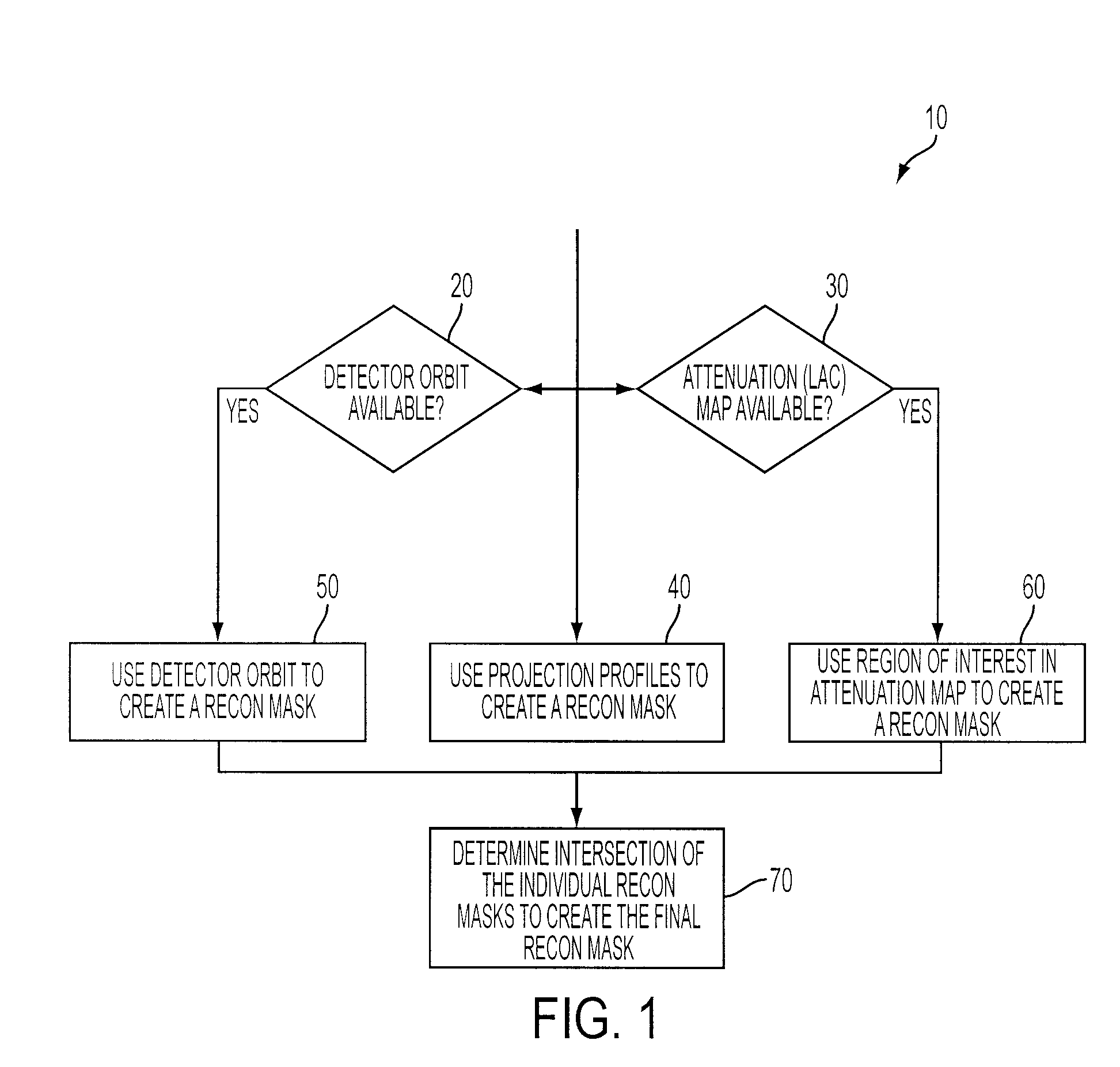
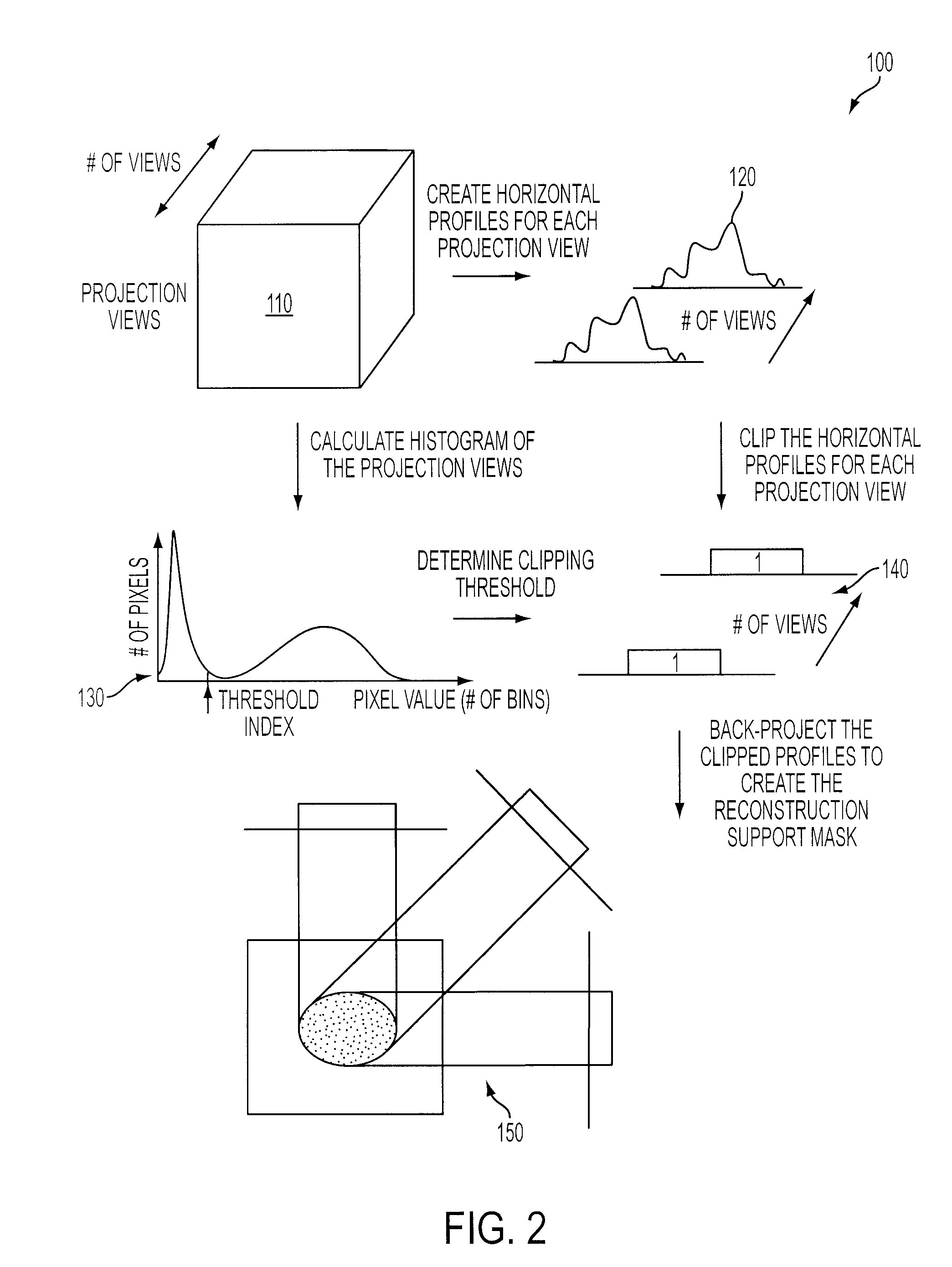
[0021]Tomographic projection images measure radioactive events emanating from an object of interest, e.g., a patient, from multiple view angles. The object of interest, e.g., a patient or portion of a patient, typically encompasses only a fraction (25-50%) of the Field of View (FOV) of a detector. However, iterative reconstruction techniques often reconstruct the entire FOV of a detector, including those regions that fall beyond the object of interest. As a result, iterative reconstr...


PUM


Abstract

A method for iterative reconstruction of tomographic images of an object of interest includes obtaining data corresponding to a field of view of a detector, processing the data to prepare a reconstruction support region corresponding to a region within the field of view encompassing the object, and reconstructing a tomographic image using the reconstruction support region. Preferably, the data corresponding to the field of view includes at least one of a projection image of the object, an orbit location of a detector relative to a center of rotation of the detector, or an attenuation coefficient map of the object, which data can be used to describe a reconstruction support region corresponding to the object.

Description

CROSS-REFERENCE TO RELATED APPLICATIONS[0001]This application claims benefit of under 35 U.S.C. § 119 of U.S. Provisional Patent Application No. 60 / 995,783, filed Sep. 28, 2007, which application is incorporated herein by reference in its entirety.FIELD OF THE INVENTION[0002]The present invention generally relates to nuclear medicine, and systems for obtaining nuclear medicine images. More specifically, the present invention relates to methods for improving iterative tomographic reconstruction techniques in nuclear medicine imaging such as SPECT (Single Photon Emission Computed Tomography).BACKGROUND OF THE INVENTION[0003]Nuclear medicine is a unique medical specialty wherein radiation is used to acquire images which show the function and anatomy of organs, bones or tissues of the body. Radiopharmaceuticals are introduced into the body, either by injection or ingestion, and are attracted to specific organs, bones or tissues of interest. Such radiopharmaceuticals produce gamma photon...

Claims


Application Information

Patent Timeline
Patent Type & Authority Applications(United States)
IPC IPC(8): G06K9/00
CPCG06T2211/424G06T11/005
Inventor CHU, YANHONGRAY, MANJIT
Owner SIEMENS MEDICAL SOLUTIONS USA INC
Learn More