Brachytherapy apparatus and method for use with minimally invasive surgeries of the lung

a brachytherapy and lung technology, applied in the field of minimally invasive lung surgery, can solve the problems of recurrence potential of diffuse proliferative disease on the resected surface, dose generally decreases exponentially, and isotope sources cannot in principle be modulated, so as to facilitate proper lung inflation, prevent or reduce pneumothorax, and eliminate over-exposure

Active Publication Date: 2009-04-30
XOFT INC
View PDF10 Cites 9 Cited by
  • Summary
  • Abstract
  • Description
  • Claims
  • Application Information

AI Technical Summary

Benefits of technology

[0010]Once the balloon is properly positioned and inflated, the lung will likely self-expand or may be gently inflated. If desired, the chest may be imaged using conventional or state-of-the-art methods to assure that the lung tissue closely conforms to and surrounds the balloon as closely as possible. Advantageously, a fluid suction circuit in the applicator leading from outside the patient to outside the shaft of the applicator in communication with the pleural cavity is helpful for preventing or reducing pneumothorax, and facilitating proper lung inflation. The port intended to open into the pleural cavity is preferably adjustable along the shaft of the applicator such that it may be properly positioned and suction can be applied as desired. Alternatively, the suction circuit can comprise a valve, and / or the circuit can be used to instill therapeutic agents.
[0011]When the apparatus is properly situated and the anatomy is in conformance with the balloon, imaging of the site can be performed, and the information used to help create the treatment plan. Such information can include the shape of the treatment cavity, and the location of the cavity relative to anatomical structures, some of which could lie within the range of target tissue, or by their anatomical position, be at risk of inadvertent exposure during delivery of the prescription. If the latter occurs, radiation sensors capable of communication with the central controller can be applied to the skin, or positioned adjacent such structures by needle means or implanted during the surgical procedure. Output from these sensors can be used to eliminate over-exposure to radiation—inadvertent or otherwise.

Problems solved by technology

As with other tumor resection procedures, and even when pathology shows “clear” margins, there is potential for disease recurrence from diffuse proliferative disease in the resected surfaces.
The absence of rib spreading greatly reduces pain and hastens patient recovery, but narrows instrument access to a few small, discrete points on the rib cage.
By contrast, isotope sources cannot in principle be modulated, and in addition require both isolation of the patient during radiotherapy and special facilities and apparatus to assure safety of personnel.
Because dose generally decreases exponentially with distance from the source, accurate dose delivery is complicated and automated treatment planning is generally employed to assure delivery of a dose to the target tissue which is at least equal to the prescription dose, but which is also within allowable limits, thus avoiding substantial necrosis of normal tissue.

Method used


Image

  • Brachytherapy apparatus and method for use with minimally invasive surgeries of the lung
  • Brachytherapy apparatus and method for use with minimally invasive surgeries of the lung
  • Brachytherapy apparatus and method for use with minimally invasive surgeries of the lung

Examples


Embodiment Construction

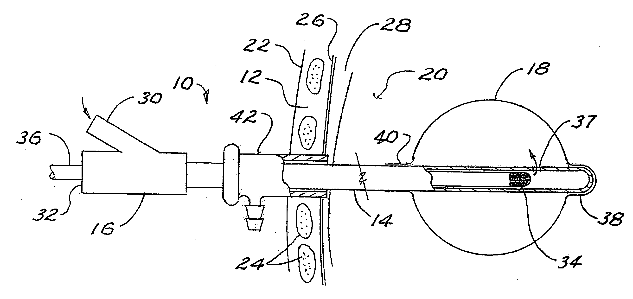
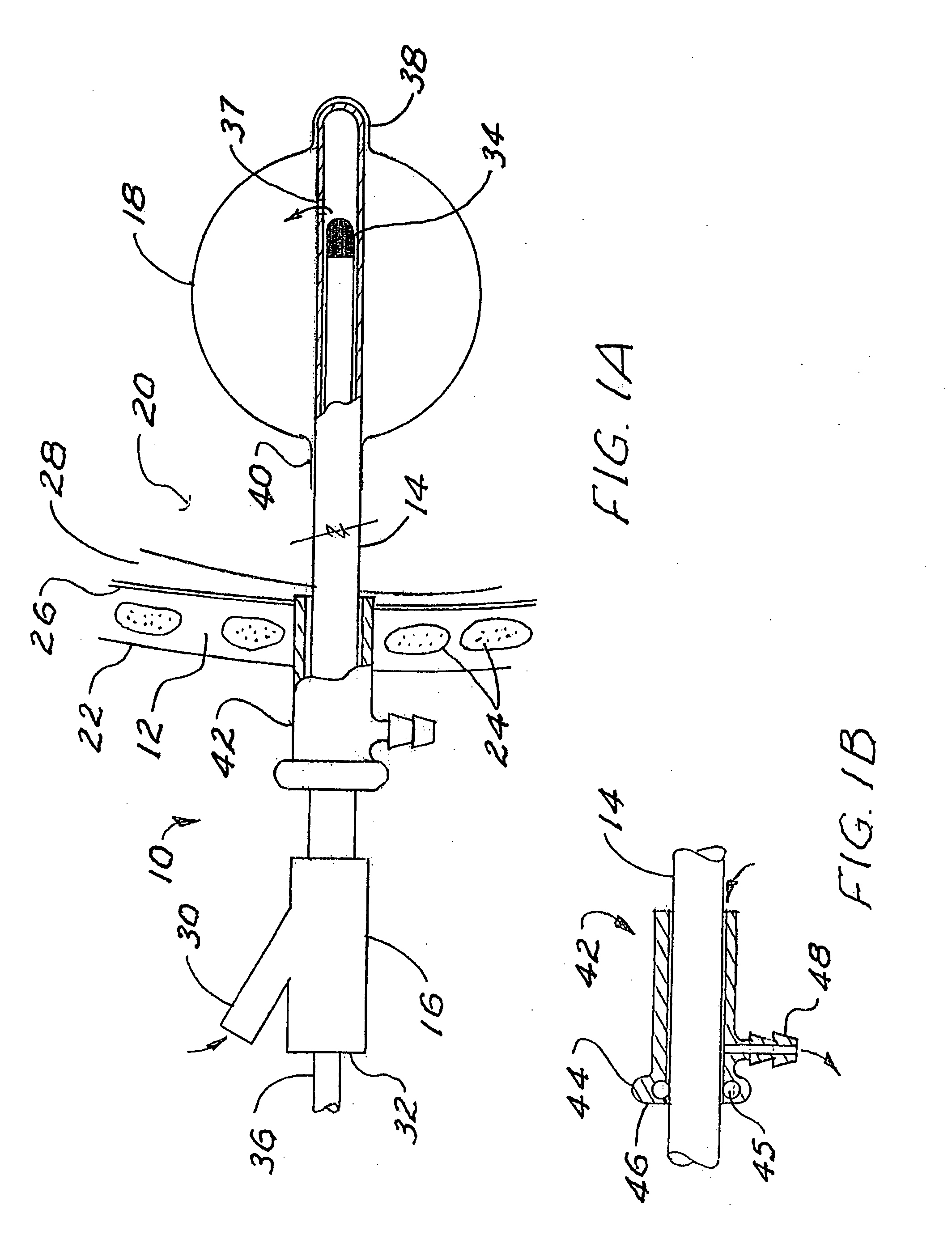
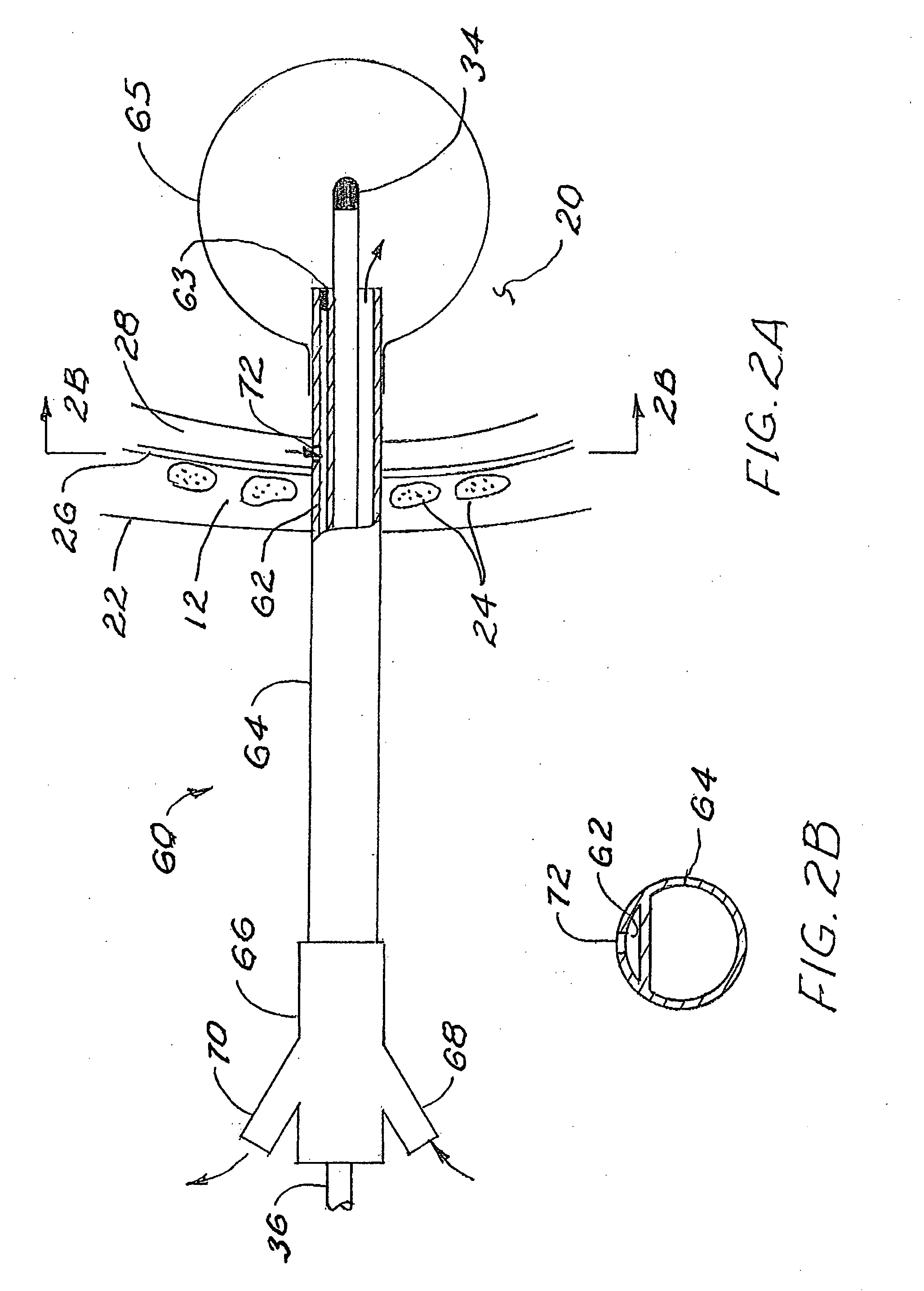
[0022]FIG. 1A is a side view in partial section showing an applicator 10 of the invention passing through the chest wall 12 of the patient. The shaft 14 of the applicator extends from a conventional hub 16 outside the patient into a balloon 18 positioned within a resected portion of the lung 20. The chest wall 12 includes the skin 22, ribs 24, and parietal pleura 26. The space between the parietal pleura 26 and the visceral pleura (not shown) covering the lung 20 defines the pleural cavity 28.

[0023]The hub 16 of the applicator 10 includes a side port 30 for inflating the balloon 18 and a central port 32 for advancing a radiation source 34 and source catheter 36 into the balloon 18. The central port includes a conventional seal (not shown) to avoid fluid leaks around the catheter through the hub. The shaft 14 extends from the hub into the balloon, and the balloon inflation medium passes through the shaft around the catheter 36 to a port 37 opening into the interior of the balloon 18 ...


PUM


Abstract

Brachytherapy treatment of a patient's lung tissue following resection is effected using a balloon applicator which is inserted, normally through the same opening used for the surgery, through the chest wall and into the cavity. The lung and chest openings are closed around the applicator and generally sealed around the applicator. A suction port is provided, in a suction circuit of the applicator, to withdraw fluid from the pleural cavity, at intervals as needed, to assure that the lung can be inflated. Different embodiments of suction circuits are disclosed. A bronchial applicator and method are also disclosed.

Description

BACKGROUND OF THE INVENTION[0001]This invention pertains to minimally invasive surgery of the lung. In particular, it applies to application of brachytherapy techniques directed primarily to lung or pleural tissue surfaces exposed by or created as a result of tumor resection or the presence of primary cancer of the pleura. As with other tumor resection procedures, and even when pathology shows “clear” margins, there is potential for disease recurrence from diffuse proliferative disease in the resected surfaces. Balloon brachytherapy as an adjuvant follow-up to resection has been shown to reduce the likelihood of such recurrent disease.[0002]Recent advances in surgical treatment of proliferative diseases of the lung include endoscopic procedures conducted through, and directed to lesions near the bronchi, and alternatively, video-assisted, minimally-invasive thoracic surgery directed to more peripheral disease and performed through incisions providing access to the thoracic cavity.[0...

Claims


Application Information

Patent Timeline
Patent Type & Authority Applications(United States)
IPC IPC(8): A61M36/04
CPCA61N5/1015A61B1/00094A61B1/00082A61N5/1002A61B1/2676A61M1/0023A61B1/018A61M1/71
Inventor CAROL, MARK P.JERVIS, JAMES E.LOVOI, PAUL A.
Owner XOFT INC
Why Eureka
Try Eureka