Methods and compositions for treating inflammation
Inactive Publication Date: 2009-05-07
CASE WESTERN RESERVE UNIV
3 Cites 2 Cited by
AI-Extracted Technical Summary
Problems solved by technology
Although an inflammatory response is often beneficial, in some cases, continued or excess inflammation may be detrimental to an individual.
This inflammatory response may become excessive and, eventually, become del...
View moreMethod used
[0110]Also provided herein for use in the methods are aerosol formulations, which contain PPARγ agonists and additionally one or more therapeutic agents. The additional therapeutic agents may be selected from any other suitable drug useful in inhalation therapy and which may be presented in a form, which is substantially completely insoluble in the selected propellant. Where appropriate, the PPARγ agonists may be used in the form of salts, esters or as solvates to optimize the activity and/or stability of the PPARγ agonists and/or to minimize the solubility of the PPARγ agonists in the propellant. See, for example, U.S. Pat. No. 6,613,307.
[0118]The expression and role of PPARγ in airway epithelial cells has not been elucidated. Because inflammation is an important part of the CF lung disease, and because expression of PPARγ has been shown to be reduced in organs known to express CFTR in CF mice, we tested the role of PPARγ in airway epithelial cells of CF and non-CF phenotype with respect to the inflammatory response, and in CF and non-CF mice challenged with the CF pathogen, Pseudomonas...
View moreBenefits of technology
[0007]The present invention relates to a method of treating a subject with a cystic fibrosis related disorder. In the method a therapeutically effective amount of at least one PPARγ agonist or a derivative thereof is administered to the subject...
View moreAbstract
A method of treating a subject with a cystic fibrosis related disorder includes administering a therapeutically effective amount of at least one PPARγ agonist or a derivative thereof.
Application Domain
Technology Topic
Cystic fibrosis lungsRelated disorder +5
Image
Examples
- Experimental program(1)
Example
EXAMPLE
Inflammatory Stimuli Alter the Interaction of PPARγ with Binding Partners in Airway
[0115]Epithelial Cells Comparison of Cystic Fibrosis (CF) Cells v. Non-Cystic Fibrosis Cells and Animals
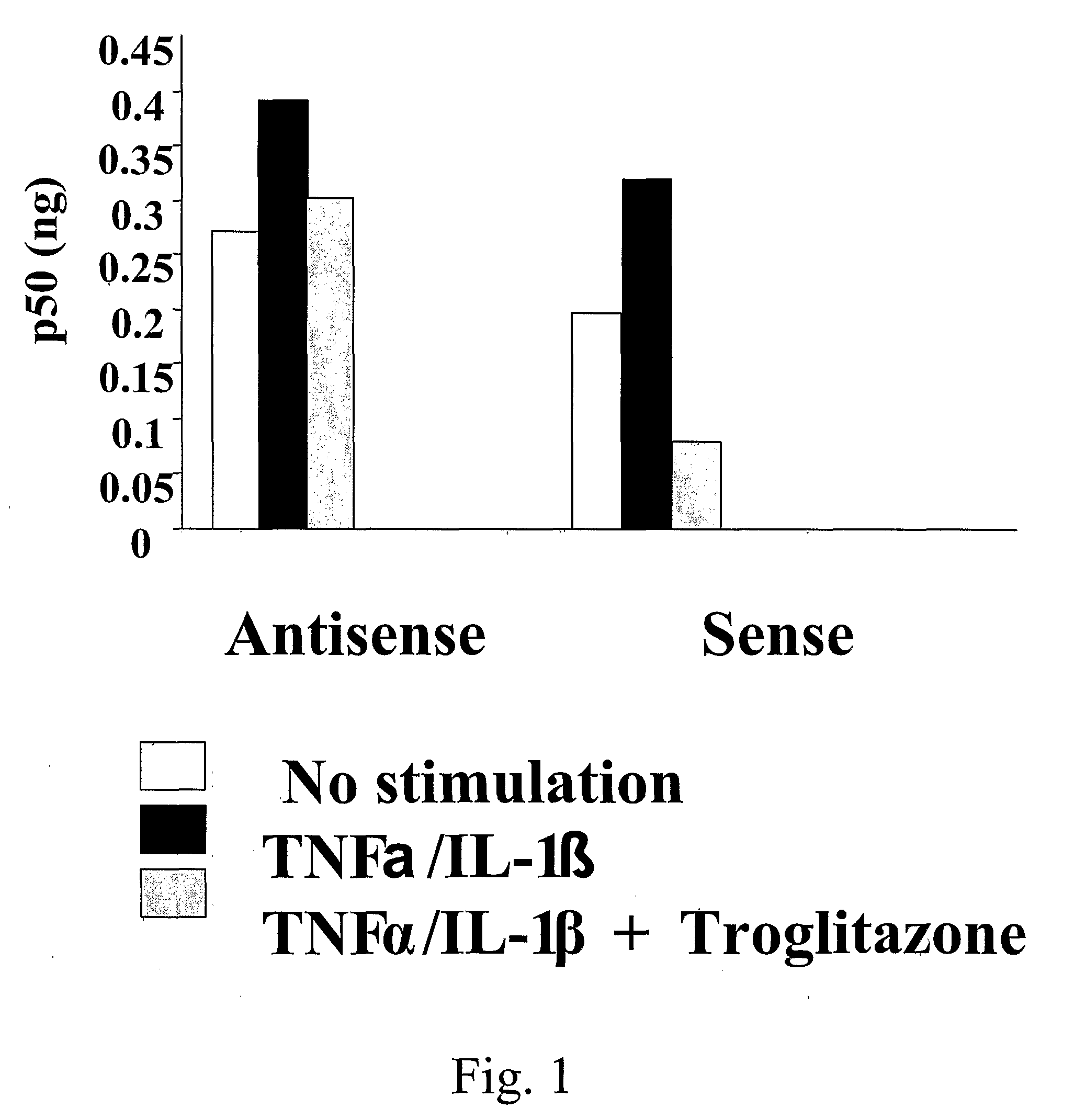
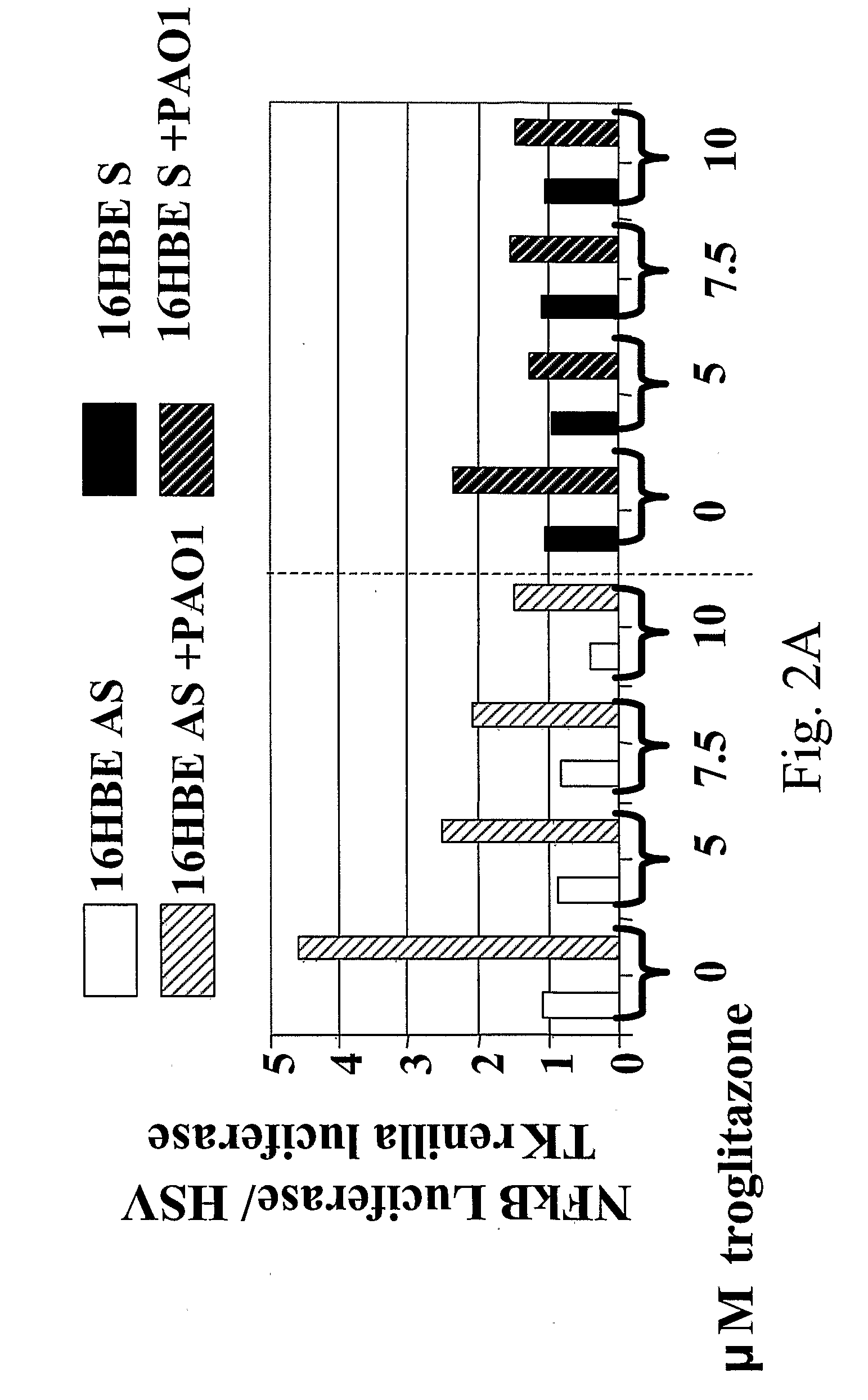
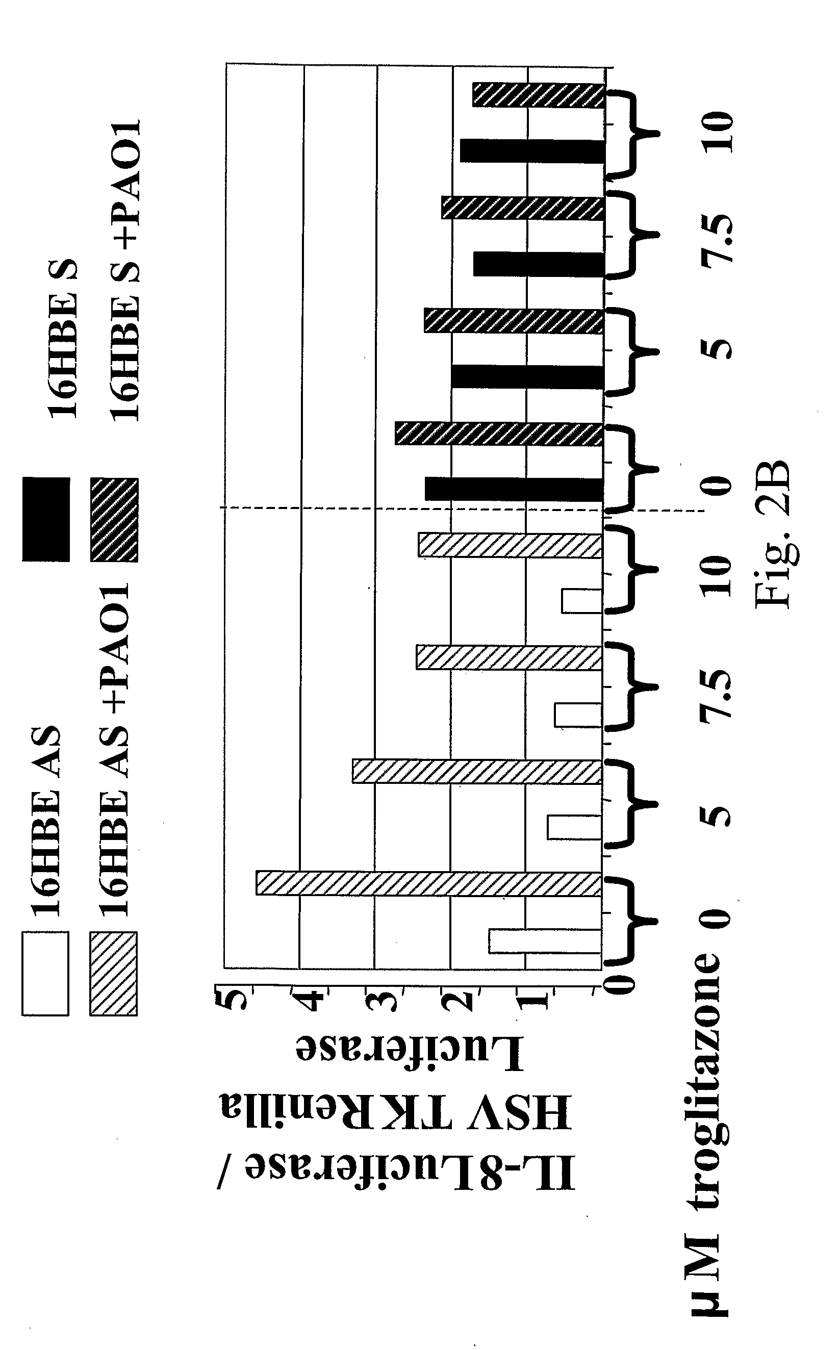
[0116]The CF airway epithelial cell responds to inflammatory stimuli with increased production of proinflammatory cytokine IL-8, as well as IL-6 and GM-CSF compared to normal controls, as a result of increased activation of NF-κB in the CF cells. In order to investigate mechanisms by which NF-κB could be activated in excess in CF, and potential therapeutic interventions to prevent this excessive activation, we assessed PPARγ in airway epithelium. In CF, PPARγ function is reduced. This may contribute to the excess NF-κB activation because PPARγ interacts with NF-κB to prevent its function as a transcription factor. Under conditions of inflammatory stimulation, such as PAO1 exposure or TNFα/IL-1β treatment, the interaction between PPARγ and NF-κB is reduced, but this reduction is abrogated by administration of PPARγ agonists. In vivo, administration of PPARγ agonists results in reduced airway inflammation in response to acute administration of P. aeruginosa in CF, but not wild type, mice. Taken together these data indicate that PPARγ influences the inflammatory response at the level of NF-κB in airway epithelial cells, and it may be a therapeutic target in CF.
[0117]In cystic fibrosis (CF), inflammation is an independent contributor to the decline in pulmonary function and a valid therapeutic target. In vivo studies in infants and children, nearly all studies in CF mice, and many studies in CF airway epithelial cell cultures and cell lines show that the inflammatory response, either to TNFα and IL1β or to P. aeruginosa, occur in excess in CF. The cytokines that are most consistently in excess in CF (e.g., IL-8 or murine equivalents, IL-6, GM-CSF) require activation of NF-κB for upregulation, and several laboratories have shown increased activation of NF-κB in CF airway epithelial cell lines. Failure to appropriately modulate activation of NF-κB could account for the excess inflammatory response in CF, and control of activation of NF-κB could be therapeutic.
[0118]The expression and role of PPARγ in airway epithelial cells has not been elucidated. Because inflammation is an important part of the CF lung disease, and because expression of PPARγ has been shown to be reduced in organs known to express CFTR in CF mice, we tested the role of PPARγ in airway epithelial cells of CF and non-CF phenotype with respect to the inflammatory response, and in CF and non-CF mice challenged with the CF pathogen, Pseudomonas aeruginosa. We found that PPARγ is expressed in human airway epithelial cells in culture and in vivo in mouse airway epithelium. The DNA binding properties of PPARγ are activated in response to challenge with P. aeruginosa. However, PPARγ also interacts with other transcription factors, including NF-κB, and this interaction is reduced by inflammatory stimuli such as P. aeruginosa or TNF-α/IL-1β. Activation of PPARγ with its agonists can restore interaction with other transcription factors, including NF-κB, and also inhibits release of inflammatory mediators and proteins by airway epithelial cells. Here we test the hypothesis that the activation of NF-κB observed in CF epithelial cells can be accounted for in part by reduced binding to PPARγ, and that activation of PPARγ in airway epithelium can prevent excess activation of NF-κB. We also test whether, in an animal model of acute pseudomonas infection, PPARγ agonists will reduce the inflammatory response. Our results show that activation of PPAR-γ can be of therapeutic use in modulating the excess inflammatory response associated with CF.
Methods
[0119]Cell lines: 16HBE-S and 16HBE-AS cells were grown in EMEM supplemented with 10% fetal bovine serum, 2 mM L-glutamine, 100 units/100 μg per ml of penicillin-streptomycin and 400 μg/ml G-418 as previously described.
Well-Differentiated Human Airway Epithelial Cells Grown at the Air-Liquid Interface:
[0120]Human tracheal epithelial cells (HTE) recovered from necropsy specimens were grown in an air-liquid interface (ALI) on collagen-coated, semipermeable membranes (either 7×10⁶ cells/4.5 cm filter or 1×10⁶ cells/1 cm2 filter, transwell-clear polyester membrane, Costar, Corning, N.Y.) and allowed to differentiate in serum-containing media for three or four weeks.
[0121]At three or four weeks, on day 0, cells are switched to submerged culture (liquid-liquid interface, LLI) and treated with either DMSO 1:1000 (vehicle control, normal cells, Sigma, St. Louis, Mo.), or 20 μM CFTRinh-172 (kindly provided by Alan Verkman) prepared in DMSO, and diluted from a 1:1000 stock. Drugs are added to both the basolateral (1 or 2 ml volume, according to filter size) and the apical side (0.35 or 1.5 ml) and media replenished every 24H. Cells grown in this way with I172 have been shown to have continuous inhibition of CFTR activity >90% but no decrease in cell viability or change in cell morphology by electron microscopy. These cells do not display increased amiloride-sensitive sodium conductance. Moreover, we have shown that cells grown in this way with I172 have increased basal and stimulated secretion of IL-8, increased activated RhoA, and decreased Smad3 expression on day 3, and that these changes are not the direct result of I172 on cytokine synthesis per se, since they do not occur in CF cells. At day 3, cells were committed to inflammatory stimulation with TNF IL-1 or PAO1 as indicated below. Cells committed to PAO1 stimulation were switched to serum-free media 24H prior PAO1 stimulation, kept in serum-free media until the end of the experiment, and media replenished every 12H during that time. Serum-free media contained 1:1 DMEM-Ham's F-12, pH. 7.2, L-glutamine 2.5 mM, Penicillin/Streptomycin 100 units/100 μg per ml, gentamicin 50 μg/ml, amphotericin B 1.25 μg/ml from Gibco, Invitrogen Corporation, Carlsbad, Calif.; fluconazole 2 μg/ml (DIFLUCON®, Pfizer); transferrin 5 μg/ml, hydrocortisone 5 μM, insulin 5 μg/ml, endothelial cell growth supplement 20 μg/mil, and bovine serum albumin 1 mg/ml from Sigma, St. Louis, Mo. Serum-containing media had the same antibiotics present as the serum-free media. Antibiotics were not present during PAO1 stimulation.
[0122]In order to observe the localization of the subunits of NF-κB by immunohistochemistry in well-differentiated airway epithelial cells, the cultures were dissociated and single cells transferred to chamber slides and allowed to adhere. This transfer was performed because cells in the well-differentiated model that had been maintained for more than two weeks displayed heaping of cells so that the nuclei could not be located in a single focal plane, making assessment of nuclear translocation of the proteins difficult. Cells were then treated either with vehicle, or TNF/IL1 for 15 min, fixed, permeabilized, and stained with antibody to the p50 and p65 subunits of NF-κB, and DAPI to locate nuclei.
[0123]Zymography: Supernatants of cells were centrifuged for 10 minutes at 14,000 rpm. Supernatants were then concentrated with an Ultra-4 Filter (Amicon) to 50-70 ul. Protein assays were done with Protein Assay Reagent (Bio-Rad). Cell supernatants were mixed with 2× nonreducing SDS sample buffer. Standard SDS-PAGE gels were prepared containing 1 mg/mil gelatin. To allow MMPs to renature, gels were washed twice in 2.5% TX-100 in sterile water. Gels were incubated in activation buffer (10 mM Tris-HCl, pH 7.5, 1.25% TX-100, 5 mM CaCl2, 1 uM ZnCl2) overnight at 37° C. Staining with 0.25% Coomassie brilliant blue R-250 diluted in 40% methanol and 10% acetic acid required 1-2 hours. Gels were destained in 40% methanol and 10% acetic acid until clear zones of protease activity are visible in a blue background.
[0124]Reporter Gene Assays: Cells were seeded in 24-well tissue culture dishes 24 hours before transfection. 20 ug luciferase plasmid and 10 ug Renilla plasmid were mixed into 2 mls serum-free DMEM. NFκB luciferase and AP-1 plasmids were purchased from BD Biosciences CLONTECH. pRLTK was used as an internal control for transfection efficiency.
[0125]300 ul Lipofectamine PLUS reagent (Invitrogen) was mixed with 200 ul serum-free DMEM. The PLUS reagent and plasmid were incubated for 15 minutes at room temperature. 100 ul Lipofectamine was added to 2.4 ml serum-free DMEM.
[0126]The lipofectamine and the DNA-PLUS solutions were mixed and incubated for an additional 15 minutes. The transfection mix was diluted into 20 mls of serum-free DMEM. 250 ul of diluted transfection mix was added to each well and the cells were incubated for 3 hours. Cells were lysed and the lysates assayed for luciferase activity.
[0127]A final stock concentration of 10 mM troglitazone (Cayman Chemical) was dissolved in DMSO and diluted to various concentrations. DMSO was added to the cells as a control. Cells were lysed in 1× Passive Lysis Buffer and assayed with the Dual Luciferase Reporter Assay System (Promega) on a microplate luminometer from Berthold Detection Systems.
[0128]Transcription Factor Arrays: TranSignal TF-TF Interaction Array I (Panomics) was processed according to the manufacturer's instructions. Nuclear extracts from 16HBEo-sense and antisense cells were incubated with biotin-labeled double-stranded oligonucleotides. PPARγ was immunoprecipitated with 3 μg of monoclonal antibody and Dynabeads (Dynal), which are magnetic protein G beads. Free cis-elements and non-specific binding proteins were washed away. PPARγ associated biotin-labeled probes were eluted from the beads and hybridized to TranSignal Protein/DNA array membranes. The arrays were blocked and incubated with Streptavidin-HRP and developed with a chemiluminescent detection system.
[0129]Immunoprecipitations: Nuclear and cytoplasmic extracts were prepared with the nuclear extraction kit (Panomics). Nuclear extracts were used for the immunoprecipitations with a polyclonal antibody against the NF-κB p50 subunit (Santa Cruz). The nuclear extract was diluted to 500 ul with immunoprecipitation buffer, 1% TX-100, 150 mM NaCl, 10 mM Tris pH 7.4, 1 mM EDTA and protease inhibitor cocktail (Sigma). Extracts were incubated with antibody and rotated 1 hour or overnight at 4° C. Antibody-antigen complexes were precipitated with Protein G beads (Roche). Beads were washed three times with cold IP buffer. Beads were eluted in SDS-PAGE sample buffer and boiled. The supernatant was run on 10% SDS-PAGE and transferred to nitrocellulose by electro blotting.
[0130]PPARγ was detected using the PPARγ western blot detection kit (Panomics). Blots were blocked in 3% nonfat dry milk in 1× Wash Buffer II and rocked overnight at 4° C. Affinity purified monoclonal:antibody (1:300) was incubated for 2 hours at room temperature. Blots were washed three times with 1× Wash Buffer II for 15 minutes. Anti-mouse HRP (1:1000) was added for 1 hour at room temperature. Blots were washed 4× with 1× Wash Buffer I for 20 minutes. The blots were developed using the Panomics chemiluminescent detection system.
Results
[0131]Confirmation that NF-κB is activated in excess in CF cell lines: Because one of our hypotheses is that failure of appropriate function of PPARγ contributes to the excess activation of NF-κB, we verified that NF-κB is activated in the CF cell lines in excess in two ways. In addition, to confirm the phenomenology in the cell lines we studied, we determined the amount of activated p50 in the nucleus of 16 HBEo-sense and antisense cells, under basal conditions, and under conditions of stimulation (FIG. 1). There was an increase in activated p50 in the nucleus in response to PAO1 in both sense and antisense cell lines and the amount was greater in the antisense (CF) cell lines than the sense (non-CF). In addition, we transfected into these cells constructs containing the luciferase gene driven by NF-κB elements, or the native IL-8 promoter. The cells were then exposed to PAO1 and promoter activity assessed by measuring luciferase activity. The antisense (CF phenotype) cell lines had greater luciferase expression in response to PAO1 than did the sense (nonCF phenotype) cells (FIG. 2). Thus, with three independent assays in two different model systems, the activation of NF-κB in CF models is in excess of that observed in non CF models.
[0132]Identification of PPARγ in extracts of airway epithelial cells: Both cytoplasmic and nuclear extracts of 9HTEo- and 16HBEo-cell pairs (CF phenotype and non-CF phenotype) demonstrated the presence of PPARγ by Western blot (FIG. 3). In the 9HTEo-cell pairs, there appeared to be equivalent amounts of the protein in the CF and non-CF members of the pair. For the 16HBEo-cell pairs, however, the antisense (AS), or CF phenotype, member of the pair expressed less PPARγ than the sense congener (non-CF phenotype). This differential expression does not change if the cells are stimulated with PAO1 (FIG. 3B). EMSAs using the PPRE (FIG. 4) demonstrate DNA binding by components of the nuclear extract from these cell lines, which is markedly reduced by inclusion of cold probe, but not by cold probe of mismatched sequence, and which undergoes supershift with antibody to PPARγ, identifying the binding protein as PPARγ. For both the 9HTEo-pair and the 16HBEo-pair, the CF member of the pair displays less PPRE binding. Therefore, PPARγ is expressed in human airway epithelial cell lines, CF and non-CF, but appears to be less functional in binding its target DNA sequence in CF. Western blot confirms that PPARγ is also present in well-differentiated airway epithelial cells grown at the air-liquid interface (data not shown).
[0133]Cytokine and MMP-9 production by well-differentiated airway epithelial cells at the air-liquid interface is inhibited by agonists of PPARγ: When exposed at the apical surface to the laboratory strain of P. aeruginosa, PAO1 for one hour, or when stimulated by TNFα/IL-1β for one hour, well differentiated airway epithelial cells produced IL-8, IL-6, and GM-CSF in a dose-dependent fashion. The absolute amounts of cytokines produced varied from sample to sample, from different donors, but there was excellent agreement in the triplicate wells from a single donor. For all donors, there were measurable quantities of IL-8, but for cells from some donors, levels of IL-6, and/or GM-CSF were sometimes below the limits of detection. When PPAR agonists were added to the medium and cytokine production measured 6, 12 or 18 hr after stimulation, there was significant inhibition of cytokine production by the agonists (FIGS. 6 and 7). At or after 24 hours after stimulation, without replenishment of drug supply, inhibition was not evident (data not shown). Inhibition was dose dependent over the range of 0.1-10 mg/ml for troglitazone (data not shown).
[0134]Gelatin zymography shows that well-differentiated airway epithelial cells grown at the air-liquid interface release MMP-9, which can digest the protein in the gel. Release of MMP-9 is also inhibited by PPARγ agonists (FIG. 5).
[0135]Activation of NF-κB is inhibited by agonists of PPARγ: 16HBEo-cell pairs transfected with a construct of NF-κB binding elements driving firefly luciferase displayed activation of luciferase activity after stimulation with PAO1. This activation was significantly inhibited by troglitazone, in dose-dependent fashion (FIG. 2). To test whether the NF-κB responsive elements would be affected by PPAR agonists in the context of a native promoter, we tested the effect of troglitazone on a luciferase construct driven by the upstream regulatory elements of the IL-8 gene. Similar inhibition was seen with PPAR agonists (FIG. 2). These transfections were not performed in the 9HTEo-pair because the two cell lines were very different in their ability to be transfected (the 9HTEo-pCEP R cell line expressed over 100 fold less reporter gene than the 9HTEo-pCEP cell line), making comparative studies difficult. They were not performed in the well-differentiated airway epithelial cells because these cells are very difficult to transfect.
[0136]Interaction of NF-κB and PPARγ: In order to test whether PPARγ can interact directly with NF-κB, we conducted co-immunoprecipitation assays. Antibodies to both the p50 and the p65 subunits of NF-κB can pull down PPARγ (FIG. 8). In addition, antibodies to PPARγ also pulled down p65 and p50, though these assays had to be performed in whole cell extracts in order to recover sufficient PPARγ (FIG. 9). In a second, more sensitive assay, which capitalizes on the ability of the DNA target sequence of each transcription factor to bind specifically both to its cognate transcription factor and to its minus strand, interaction of PPARγ with NF-κB was also identified (FIGS. 10 and 11). This interaction was reduced by prior incubation of the cells with PAO1, but could be preserved in part by the inclusion of troglitazone in the incubation mix and in the subsequent culture media. These data suggest that although inflammatory stimulation causes changes in NF-κB that reduce its interaction with PPARγ, these changes can be partly abrogated by activation of PPARγ with an agonist ligand.
[0137]In order to test the impact of CFTR deficiency on the interaction of PPAR-γ with other transcription factors, we treated well differentiated airway epithelial cells grown at the ALI with I172 (20 uM), an inhibitor of CFTR activity, continuously for 72 hr prior to preparation of nuclear extracts or stimulation. This treatment has been shown to inhibit CFTR activity continuously by over 90%, as assessed by using chamber estimates of ion currents, without compromising cell viability. This model allows one to compare, in well-differentiated airway epithelial cells that have identical genetic endowment at all loci, the effect of CFTR inhibition on various cellular processes. Cells treated with I172 displayed less interaction between PPARγ and other transcription factors, particularly following stimulation with TNFα/IL1β. During the course of these experiments, cells from the airways of a patient with CF of genotype ΔF508/ΔF508, obtained at transplant, became available and were cultured at the air-liquid interface. These cells displayed vigorous stimulation of IL-8 in response to PAO1 or TNFα/IL-1β. Nuclear extracts of these cells showed very limited interaction between PPARγ and other transcription factors, including NF-κB. While this represents only a single sample, with the attendant possibility that variation at other genetic loci could produce the observed results, these results support the concept that in CF, reduced interaction of PPARγ and NF-κB may contribute to the excess activation of genes driven by NF-κB.
[0138]Pioglitazone inhibits the inflammatory response in CF mice to acute administration of Pseudomonas: Mice pretreated with pioglitazone or vehicle by gavage, then challenged with prior to challenge with M57-15 P. aeruginosa, underwent BAL for inflammatory response outcome measures 24 hours after challenge. Cell counts, cytokine values, and body weight were recorded. WT mice had similar inflammatory parameters and weight loss whether they received pioglitazone or vehicle. CF mice treated with vehicle had marked increase in inflammatory response compared to WT mice treated with vehicle, as previously reported for untreated mice (FIGS. 12-14). However CF mice treated with pioglitazone had significant reduction of the inflammatory response by pioglitazone.
[0139]PPARγ expression in airway epithelium of mice: Immunostaining for PPARγ is observed in airway epithelial cells in sections of mouse lung, whereas sections treated with the secondary antibody with no primary antibody show no signal. Expression is indistinguishable in airways from CF and WT mice, is present in both cytoplasm and nucleus, and does not change in intensity or location following acute infection with P. aeruginosa in either CF or WT mice, even in areas in which an inflammatory infiltrate is identified. Therefore, in contrast to findings described for intestinal epithelium, we cannot ascribe the differential anti-inflammatory response of CF and WT mice to pioglitazone to differences is subcellular localization of the protein following drug administration or infection.
Discussion
[0140]In the lungs of CF children, exposure to bacteria results in neutrophil and IL-8 recruitment into the BAL fluid in excess of what is seen in infected non-CF control young children, even when controlled for burden of organisms in the lungs. Some studies in CF infants suggest that inflammation may even precede infection, though it is difficult to exclude the possibility that those infants were infected earlier and the inflammatory response simply persists well after the infectious agents can no longer be detected. CF mice of various genotypes (G551D, S489X, ΔF508, Y122X, R117H) on different genetic backgrounds (CD-1, C57BL/6, mixed C57BL/6 and 129, and mixed C57BL/6, 129, and FV/B) studied in at least three different laboratories around the world, challenged with pseudomonas embedded in agarose beads, have excess cytokines and inflammatory cells in BAL fluid. In addition, in response to acute challenge with pseudomonas, CF mice have greater cell and cytokine response, even though they kill the bacteria at least as well as their wild type counterparts. This inflammatory response is itself an independent contributor to the progression of the CF lung disease, because when inflammation is inhibited by alternate-day steroids or high dose ibuprofen, the rate of decline of pulmonary function is slowed. However, adverse effects from alternate-day steroids are prohibitive in CF, and the increased incidence of the rare complication of gastrointestinal hemorrhage with high dose ibuprofen has made many clinicians avoid its use, despite unequivocal evidence of benefit. Understanding and controlling the inflammatory response without harming the host defenses against bacteria and without incurring adverse effects could be of great benefit to CF patients.
[0141]Most, but not all, published data suggest that airway epithelial cells may contribute to the excess inflammatory response in CF. These cells are good candidates to contribute to the inflammatory response because they are the initial site of contact with the outside world and often the first cells to contact inhaled bacteria, they are known to express CFTR and to manifest its lack by altered salt transport and other abnormalities, such as reduced NOS-2 expression, and CF mice whose airway epithelial cells have been corrected by expression of the CFTR transgene driven by the K18 promoter only in epithelial cells only lack the excess inflammation in response to agarose containing agar beads. Human airway epithelial cells in culture with the CF phenotype usually, but not invariably, produce more IL-8 and sometimes other cytokines in response to PAO1 or its products, or TNF-α and IL-1β. Data from several laboratories indicate that activation of NF-κB occurs in excess in CF airway epithelial cells. Increased NF-κB driven transcription could account for the increased IL-8, IL-6, GM-CSF, ICAM-1 and other inflammatory proteins that have been detected in the surface or media from CF airway epithelial cells. Our data in our well-matched cell lines and in WD AECs treated or not with the CFTR inhibitor, I172, confirm the reports of increased IL-8, IL-6, and/or GM-CSF in CF phenotype cells in response to PAO1 or TNF-α plus IL-1β. Our data also indicates that increased activation of NF-κB is associated with this increase in proinflammatory mediator production in both cell lines and well differentiated cells grown at the air-liquid interface.
[0142]The nuclear receptor, PPARγ, is expressed in airway epithelial cells. When PPARγ ligands are administered along with or prior to inflammatory stimuli, NF-κB driven processes are inhibited, including the production of IL-8, IL-6, and GM-CSF and the release of matrix metalloproteinase 9 (MMP9) in response to pseudomonas or cytokine stimulation. Transcription from an NF-κB luciferase construct or one in which the IL-8 promoter is used to drive luciferase is reduced by agonists of PPARγ in airway epithelial cells, indicating that these agonists may exert at least a portion of their activity at the level of gene transcription. Here we show that the mechanism by which this occurs is could be by direct interaction with NF-κB or by interaction with a third protein, possibly a DNA helicase, to which both NF-κB and PPARγ bind. Both the p50 and the p65 subunits co-immunoprecipitated with PPARγ, and PPARγ co-immunoprecipitated with specific antibodies to both p50 and p65. This interaction was confirmed by another technique of recognizing interaction, which is much more sensitive because it recognizes transcription factors by DNA base pairing in their target sequences. These data indicate that the failure of interaction between NF-κB and PPARγ in CF, especially under conditions of inflammatory stimulation, is mirrored by the reduced interactions of PPARγ with many other transcription factors, some of which also drive or promote inflammatory processes. Thus, the interactions of PPARγ with transcription factors could have broad implications for regulation of the inflammatory process. PPARγ interacts with AP-1 and AP-2, which are required for transcription of MMP-9. Other transcription factors identified in these arrays are: RXR, the known binding partner of PPARγ as well as Stat 1 and Stat 4. The interaction of PPARγ with these other transcription factors, including AP-1 and AP-2, is also attenuated when the cells are stimulated with PAO1 or TNFα/IL-1β, and the attenuation is rescued by troglitazone, both in the CF and the non-CF cell lines. The specific mechanisms by which PPAR Y interaction is reduced by proinflammatory stimuli are not clear. However, the most parsimonious explanation for the near-universal concomitant decrease in interaction of PPARγ with other transcription factors as well as the rescue of interaction with nearly all these factors by troglitazone is that a conformational change has taken place in PPARγ in the face of inflammatory stimuli, probably by post-translational modification (e.g., by phosphorylation) which reduces its ability to interact with other transcription factors. Binding to troglitazone could then either protect PPARγ from the post-translational modification, or change its conformation so that, even if it is modified, it can still interact with transcription factors. It seems unlikely that concomitant changes take place in all the other transcription factors in the array that alter their ability to interact with PPARγ. However, it is possible that the inflammatory process alters a common binding partner of all the transcription factors, such as a helicase, and it is this change, rather than changes in PPARγ, that alters the interactions we observe. However, the fact that we also observe that inflammatory stimulation increases binding of PPARγ to its target DNA sequence in the EMSA assay, together with the ELISA data indicating increased PPRE binding of PPARγ in nuclear extracts following inflammatory stimulation suggests that there the conformational changes in PPAR that reduce its ability to interact with other transcription factors, may increase its propensity to bind to its DNA target sequence. One possible explanation is that activation of kinases such as JNK and ERK following inflammatory stimuli can phosphorylate PPARγ in such a way as to promote its inactivation and degradation. If PPARγ is bound to its ligand, it may remain in a conformation less favorable for phosphorylation and subsequent accelerated degradation.
[0143]The DNA binding activity of PPARγ is reduced in CF airway epithelial cells. EMSAs indicate less interaction of PPARγ with its target DNA sequence in two CF model systems compared to matched controls. For the 16HBEo-cells, this could be due, at least in part, to reduced expression of PPARγ in the CF member of the pair, as demonstrated by Western blot, but in the 9HTEo-cell pair, expression is comparable in the CF and the non-CF members of the pair. It seems most likely that the ability of PPARγ to bind to its target DNA sequence is reduced. The 9HTEo-cell pair differs from the 16HBEo-cell pair in that the 16HBEo-pair displays activation of IL-8 and IL-6 production at baseline, but the 9HTEo-cell lines are quiescent until a stimulus is applied, and the basal production of cytokines is minimal. If the continuous activation in the 16HBEo-cells results in more rapid degradation of PPARγ, this might account for the greater deficit in CF cells in this cell line. It is possible that the CF cell lines exist in a heightened inflammatory state and PPARγ is sensitive to this constitutive activation. In this CP mouse model, application of troglitazone results in the proper nuclear translocation of the PPARγ in the gut, which is not observed in the absence of ligand. However, we did not observe these changes in localization of PPAR immunostaining in the lungs of CF knockout mice compared to wild type. It may be that in the lung, the expression of PPARγ is quite sensitive to the inflammatory environment, and any changes we observe in CF may be due to heightened inflammatory tendencies. This suggestion is supported by the reduction of PPARγ in patients with asthma or alveolar proteinosis, diseases characterized by inflammation, but normal CFTR. Even if the changes in PPARγ are associated with changes in the inflammatory milieu in CF and are not related to the CF defect itself, they could in turn contribute to the excess inflammatory response and could represent a valid therapeutic target. It now appears that some, but not all, nonsteroidal anti-inflammatory drugs (NSAIDs) can ligate PPARγ. Ibuprofen, at the concentration required to observe the therapeutic effect in CF, is one of those drugs. Ligation of PPARγ might, therefore, be the mechanism of action of one of the proven anti-inflammatory therapeutic agents in CF.
[0144]In order to test the therapeutic potential of PPARγ agonists in CF animals, we utilized the acute pseudomonas challenge model in CF and non-CF mice, because it was the closest mimic of the acute pseudomonas challenge applied to the epithelial cells in culture. We administered pioglitazone by gavage because this is one of the two PPARγ agonists available for human use. In two of the three experiments, pioglitazone limited the inflammatory response in the CF mice. However, the dose used in these studies was high compared to conventional human doses, on a weight basis, and the drug was administered prior to challenge, a luxury that may not be available for many patients with CF.
Login to view more PUM


Description & Claims & Application Information
We can also present the details of the Description, Claims and Application information to help users get a comprehensive understanding of the technical details of the patent, such as background art, summary of invention, brief description of drawings, description of embodiments, and other original content. On the other hand, users can also determine the specific scope of protection of the technology through the list of claims; as well as understand the changes in the life cycle of the technology with the presentation of the patent timeline. Login to view more.
Login to view more Similar technology patents
Adam-15 antibodies and immunogenic peptides
ActiveUS20110117090A1Inhibit activationEnhance activityOrganic active ingredientsSugar derivativesMetalloproteinasePeptide
Owner:VASGEN
Methods for identifying autoimmune arthritis and for screening for inhibitor of activation of autoimmune arthritogenic t cells
InactiveUS20160169906A1Inhibit activationCompound screeningApoptosis detectionCytokine secretionRibosomal protein E-L30
Owner:KYOTO UNIV
Polymer compound for medical use and composition and device using same
Owner:YAMAGATA UNIVERSITY +1
Immune cell activation inhibitor and use thereof
InactiveUS20150202170A1Inhibit activationAntibacterial agentsUrea derivatives preparationInflammatory skin diseaseDrug
Owner:NAT UNIV CORP EHIME UNIV
Pharmaceutical composition for preventing or treating inflammatory diseases, allergic diseases or asthma, containing diospyros blancoi a. dc. extract as active ingredient
ActiveUS20130164395A1Inhibit activationSuppress secretionBiocideAntipyreticInflammatory skin diseaseBronchoalveolar lavage
Owner:KOREA RES INST OF BIOSCI & BIOTECH
Classification and recommendation of technical efficacy words
- Inhibit activation
Thiazole and thiadiazole compounds for inflammation and immune-related uses
InactiveUS20070254925A1Suppress immune systemInhibit activationBiocideSenses disorderHypersensitivity DiseasesImmune disorder
Owner:SYNTA PHARMA
Use of specific T2R taste receptors to identify compounds that block bitter taste
ActiveUS20030170608A1Block bitter tasteInhibit activationCompound screeningApoptosis detectionDrug6-nitrosaccharin
Owner:SENOMYX INC
Vehicle AC Ground Fault Detection System
InactiveUS20090091868A1Prevent shockInhibit activationElectric devicesShort-circuit testingElectrical switchingElectricity
Owner:LEAR CORP
Uses of inhibitors for the activation of CXCR4 receptor by SDF-1 in treating rheumatoid arthritis
InactiveUS20050202005A1Inhibit activationOrganic active ingredientsAntibody ingredientsInflammatory arthropathyMedicine
Owner:THE TRUSTEES OF COLUMBIA UNIV IN THE CITY OF NEW YORK
Protection of cells from degeneration and treatment of geographic atrophy
ActiveUS9707235B1Increase levelInhibit activationOrganic active ingredientsAgainst vector-borne diseasesCancer researchScreening method
Owner:UNIV OF KENTUCKY RES FOUND