Immune cell activation inhibitor and use thereof

Inactive Publication Date: 2015-07-23
NAT UNIV CORP EHIME UNIV
1 Cites 0 Cited by

AI-Extracted Technical Summary

Problems solved by technology

In recent years, increase in the number of patients with these diseases is seen as a problem, and the prevention and trea...

Benefits of technology

[0013]According to the present invention, activation of immune cells can be inhibited by the presence of bromovalerylurea or a derivative thereof.
[0014]Because bromovalerylurea or a derivative thereof can inhibit the activation of immune cells ...

Abstract

The present invention provides a pharmaceutical that can be used widely for intractable neurological diseases, inflammatory diseases, etc. The immune cell activation inhibitor of the present invention contains bromovalerylurea or a derivative thereof. The immune cell activation inhibitor of the present invention can be used as a pharmaceutical for, for example: chronic neurological diseases such as Parkinson's disease and Alzheimer's disease; acute neurological diseases such as cerebral infarction and brain injury; and inflammatory diseases such as systemic inflammatory response syndromes.

Application Domain

Antibacterial agentsUrea derivatives preparation +8

Technology Topic

Inflammatory skin diseaseDrug +9

Image

  • Immune cell activation inhibitor and use thereof
  • Immune cell activation inhibitor and use thereof
  • Immune cell activation inhibitor and use thereof

Examples

  • Experimental program(5)

Example

Example 1
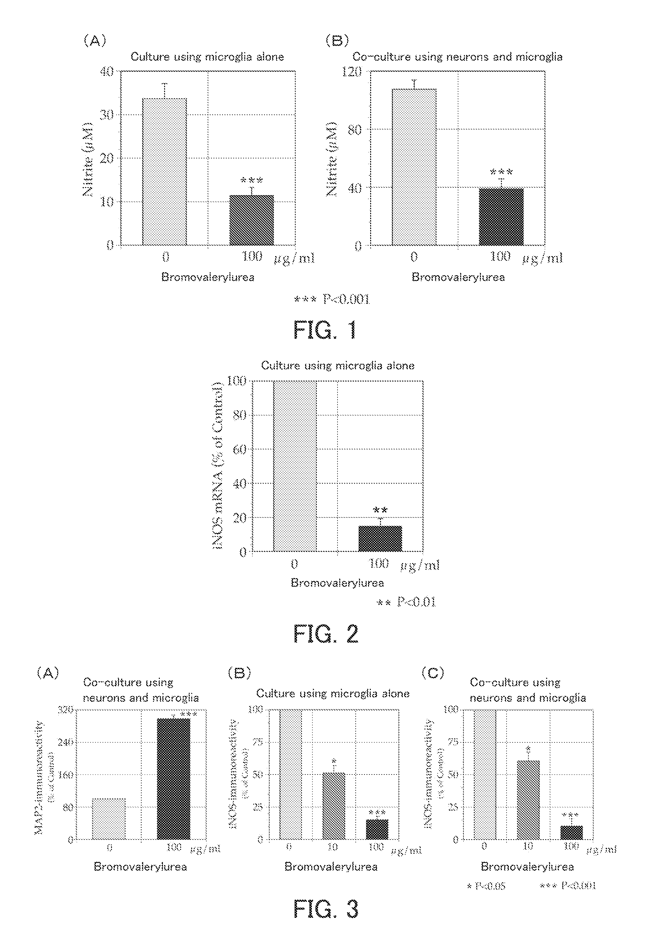
[0076]The present example examined the effect of bromovalerylurea on Parkinson's disease.
[0077](1) Inhibition of Release of Nitric Oxide
[0078]As described below, newborn rat-derived primary culture microglia (MG) were cultured in the presence of bromovalerylurea and LPS (lipopolysaccharide), and the amount of nitric oxide, which is a neurocytotoxic factor, released from the MG was measured.
[0079]As culture systems, a culture system containing the MG alone and a co-culture system containing the MG and rat-derived primary culture cerebral cortical neurons were provided to carry out culture. The medium used was a serum-free medium (pH 7.4) containing: a Dulbecco's modified Eagle's medium (DMEM) as a basal medium; LPS at a final concentration of 1 μg/ml; and bromovalerylurea at a final concentration of 100 μg/ml. The culture conditions were set to 37° C., 48 hours, and 5% CO2. The concentration of nitrite ions in each of the culture solutions at the end of the culture was measured using a Griess reagent. Because nitric oxide released from the MG was oxidized in the culture solution and turned into nitrite ions, the released nitric oxide was measured indirectly by measuring the nitrite ions.
[0080]The results thereof are shown in FIG. 1. FIG. 1 shows graphs showing the nitrite ion concentrations (μmol/L) in the respective culture systems. FIG. 1A shows the result obtained when the culture system containing the MG alone was used. FIG. 1B shows the result obtained when the co-culture system containing the MG and the neurons was used. As can be seen from FIG. 1A, by adding the bromovalerylurea (100 μg/ml), the concentration of the nitrite ions released from the MG was lowered significantly. Normally, when MG are co-cultured with neurons, they are activated strongly and thus release a large amount of nitric oxide. However, as can be seen from FIG. 1B, by adding the bromovalerylurea, the concentration of the released nitrite ions could be inhibited significantly. These results demonstrate that bromovalerylurea can inhibit the release of nitric oxide as a cytotoxic factor from MG.
[0081](2) Inhibition of Transcription of Nitric Oxide Synthase
[0082]The present experiment examined whether the inhibition of the release of the nitric oxide described in the item (1) above occurs at the transcription level of nitric oxide synthase (inducible; iNOS) induced by the LPS. Specifically, culture was carried out using a culture system containing MG alone in the same manner as in the item (1), and the amount of transcribed iNOS mRNA was measured by real-time reverse transcription (RT)-PCR. The real-time RT-PCR was carried out by a routine method. Then, assuming that the amount of transcribed iNOS mRNA in a culture system containing no bromovalerylurea was 100%, the relative value (%) of the amount of transcribed iNOS mRNA in the above culture system was determined.
[0083]The result thereof is shown in FIG. 2. FIG. 2 is a graph showing the relative value (%) of the amount of the transcribed iNOS mRNA in the culture system. As can be seen from FIG. 2, by adding the bromovalerylurea, the amount of the transcribed iNOS mRNA was decreased significantly. This result demonstrates that inhibition of nitric oxide production by bromovalerylurea occurs at the transcriptional level of iNOS.
[0084](3) MAP2 expression and inhibition of iNOS expression
[0085]Regarding each of the culture system containing the MG alone and the co-culture system containing the MG and the neurons, the expression of a neuron-specific protein MAP2 and the expression of an iNOS protein producing nitric oxide were measured.
[0086]Culture was carried out in the same manner as in the item (1) above.
[0087]Then, by Western blotting using an antibody against mouse-derived MAP2 (trade name: Mouse monoclonal anti-MAP2 antibody (clone AP20), commercially available from Sternberger Monoclonals) and an antibody against mouse-derived iNOS (trade name: Mouse monoclonal anti-iNOS antibody (clone 6), commercially available from BD Biosciences), the MAP2 immunoreactivity and the iNOS immunoreactivity corresponding to the expression levels of the proteins were measured. Then, assuming that the MAP2 immunoreactivity and the iNOS immunoreactivity in a culture system containing no bromovalerylurea were 100%, the relative value (%) of each immunoreactivity in each culture system was determined.
[0088]The results thereof are shown in FIG. 3. FIG. 3A is a graph showing the MAP2 immunoreactivity in the co-culture system. FIG. 3B is a graph showing the iNOS immunoreactivity in the culture system containing the MG alone. FIG. 3C is a graph showing the iNOS immunoreactivity in the co-culture system. As can be seen from FIG. 3A, by adding the bromovalerylurea, the expression level of the neuron-specific protein MAP2 was increased greatly as compared with the case where bromovalerylurea was not added. This result demonstrates that neuron death was inhibited in the co-culture system. Also, as can be seen from FIGS. 3B and 3C, by adding the bromovalerylurea, the expression level of the iNOS was decreased in a bromovalerylurea concentration dependent manner. These results demonstrate that bromovalerylurea can inhibit the expression of iNOS induced by LPS and thus can inhibit neuron death.
[0089](4) Inhibition of Death of Dopamine Neurons in Parkinson's Disease Model Rats
[0090]The present experiment examined whether bromovalerylurea inhibits neuron death in rat Parkinson's disease models in which the Parkinson's disease was induced by 6-hydroxydopamine (6-OHDA).
[0091]The model rats used were those having dopamine neuron injury in the substantia nigra on the right side of the midbrain, induced by injecting 6-OHDA to the right striatum (see Choudhury et al., Brain and Behavior, 1: pp. 26-43, 2011). To the model rats (n=5), drinking water containing 500 mg/l bromovalerylurea was administered orally for 7 days in such a manner that each rat drank an average of 25 ml of the drinking water per day. Then, on the last day of the oral administration, each model rat was dissected, and the substantiae nigra on the right and left sides of the midbrain were collected. Then, the thus-collected midbrain substantiae nigra were subjected to immunoblot to measure tyrosine hydroxylase (TH) as a dopamine neuron marker. Also, as a control, TH was measured in the same manner, except that drinking water not containing bromovalerylurea was used instead of the drinking water containing bromovalerylurea. Then, regarding each rat group, the ratio (%) of the TH immunoreactivity (R) in the substantia nigra on the right side of the midbrain where the injury was induced to the TH immunoreactivity (L) in the substantia nigra on the left side of the midbrain where injury was not induced was calculated according to the following equation.
Relative value (%)=100×R/L
[0092]Furthermore, the model rats to which the bromovalerylurea had been administered orally for 7 days were reared for further 3 days. Thereafter, the motor function of each model rat was measured by a rotarod test.
[0093]The results thereof are shown in FIG. 4. FIG. 4A is a graph showing the TH immunoreactivity. As can be seen from FIG. 4A, in the control where the bromovalerylurea had not been administered, the TH immunoreactivity in the substantia nigra on the right side of the midbrain was deteriorated greatly, whereas, in the example where the bromovalerylurea had been administered, the deterioration of the TH immunoreactivity in the substantia nigra on the right side of the midbrain was inhibited sufficiently. These results demonstrate that bromovalerylurea can inhibit the death of dopamine neurons in the midbrain substantia nigra. FIG. 4B is a graph showing the results of the rotarod test. FIG. 4B shows the total number of revolutions of a rotating rod until the model rat fell from the rotating rod. The model rats to which the bromovalerylurea had been administered could stay on the rotating rod longer than the model rats to which the bromovalerylurea had not been administered. This result demonstrates that bromovalerylurea can inhibit the movement disorder in Parkinson's disease.

Example

Example 2
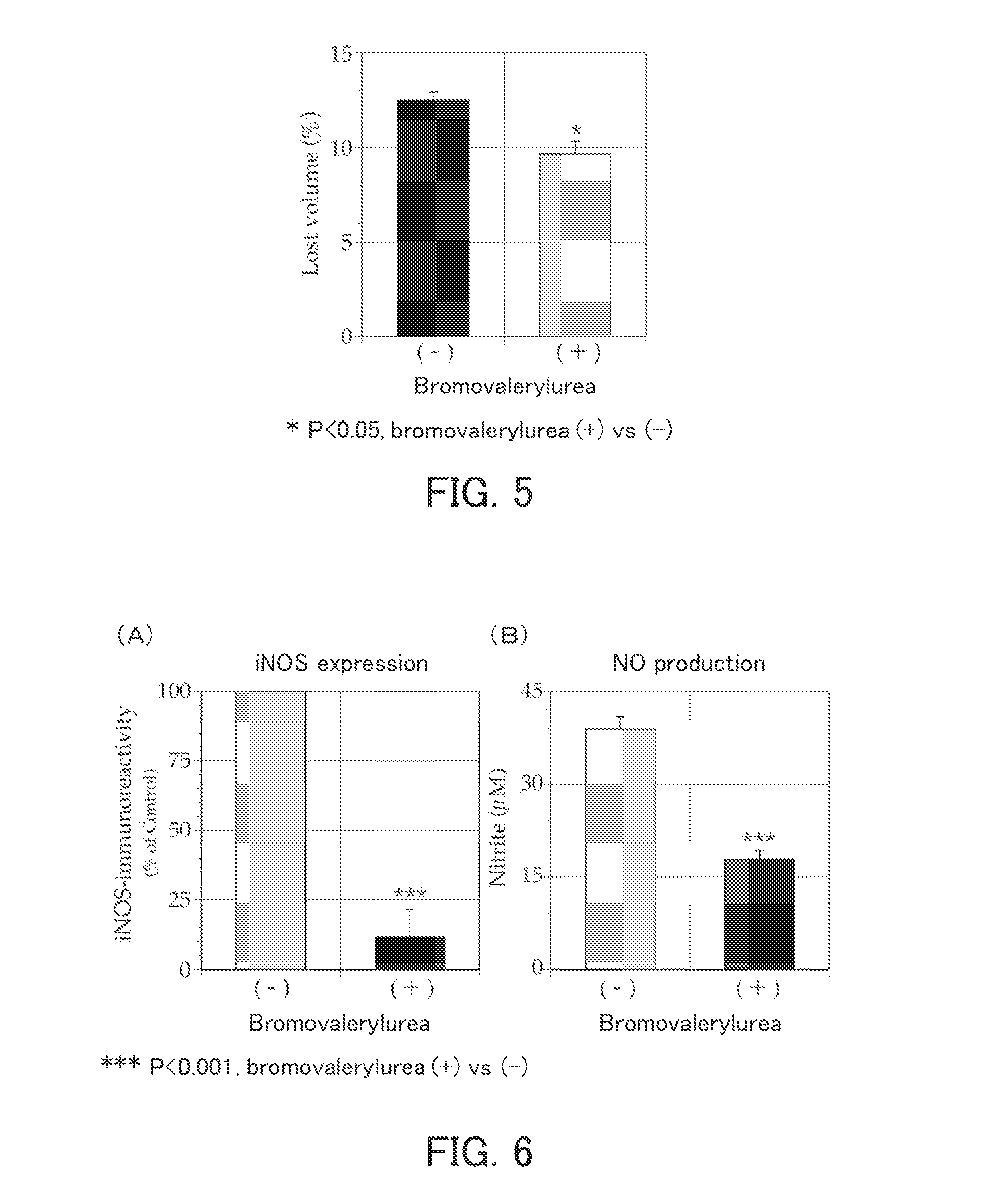
[0094]The present example examined the effect of bromovalerylurea on cerebral infarction.
[0095](1) Inhibition of tissue loss in cerebral infarction model rats
[0096]Cerebral infarction model rats each having cerebral infarction induced by 90-minute transient occlusion of the right middle cerebral artery were used to examine whether bromovalerylurea inhibits tissue loss.
[0097]As the model rats, male Wistar rats were used (Matsumoto et al., Journal of Cerebral Blood Flow & Metabolism (2008) 28: pp. 149-163). By magnetic resonance imaging (MRI), only the model rats in which cerebral infarction had been induced sufficiently were selected (n=4). To these model rats, drinking water containing 500 mg/l bromovalerylurea was administered orally for 14 days in such a manner that each rat drank an average of 25 ml of the drinking water per day. On Day 16 from the last day of oral administration, each model rat was dissected, and a middle cerebral artery perfusion region in the brain was collected. The collected perfusion region was sliced from the anterior side to the posterior side so as to provide seven layers of slices. Then, regarding the 2nd, 3rd, and 4th layers from the top, the total volume of the left hemisphere where infarction was not caused and the total volume of the right hemisphere where infarction was caused were measured separately, and the mean values were determined (L: the total volume of the left hemisphere, R: the total volume of the right hemisphere). Then, the proportion of the tissue loss (%) was calculated according to the following equation.
Proportion of tissue loss (%)=100×(L−R)/L
[0098]The results thereof are shown in FIG. 5. FIG. 5 is a graph showing the proportion of the brain tissue loss. As can be seen from FIG. 5, in the example where the bromovalerylurea had been administered, the proportion of the brain tissue loss could be lowered as compared with the control where the bromovalerylurea had not been administered. These results demonstrate that bromovalerylurea can inhibit the brain tissue loss in cerebral infarction.
[0099](2) Inhibition of iNOS Expression and Inhibition of Nitric Oxide Production
[0100]The present experiment examined whether bromovalerylurea inhibits iNOS expression and nitric oxide production in macrophages BINCs accumulated in a cerebral infarction nest. Regarding the BINCs, Matsumoto et al., Journal of Cerebral Blood Flow & Metabolism (2008) 28: pp. 149-163 was referenced to.
[0101]Culture was carried out using, as the macrophages, primary culture brain macrophages (BINCs) derived from the core lesion of rat cerebral infarction caused by transient occlusion of the middle cerebral artery. The medium used was a serum-free medium (pH 7.4) containing: the DMEM as a basal medium; and bromovalerylurea at a final concentration of 0 or 100 μg/ml. The culture conditions were set to 37° C., 24 hours, and 5% CO2. Nitrite ions in the culture solution at the end of the culture were measured. Also, iNOS and β-actin were measured by immunoblot, and the ratio between the immunoreactivities of iNOS and β-actin (iNOS/β-actin) was calculated. Then, assuming that the ratio when the culture system not containing bromovalerylurea was used was 100%, the relative value (%) of the ratio when the above culture system was used was determined.
[0102]The results thereof are shown in FIG. 6. FIG. 6A is a graph showing the iNOS immunoreactivity. FIG. 6B is a graph showing the concentration of nitrite ions. As can be seen from FIG. 6A, by adding the bromovalerylurea, the expression level of iNOS was lowered. Also, as can be seen from FIG. 6B, by adding the bromovalerylurea, the amount of the produced nitrite ions, i.e., the amount of the produced nitric oxide, was decreased. These results demonstrate that bromovalerylurea can inhibit the iNOS expression in macrophages BINCs accumulated in a cerebral infarction core lesion and thus can inhibit the production of nitric oxide.
[0103](3) Inhibition of Inflammatory Reaction
[0104]The present experiment examined whether bromovalerylurea inhibits an inflammatory reaction in macrophages BINCs accumulated in a cerebral infarction core lesion.
[0105]Macrophages were cultured in the same manner as in the item (2) above. Then, the amounts of transcribed mRNAs of various factors in the medium after the end of the culture were measured by RT-PCR. Then, assuming that the amounts of the mRNAs transcribed in a culture system not containing bromovalerylurea were 100%, the relative values (%) of the amounts of the mRNAs transcribed in each culture system were determined.
[0106]The results thereof are shown in FIG. 7. FIG. 7 shows graphs showing the amounts of the transcribed mRNAs of the various factors. As can be seen from FIG. 7, the amounts of the transcribed mRNAs of neurocytotoxic factors IL-1β, IL-6, IFNIβ, and Bak and the amount of the transcribed mRNA of the iNOS, which is an enzyme that produces nitric oxide as a neurocytotoxic factor, were decreased in a bromovalerylurea concentration dependent manner. In contrast, the amounts of the transcribed mRNAs of HGF and IGF-1, which are factors useful for cell growth, were not decreased by the addition of the bromovalerylurea and were not influenced by the bromovalerylurea.
[0107](4) Inhibition of brain tissue loss caused by needlestick injury
[0108]Brain injury model rats each having brain injury caused by inserting an injection needle from the outside of the cranium were used to examine whether bromovalerylurea inhibits tissue loss.
[0109]As the rats, male Wistar rats were used. In each rat, a needle (18-gauge injection needle) was inserted into the cerebrum at a site 2 mm posterior and 2 mm lateral to the right from the anterior fontanelle, and a fan-shaped injury extending in an anteroposterior direction and having an angle of about 120° was formed. To the rats (n=5), drinking water containing 500 mg/l bromovalerylurea was administered orally for 60 days in such a manner that each rat drank an average of 25 ml of the drinking water per day. Then, on Day 3 from the last day of oral administration, each model rat was dissected, and the cerebrum was collected. The collected cerebrum was sliced from the anterior side to the posterior side so as to provide seven layers of slices. Then, regarding the 2nd to 5th layers from the top, the volume of the left hemisphere and the volume of the right hemisphere were measured separately, and the mean values were determined (L: the volume of the left hemisphere, R: the volume of the right hemisphere). As a control experiment, drinking water not containing bromovalerylurea was administered orally to rats having brain injury caused in the same manner, and the volumes of the right and left hemispheres of the brain were measured in the same manner. Then, the proportion of tissue loss (%) was calculated according to the following equation.
Proportion of tissue loss (%)=100×(L−R)/L
[0110]The results thereof are shown in FIG. 8. FIG. 8A shows photographs showing the brain tissue loss. The photograph on the left shows the results obtained regarding the group to which the drinking water not containing bromovalerylurea had been administered, and the photograph on the right shows the results obtained regarding the group to which the drinking water containing bromovalerylurea had been administered. As can be seen from FIG. 8A, considerable brain tissue loss was seen in the group to which bromovalerylurea had not been administered, whereas the brain tissue loss was inhibited in the group to which the bromovalerylurea had been administered. FIG. 8B is a graph showing the proportion of the brain tissue loss. As can be seen from FIG. 8B, the proportion of the brain tissue loss in the group to which the bromovalerylurea had been administered (+) could be lowered as compared with that in the group to which bromovalerylurea had not been administered (−). These results demonstrate that bromovalerylurea can inhibit brain tissue loss caused by brain injury.

Example

Example 3
[0111]The present example examined the effect of bromovalerylurea on sepsis.
[0112](1) Effect of improving survival rate
[0113]The cecum of each of 8-week old male Wistar rats was ligated, and two perforations were formed with an 18-gauge injection needle in the ligated part of the cecum. Thus, perforative peritonitis was caused to prepare rat sepsis models. Immediately after the preparation of the model rats, 10 ml of a commercially available maintenance infusion solution (SOLITA T3, Ajinomoto Pharmaceuticals Co., Ltd.) in which bromovalerylurea had been dissolved at a final concentration of 500 μg/ml was injected subcutaneously to the model rats. As a control group, the same amount of the maintenance infusion solution not containing bromovalerylurea was injected subcutaneously. Thereafter, the injection was performed in the same manner about every 12 hours. Also, rats subjected only to laparotomy, i.e., the incision of the skin and the peritoneum with no injury to the intestinal tract, were provided as a sham operation group. To the sham operation group, subcutaneous injection was not performed. The results obtained regarding the model rats of the respective groups were examined over 8 days after the incision.
[0114]The results thereof are shown in FIG. 9. In FIG. 9, the filled square (▪) shows the results obtained regarding the sham operation group, the filled circle () shows the results obtained regarding the control group to which bromovalerylurea had not been administered, and the filled triangle (▴) shows the results obtained regarding the example group to which the bromovalerylurea had been administered. As can be seen from FIG. 9, in the sham operation group (Sham), the mortality rate was 0%. In the group to which bromovalerylurea had not been administered (Sepsis/control), the mortality rate was 80% on Day 8. In contrast, in the example group to which the bromovalerylurea had been administered (Sepsis/BU), the mortality rate could be lowered to 47% with the significant difference of P<0. 0001. These results demonstrate that bromovalerylurea exhibits significant lifesaving effect.
[0115](2) Inhibition of Production of Proinflammatory Interleukins
[0116]The present experiment examined whether bromovalerylurea inhibits the production of interleukins induced by peritoneal macrophages.
[0117]Culture was carried out using, as macrophages, macrophages collected in the abdominal cavities of Wistar rats. The medium used was a serum-free medium (pH 7.4) containing: the DMEM as a basal medium; and bromovalerylurea at a final concentration of 0, 30, or 100 μg/ml. The culture conditions were set to 37° C., 18 hours, and 5% CO2. The concentrations of IL-6 and IL-1β in each culture solution at the end of the culture were measured by ELISA.
[0118]The results thereof are shown in FIG. 10. FIG. 10A is a graph showing the amount of the produced IL-1β. FIG. 10B is a graph showing the amount of the produced IL-6. As can be seen from FIGS. 10A and 10B, by adding the bromovalerylurea, the amounts of the produced IL-6 and IL-1β were decreased in a bromovalerylurea concentration dependent manner. These results demonstrate that bromovalerylurea can inhibit inflammation.
[0119](3) Inhibition of IL-6 and inhibition of renal failure in sepsis model rats
[0120]Sepsis model rats were used to examine whether bromovalerylurea inhibits IL-6.
[0121]The sepsis model rats were prepared by ligating the cecum of each of male Wistar rats and then forming two perforations with an 18-gauge injection needle. To the model rats (n=10), a commercially available maintenance infusion solution (SOLITA T3, Ajinomoto Pharmaceuticals Co., Ltd.) in which bromovalerylurea had been dissolved at a final concentration of 500 μg/ml was injected subcutaneously three times in total for about one day, in such a manner that the subcutaneous injection was performed twice a day (once before noon and once in the afternoon) with the single dose being 10 ml and the total dose per day being 20 ml. After the third subcutaneous injection, i.e., on the following day of the preparation of the sepsis model by causing perforative peritonitis, serum of each model rat was collected, and serum IL-6 was measured by ELISA. As a comparative example, the maintenance infusion solution not containing bromovalerylurea was injected subcutaneously to the sepsis rats, and serum IL-6 was measured in the same manner. Also, as a control, regarding non-sepsis rats subjected only to laparotomy (sham operation group), serum IL-6 was measured in the same manner. Furthermore, regarding each serum, the creatinine concentration in serum as a renal failure indicator also was measured.
[0122]The results thereof are shown in FIG. 11. FIG. 11A is a graph showing serum IL-6. As can be seen from FIG. 11, in the example where the bromovalerylurea had been administered, serum IL-6 could be decreased greatly as compared with the comparative example (−) where bromovalerylurea had not been administered. This result demonstrates that bromovalerylurea can inhibit cytokine storm in sepsis.

PUM

no PUM

Description & Claims & Application Information

We can also present the details of the Description, Claims and Application information to help users get a comprehensive understanding of the technical details of the patent, such as background art, summary of invention, brief description of drawings, description of embodiments, and other original content. On the other hand, users can also determine the specific scope of protection of the technology through the list of claims; as well as understand the changes in the life cycle of the technology with the presentation of the patent timeline. Login to view more.

Similar technology patents

Adam-15 antibodies and immunogenic peptides

ActiveUS20110117090A1inhibit activationenhance activity
Owner:VASGEN

Fc mutants

ActiveUS8618251B1inhibit activation
Owner:THE ROCKEFELLER UNIV

Methods and compositions for treating inflammation

InactiveUS20090118337A1inhibit activationsuppress airway inflammation
Owner:CASE WESTERN RESERVE UNIV

Classification and recommendation of technical efficacy words

  • inhibit activation

Use of specific T2R taste receptors to identify compounds that block bitter taste

ActiveUS20030170608A1block bitter tasteinhibit activation
Owner:SENOMYX INC

Vehicle AC Ground Fault Detection System

InactiveUS20090091868A1prevent shockinhibit activation
Owner:LEAR CORP

Uses of inhibitors for the activation of CXCR4 receptor by SDF-1 in treating rheumatoid arthritis

InactiveUS20050202005A1inhibit activation
Owner:THE TRUSTEES OF COLUMBIA UNIV IN THE CITY OF NEW YORK

Protection of cells from degeneration and treatment of geographic atrophy

ActiveUS9707235B1increase levelinhibit activation
Owner:UNIV OF KENTUCKY RES FOUND

Methods and compositions for treating inflammation

InactiveUS20090118337A1inhibit activationsuppress airway inflammation
Owner:CASE WESTERN RESERVE UNIV
Why Eureka
Try Eureka