Fracture Fixation Plate for the Coronoid of the Proximal Ulna

a technology of proximal ulna and fixation plate, which is applied in the field of surgical devices and methods for the internal fixation of fractured bones, can solve problems such as patient discomfort and complications, and achieve the effect of easy and safe reconfiguration inside the patien

Active Publication Date: 2009-05-14
BIOMET CV
View PDF31 Cites 33 Cited by
  • Summary
  • Abstract
  • Description
  • Claims
  • Application Information

AI Technical Summary

Benefits of technology

[0011]A system of bendable plates is provided that may be easily and safely reconfigured inside the patient's body (in situ) during the surgical procedure. The system can be reconfigured without distorting the shape of bone fastener holes in the plate, and any threads within the holes. The system includes and is adapted for use with in situ bending tools to reconfigure the plate inside the patient's body during the surgical procedure.
[0013]An elbow fracture fixation system is provided that also includes locking fasteners for attachment of the bone plate to the fractured bone. In general, the primary functions of various bone plates of the system (which are all adjacent near the elbow joint) include not only holding the bone fragments together in healing alignment, but also the transfer of forces from the metaphysis to the diaphysis of the fractured bone while the bone is mending. The system allows the distal tip of a fastener to be anchored into healthy, cortical bone, and the transfer of force from the healthy bone to the plate, such that the plate properly accomplishes load sharing.
[0019]According to another aspect of the system, bone plates for the lateral and medial surfaces of the distal humerus each have a rigid body portion with substantially the same thickness. The rigid portion of each of the medial and lateral plates has a distal end, a proximal end, a top surface, a bottom surface, a medial edge and an opposing lateral edge. The plates also have a plurality of holes extending between the top and bottom surfaces, each of the holes for receiving a fastener for attachment of the bone plate to the bone. The lateral bone plate also has at least one positioning foot extending from an edge downwardly towards the bone surface to aid in the positioning of the bone plate on the bone surface.
[0021]According to another aspect of the system, a bone plate for the posterolateral surface of the distal humerus has a body with a thickness substantially greater than the medial plate (greater than fifty percent thicker). The body has a proximal end, a distal end and a curvilinear, longitudinal axis extending therebetween. A first arm and a second arm extend from proximal end on opposing sides of the longitudinal axis, thereby forming a Y-shape, and a third arm extends transversely away from the longitudinal axis to extend partially around the lateral side of the distal humerus. The first, second, and third arms each includes a ring element having a hole and are attached to the body by respective bendable bridge elements. The body includes threaded holes and an elongated slot, each of which may be located along the longitudinal axis. The slot may be configured to receive a compression fastener. Each of threaded holes is configured for receiving one of the fasteners. The threaded holes may be preassembled with a plurality of drill guides, with a proximal hole receiving a short drill guide. In the same manner as with the lateral and medial plates, the surgeon may closely match the shape of posterolateral plate to the bone surface and redirect the trajectories of the fasteners to capture bone fragments and avoid fracture lines and other fasteners.
[0029]According to another aspect of the system, the system has a bone plate having a threaded hole defining a thread axis for receiving a fixed angle, locking fastener. The system also has a drill guide preassembled into the threaded hole, the drill guide including a drill guide bore sized to guide a bone drill. The system also has an insertion tool having a cylindrical body with distal and proximal ends and a longitudinal axis extending therebetween. The cylindrical body has a grip surface for holding the insertion tool during use. The cylindrical body also has a longitudinal bore extending between the distal and proximal ends and sized for guiding a K-wire, and the distal end is configured to be removably attached to the drill guide so that the longitudinal bore aligns with the thread axis. The distal end of the insertion tool may also fit securely into the drill guide, such that the user may use the cylindrical body as a handle to manipulate the bone plate during the surgical procedure.

Problems solved by technology

A “proud” fastener head may lead to soft tissue irritation, inflammation or other types of trauma that may cause complications and patient discomfort.

Method used


Image

  • Fracture Fixation Plate for the Coronoid of the Proximal Ulna
  • Fracture Fixation Plate for the Coronoid of the Proximal Ulna
  • Fracture Fixation Plate for the Coronoid of the Proximal Ulna

Examples


Embodiment Construction

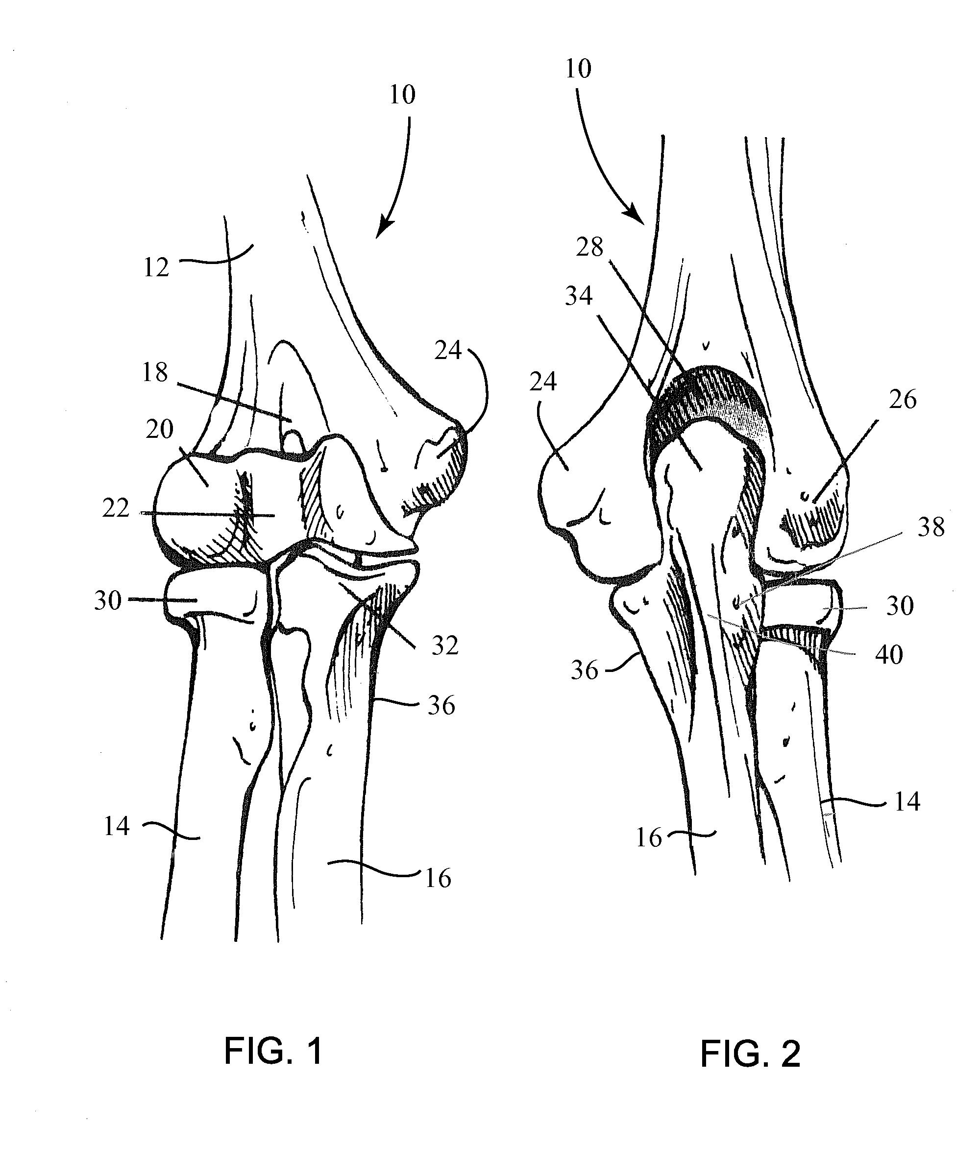
[0090]FIG. 1 is an anterior (front) view and FIG. 2 is a posterior (back) view of the bones of the human elbow joint 10: the distal humerus 12, the proximal radius 14 and the proximal ulna 16. The distal humerus 12 includes the coronoid fossa 18, the capitellum 20, the trochlea 22, the medial epicondyle 24 and the lateral epicondyle 26, and the olecranon fossa 28 therebetween. The proximal radius 14 includes the radial head 30. The proximal ulna 16 includes the coronoid process 32 (FIG. 1) and the olecranon 34 (FIG. 2) which articulates within the olecranon fossa 28 between the lateral and medial epicondyles 24, 26 of distal humerus 12. Each of the distal humerus 12, proximal radius 14 and proximal ulna 16 are susceptible to a large variety of fractures, such as during a fall.

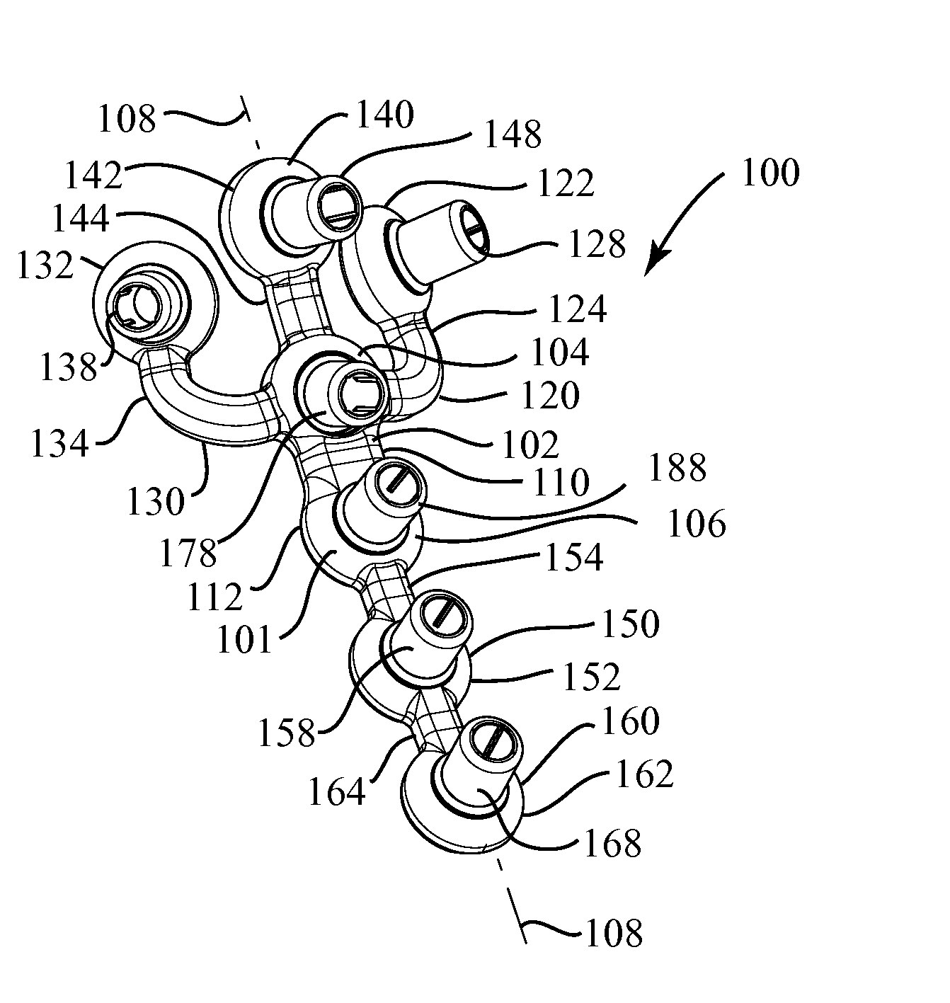
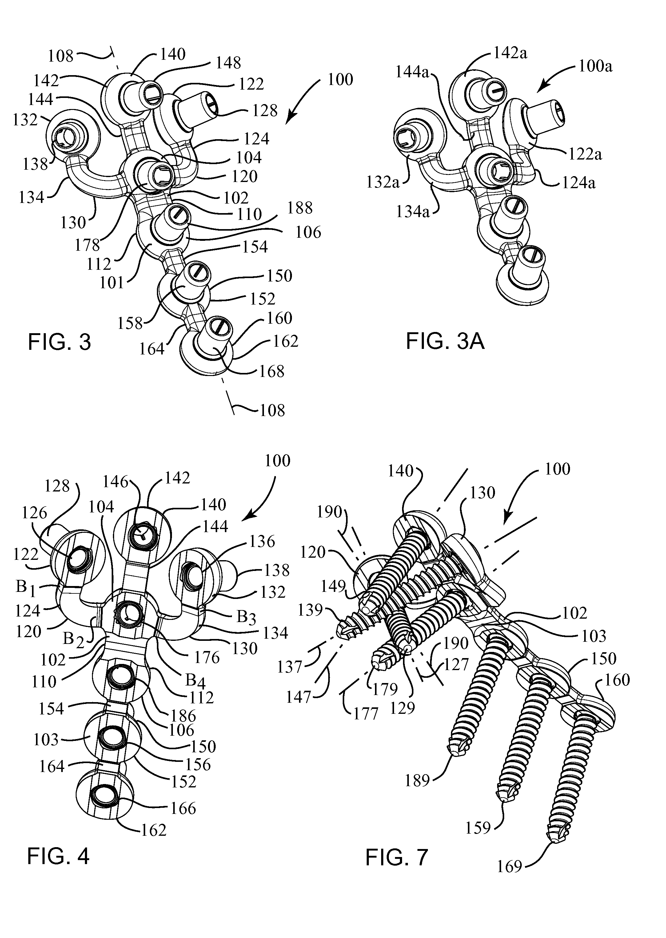
[0091]The present system for the repair of elbow fractures may include a plurality of anatomically specific bone plates and a plurality of fasteners for the attachment of the plates to the bone. The system may ...


PUM


Abstract

A system for the internal fixation of a fractured bone of an elbow joint of a patient includes at least one bone plate, each bone plate having a plurality of holes and generally configured to fit an anatomical surface of the fractured bone. The at least one plate is adapted to be customized to the shape of a patient's bone. The system also includes a plurality of fasteners including at least one locking fastener for attaching the bone plate to the bone. At least one of the holes is a threaded hole. Guides for plate benders, drills, and / or K-wires can be pre-assembled to the threaded holes, and the locking fastener can lock into any of the threaded holes after the guides are removed.

Description

CROSS-REFERENCE TO RELATED APPLICATION[0001]This application claims benefit of Ser. No. 60 / 985,000, filed Nov. 2, 2007, which is hereby incorporated by reference herein in its entirety.FIELD OF THE INVENTION[0002]The subject matter of this disclosure relates broadly to surgical devices and methods for the internal fixation of fractured bones, and more particularly, to bone plates and fasteners.BACKGROUND OF THE INVENTION[0003]The three long bones of the upper extremity are the humerus, radius and ulna. The distal portion of the humerus and the proximal portions of the radius and the ulna form the elbow joint. Elbow fractures account for only about 5-8% of all fractures and occur most commonly in older people as a result of a fall. The functional outcomes of elbow fractures often include high rates of joint stiffness, loss of range of motion and non-union.[0004]Orthopedic surgeons generally follow certain principles for the proper internal fixation of the bones of the elbow joint. Ea...

Claims


Application Information

Patent Timeline
Patent Type & Authority Applications(United States)
IPC IPC(8): A61B17/70A61B17/04A61B19/00
CPCA61B17/1728A61B17/8061A61B17/8085A61B17/809A61B17/864B25B27/026B25B27/064A61B17/1739Y10S606/902A61B17/8863A61B17/8047A61B17/17A61B17/80
Inventor SIXTO, JR., ROBERTKORTENBACH, JUERGEN A.STEINMANN, SCOTT
Owner BIOMET CV
Why Eureka
Try Eureka