
System and Method for Additive Spatial/Intensity Decomposition of Medical Images

a technology of spatial/intensity decomposition and medical images, applied in image enhancement, image analysis, instruments, etc., can solve the problems of difficult to distinguish between two different objects that have similar intensity values in imaged data, and the 3-dimensional dataset obtained from scanning modalities such as computed tomography (ct), is usually quite complex and difficult to achieve the effect of achieving the effect of reducing the number of images

Active Publication Date: 2009-05-21
SIEMENS HEALTHCARE GMBH
View PDF4 Cites 28 Cited by
  • Summary
  • Abstract
  • Description
  • Claims
  • Application Information

AI Technical Summary

Benefits of technology

[0008]Exemplary embodiments of the invention as described herein generally include methods and systems for decomposing an image into two or more image components where each contains a portion of the original image. Methods and systems according to embodiments of th...

Problems solved by technology

The 3-dimensional datasets obtained from scanning modalities such as computed tomography (CT), magnetic resonance imaging (MRI), positron emission tomography (PET), ultrasound (US), etc., are usually quite complex, and contain many different objects and structures.
In many instances, it is difficult to distinguish between two different objects that have similar intensity values in the imaged data.
This technique is computationally fast, but the 2D results do not provide a good sense of depth of the original data.
However, since the projection is orthographic, the viewer cannot distinguish between left or right, front or back and even if the object is rotating clockwise or counter-clockwise.
However, bones and other high intensity structures can create challenges for these types of images, as the intensity of bone voxels in a CT image volume is such that bone voxels can occlude tissue voxels of greater interest for analysis.

Method used


Image

  • System and Method for Additive Spatial/Intensity Decomposition of Medical Images
  • System and Method for Additive Spatial/Intensity Decomposition of Medical Images
  • System and Method for Additive Spatial/Intensity Decomposition of Medical Images

Examples


Embodiment Construction

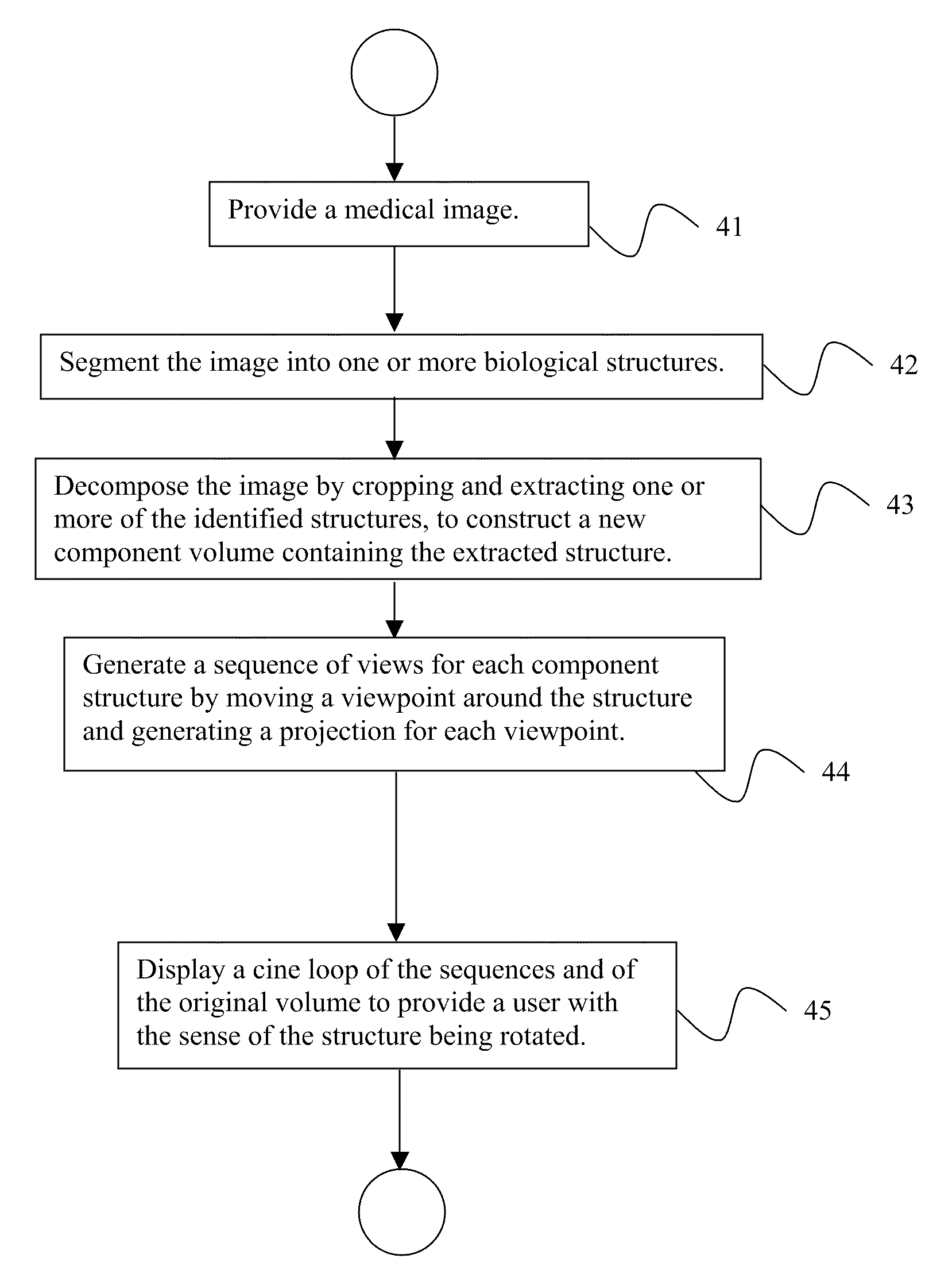
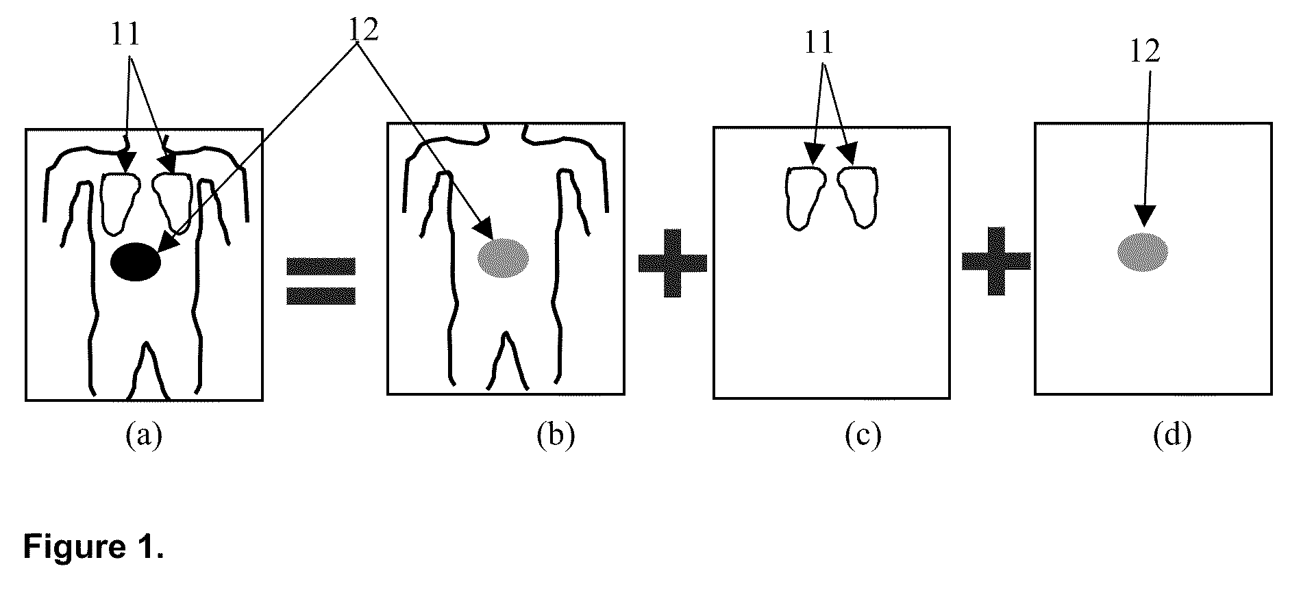
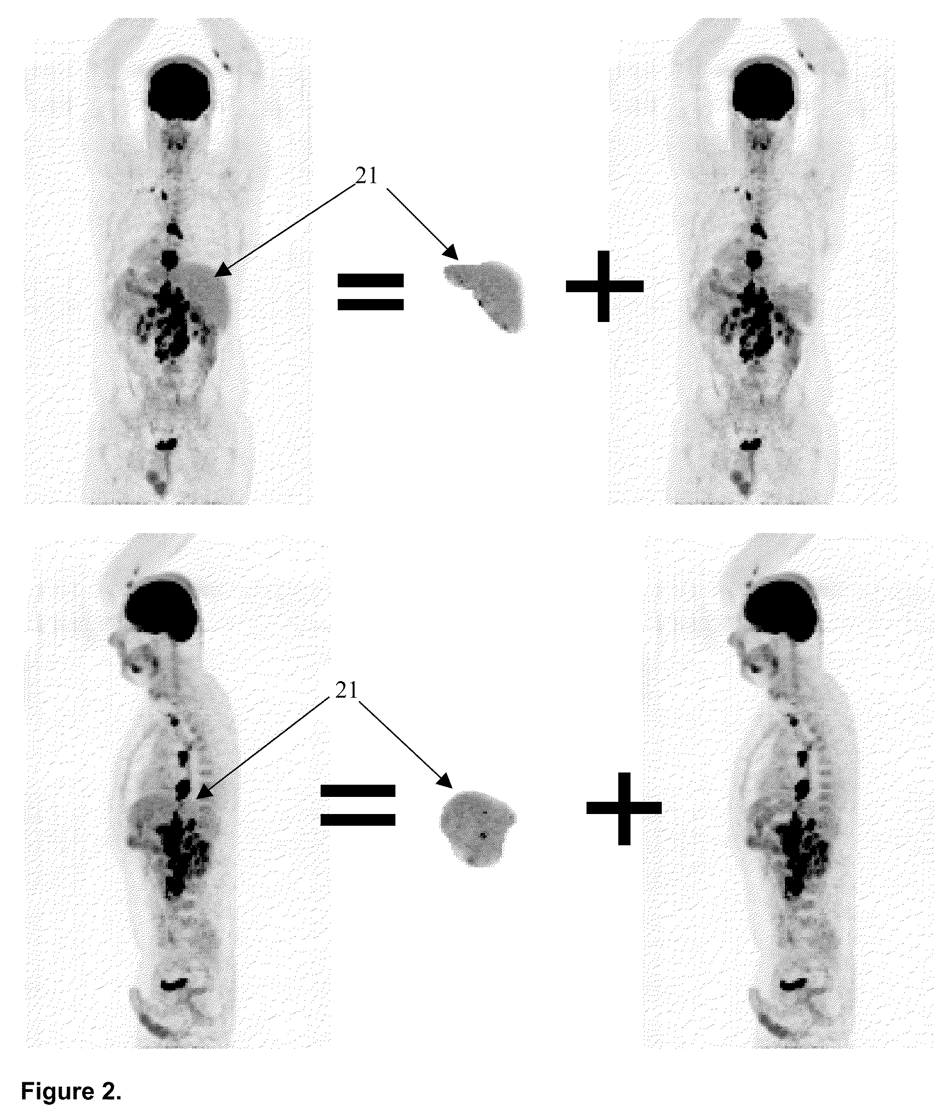
[0030]Exemplary embodiments of the invention as described herein generally include systems and methods for additive spatial / intensity decomposition of medical images based on parsed anatomical structures. Accordingly, while the invention is susceptible to various modifications and alternative forms, specific embodiments thereof are shown by way of example in the drawings and will herein be described in detail. It should be understood, however, that there is no intent to limit the invention to the particular forms disclosed, but on the contrary, the invention is to cover all modifications, equivalents, and alternatives falling within the spirit and scope of the invention.

[0031]As used herein, the term “image” refers to multi-dimensional data composed of discrete image elements (e.g., pixels for 2-D images and voxels for 3-D images). The image may be, for example, a medical image of a subject collected by computer tomography, magnetic resonance imaging, ultrasound, or any other medica...


PUM


Abstract

A method for decomposing digital medical images includes providing a digital medical image, segmenting the image into one or more biological structures, extracting one or more segmented biological structures from the image by extracting all voxels within a spatial extent of each of the biological structures to construct one or more new component volumes of the biological structures. For each of the one or more new component volumes, generate a sequence of 2-dimensional projective views by moving a projection viewpoint around each the biological structure in the one or more new component images, and generate a 2-dimensional projective view from each viewpoint, and display a cine loop of the sequence of projective views where the biological structures appear to be rotating in the display.

Description

CROSS REFERENCE TO RELATED UNITED STATES APPLICATIONS[0001]This application claims priority from “Additive Spatial / Intensity Decomposition of Medical Images based on Parsed Anatomical Structures and Its Presentation Using Projective Views”, Provisional Application No. 60 / 989,642 of Xiang Zhou, filed Nov. 21, 2007, the contents of which are herein incorporated by reference in their entireties.TECHNICAL FIELD[0002]This disclosure is directed to new ways of separating and presenting information a contained in medical images, such as positron emission tomography (PET), computed tomography (CT), and magnetic resonance imaging (MRI), etc., to the user.DISCUSSION OF THE RELATED ART[0003]The diagnostically superior information available from data acquired from current imaging systems enables the detection of potential problems at earlier and more treatable stages. Given the vast quantity of detailed data acquirable from imaging systems, various algorithms must be developed to efficiently an...

Claims


Application Information

Patent Timeline
IPC IPC(8): G06T7/00
CPCG06T7/0081G06T19/00G06T15/20G06T2207/30004G06T2210/41G06T2207/10072G06T7/11
Inventor ZHOU, XIANG
Owner SIEMENS HEALTHCARE GMBH
Learn More