
Ultrasound system and method of forming an ultrasound image

a technology of ultrasound image and ultrasonic system, which is applied in the field of ultrasonic system, can solve the problems of inability to provide accurate spectral doppler image and difficulty for users to set sample volume on a desirable vessel

Inactive Publication Date: 2009-06-11
MEDISON CO LTD
View PDF13 Cites 6 Cited by
  • Summary
  • Abstract
  • Description
  • Claims
  • Application Information

AI Technical Summary

Problems solved by technology

However, since the B-mode image is a 2-dimensional image of a grey scale, it may be difficult for the user to set the sample volume on a desirable vessel or a cardiac valve on the B-mode image.
Thus, an accurate spectral Doppler image may not be provided.

Method used


Image

  • Ultrasound system and method of forming an ultrasound image
  • Ultrasound system and method of forming an ultrasound image
  • Ultrasound system and method of forming an ultrasound image

Examples


Embodiment Construction

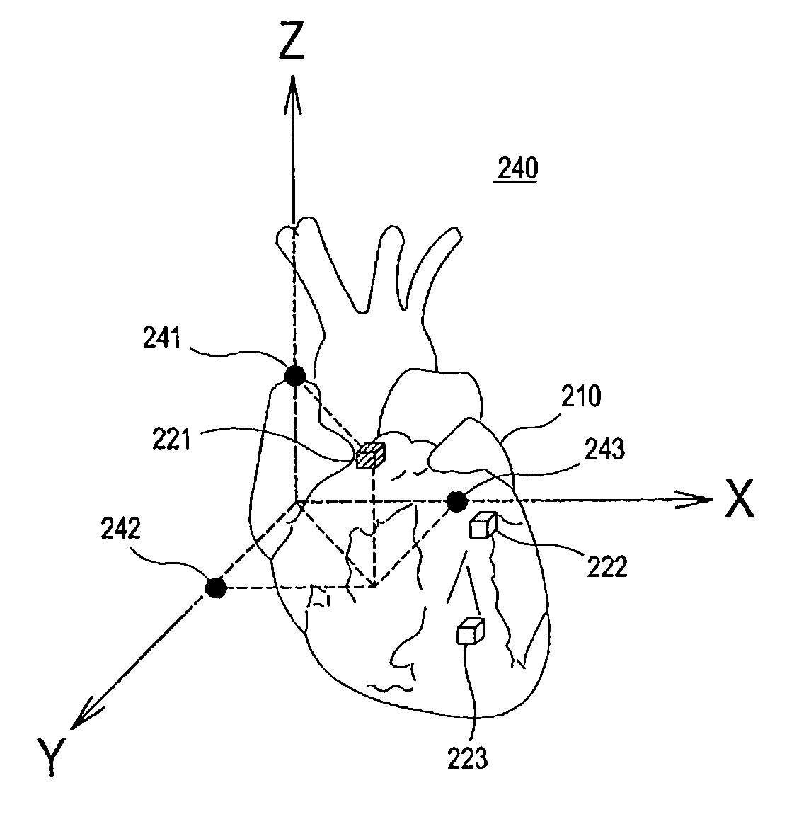
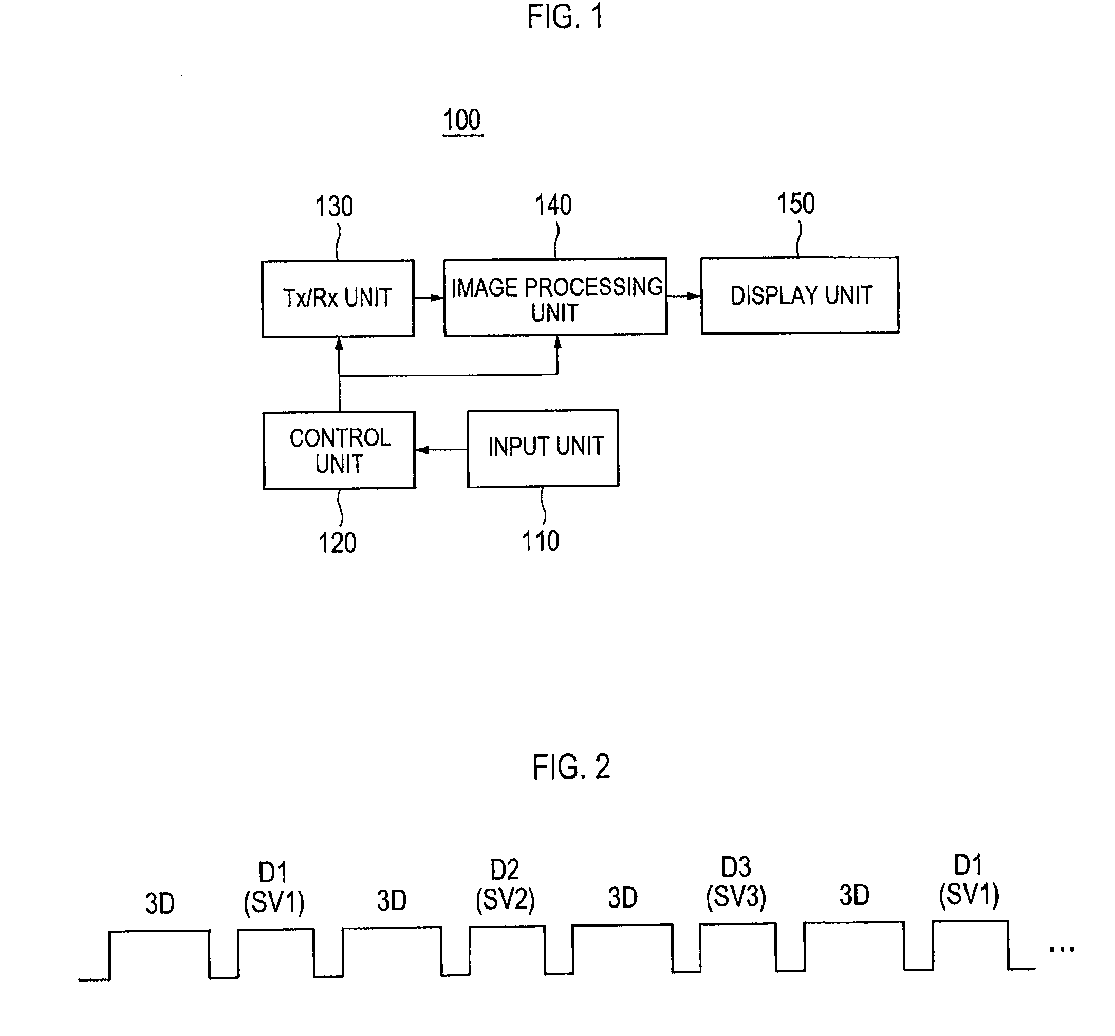
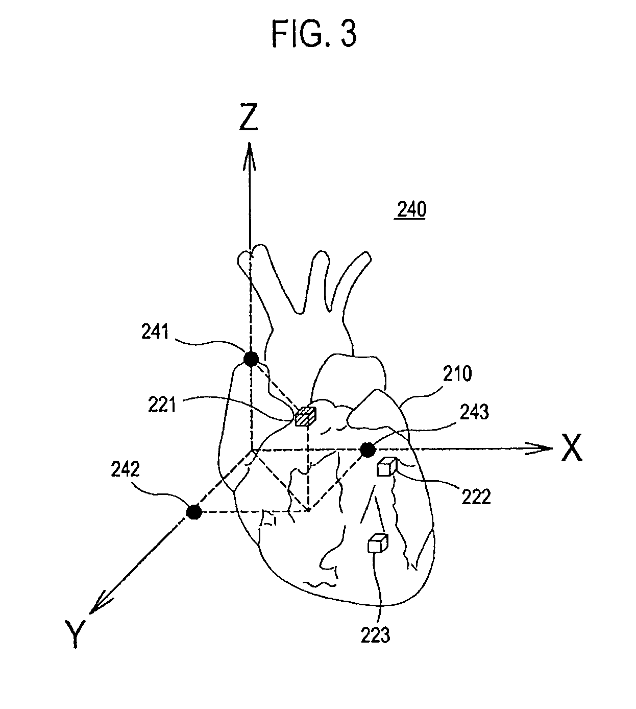
[0012]FIG. 1 is a block diagram illustrating one embodiment of an ultrasound system. Referring to FIG. 1, the ultrasound system 100 may include an input unit 110 for receiving user instructions. The user instructions may include a setup instruction for setting the number, size and position of sample volumes, a selection instruction for selecting at least one of the sample volumes, and a moving instruction for moving the sample volume. The sample volume may be formed to have a 3-dimensional figure such as a predefined hexahedron, sphere or the like.

[0013]The ultrasound system 100 may further include a control unit 120, which may be configured to control the transmission / reception of an ultrasound beam according to image modes of the ultrasound system 100. In a 3-dimensional image mode of the ultrasound system 100, the control unit 120 may generate a first control signal for controlling the transmission of a first ultrasound beam for forming a 3-dimensional ultrasound image of a targe...


PUM


Abstract

The present invention relates to an ultrasound system. The ultrasound system includes: an image processing unit for forming at least one 3-dimensional ultrasound image based on ultrasound echoes reflected from a target object; an input unit for receiving a setup instruction; and a control unit for outputting a first control signal for setting at least one predefined sample volume on the 3-dimensional ultrasound image in response to the setup instruction. The image processing unit is further configured to form at least one spectral Doppler image corresponding to the sample volume in response to the first control signal.

Description

[0001]The present application claims priority from Korean Patent Application Nos. 10-2007-0125376 and 10-2008-0078392 filed on Dec. 5, 2007 and Aug. 11, 2008, respectively, the entire subject matters of which are incorporated herein by reference.BACKGROUND OF THE INVENTION[0002]1. Technical Field[0003]The present invention generally relates to ultrasound systems, and more particularly to an ultrasound system and a method of forming an ultrasound image.[0004]2. Background Art[0005]An ultrasound system has become an important and popular diagnostic tool since it has a wide range of applications. Specifically, due to its non-invasive and non-destructive nature, the ultrasound has been extensively used in the medical profession. Modern high-performance ultrasound diagnostic systems and techniques are commonly used to produce two or three-dimensional diagnostic images of internal features of an object (e.g., human organs).[0006]The ultrasound system may provide a Doppler mode (D mode) fo...

Claims


Application Information

Patent Timeline
IPC IPC(8): A61B8/14
CPCG01S7/52063G01S7/52066G01S15/8993G01S15/8979G01S7/52074
Inventor AHN, CHI YOUNGHYUN, DONG GYU
Owner MEDISON CO LTD
Learn More