Image processing apparatus and method thereof
- Summary
- Abstract
- Description
- Claims
- Application Information
AI Technical Summary
Benefits of technology
Problems solved by technology
Method used
Image


Examples
first embodiment
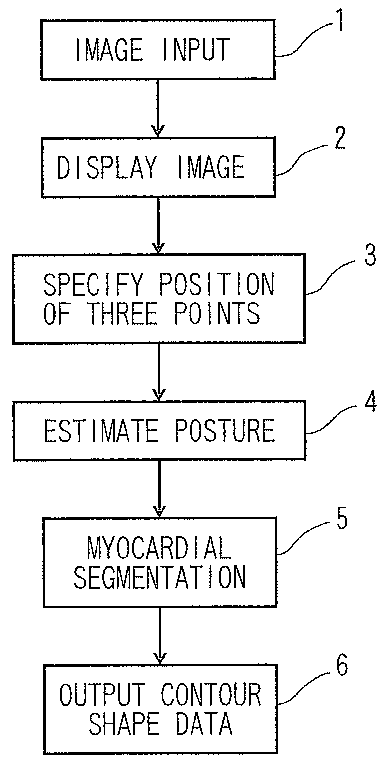
[0020]Referring now to FIG. 1 to FIG. 5, an image processing apparatus 10 as a myocardial segmentation apparatus according to a first embodiment of the invention will be described.
[0021]FIG. 1 is a block diagram showing the image processing apparatus 10 configured to perform a heart analysis according to the first embodiment. The image processing apparatus 10 may include an image input unit 22 configured to accept an entry of an image; a memory 24 configured to store the image; a display unit 26 configured to display the image; a position specifying unit 12 configured to specify positions on the image; a posture estimating unit 14 configured to estimate the posture of a heart on the image using the specified positions; a first calculating unit 16 configured to extract the shapes of inner and outer myocardial boundaries using training data which is learned in advance; an output unit 18 configured to output segmented boundary shape data; and a dictionary 20 configured to store the tra...
second embodiment
[0063]Referring now to FIG. 6, the image processing apparatus 10 according to a second embodiment will be described.
[0064]According to the second embodiment, the myocardial segmentation and the positional specification of the partial area boundaries are achieved simultaneously by specifying the positions of the three points by training also the positions of the myocardial partial area boundaries in advance at the time of training the myocardial contour shape (see FIG. 6).
[0065]The term “myocardial partial areas” represents areas which are used anatomically, and includes, for example, areas referred to as “anterior wall of heart base” or “posterior wall of heart base”.
[0066]A training process will now be described.
[0067]For the training process of the myocardial contour shape, the training contour shape data, the training image, and the training partial area boundary position data are used.
[0068]In the same manner as the first embodiment, the user specifies three training positions s...
third embodiment
[0080]Referring now to FIG. 8, the image processing apparatus 10 according to a third embodiment will be described.
[0081]In the third embodiment, the myocardial segmentation is performed by specifying the positions of two points on the contour using a reference straight line instead of the reference triangle.
[0082]Different points of the image processing apparatus 10 from the above-described embodiments will be mainly described.
[0083]A training process which is to be performed in advance will be described.
[0084]In this training process, a reference straight line which indicates the inclination of the straight line passing through the two points specified on the myocardial boundary is specified. For example, when specifying the uppermost point and the lowermost point on the contour, the reference straight line is determined to be a vertical straight line.
[0085]First of all, when the reference straight line is arranged so as to pass through an estimated center of gravity of the traini...
PUM
 Abstract
Description
Claims
Application Information