Medical Image Processing Apparatus, Luminal Image Processing Apparatus, Luminal Image Processing Method, and Programs for the Same

a technology of luminal image and processing apparatus, applied in the field of medical image processing apparatus, luminal image processing apparatus, luminal image processing method, and programs for the same, can solve the problems of long time required, need to make much effort, and need to wait a long time, etc., to obtain appropriate processing results

Active Publication Date: 2009-08-20
OLYMPUS CORP
View PDF4 Cites 33 Cited by
  • Summary
  • Abstract
  • Description
  • Claims
  • Application Information

AI Technical Summary

Benefits of technology

[0025]A program in accordance with the fourth aspect of the present invention allows a computer to execute a function of calculating a predetermined feature value from one or a plurality of ...

Problems solved by technology

However, when the physician views motion pictures after endoscopic examinations or views motion pictures picked up by an in-capsule endoscope, the enormous number of frame images contained in the motion pictures results in the need to make much effort in finding a point on the motion pictures at which a suspected lesion is shown, extracting each frame image showing the lesion and apply an image analysis process to the image, and making diagnosis on the basis of each image analysis result.
However, the application of the same image analysis process to all the frame images increases processing time, resulting in the need to wait a lon...

Method used


Image

  • Medical Image Processing Apparatus, Luminal Image Processing Apparatus, Luminal Image Processing Method, and Programs for the Same
  • Medical Image Processing Apparatus, Luminal Image Processing Apparatus, Luminal Image Processing Method, and Programs for the Same
  • Medical Image Processing Apparatus, Luminal Image Processing Apparatus, Luminal Image Processing Method, and Programs for the Same

Examples


first embodiment

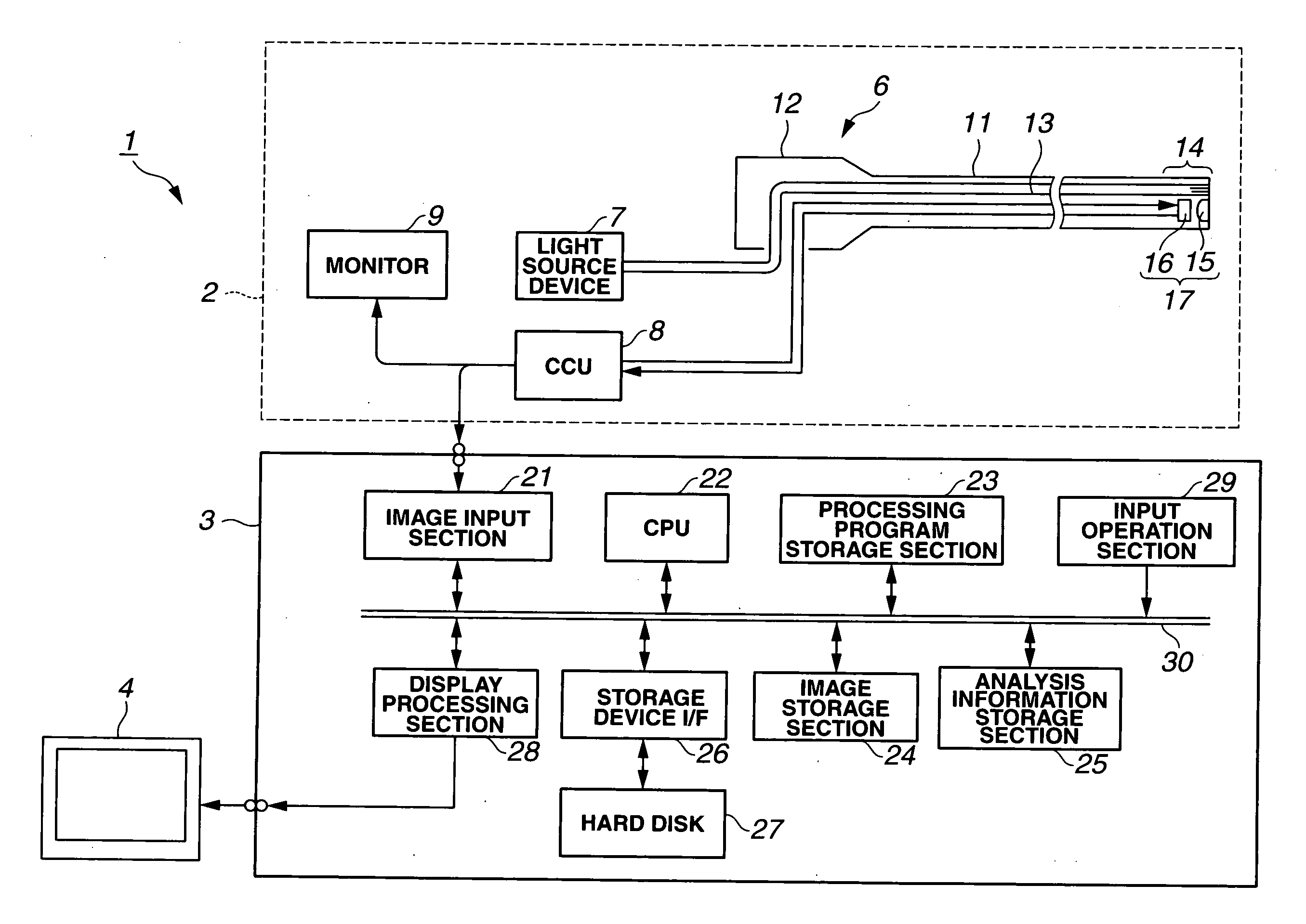
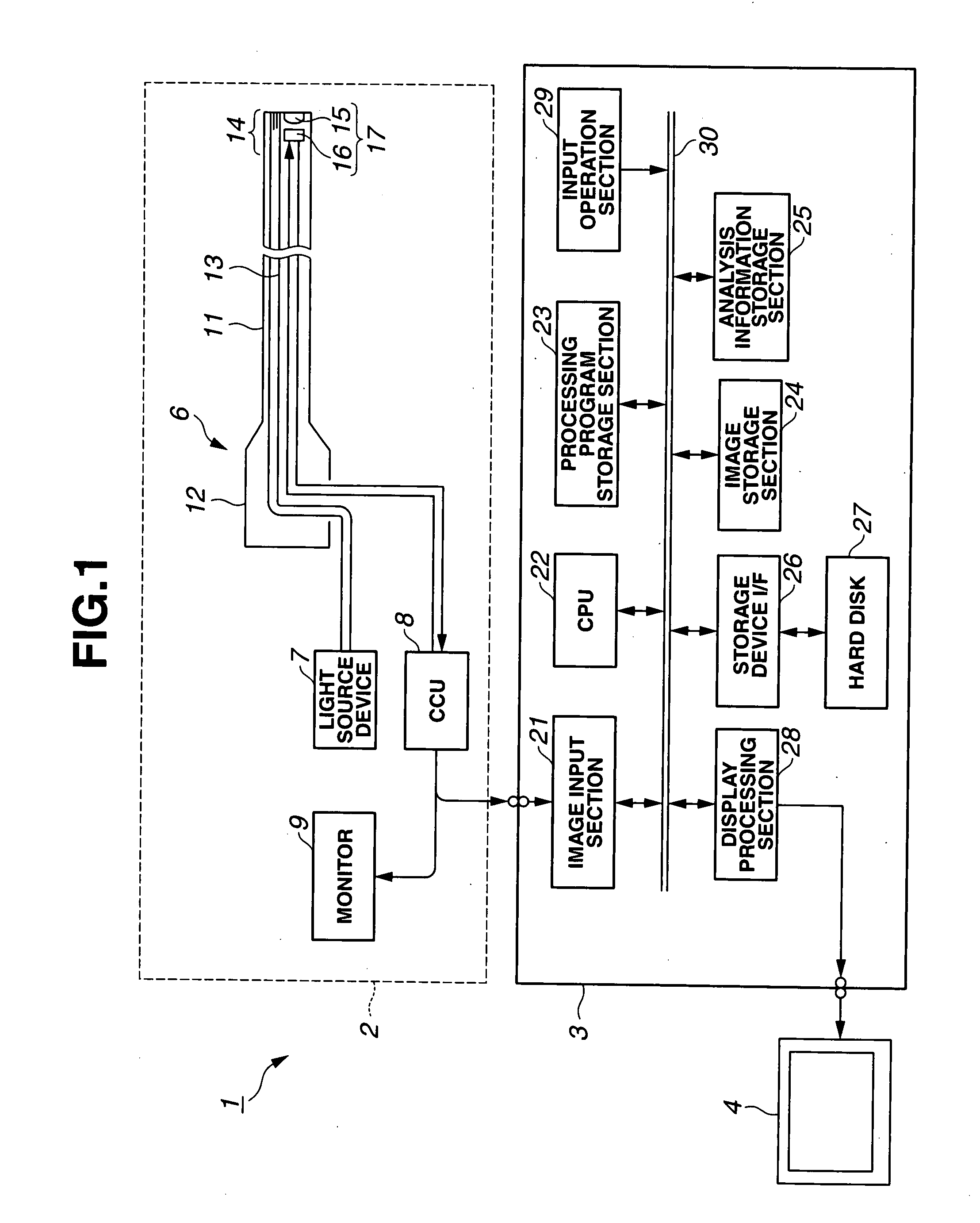
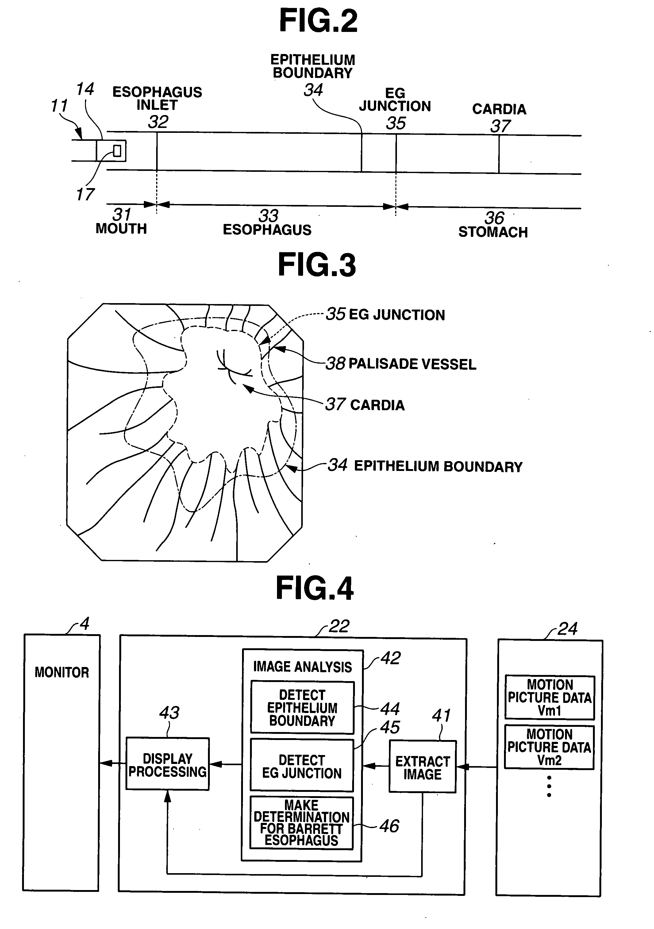
[0099]FIGS. 1 to 16 relate to a first embodiment. FIG. 1 shows the entire configuration of an endoscopic system comprising the present embodiment. FIG. 2 schematically shows the parts of the upper gastrointestinal tract endoscopically examined by orally inserting an endoscope. FIG. 3 shows an example of an endoscopic image of the vicinity of the boundary between the esophagus and the stomach. FIG. 4 shows the functional configuration of an image processing apparatus in accordance with the present embodiment. FIG. 5 shows that motion picture data stored in an image storage section is stored as sets of still image data.

[0100]FIGS. 6A and 6B show analysis results stored in an analysis information storage section, information stored in a processing program storage section, and the like. FIG. 7 shows an example of a monitor display showing an analysis result together with an endoscopic image. FIG. 8 is a flowchart of a process procedure for determining the Barrett esophagus condition in ...

second embodiment

[0221]Now, a second embodiment will be described with reference to FIGS. 17 to 26. In the above first embodiment, to determine whether or not the target site is the Barrett esophagus, the process of detecting the EG junction 35 is first executed to detect an image containing the EG junction 35.

[0222]The process of detecting the EG junction 35 imposes a heavy load, reducing processing speed. Accordingly, the processing speed or the detection speed needs to be improved. A cardia detection process is possible which can be executed more quickly than the detection of the EG junction 35 and which deals with the biological site expected to be accurately detected.

[0223]The present embodiment focuses on this to improve the processing speed and the like.

[0224]The configuration of the hardware of an image processing apparatus in accordance with the present embodiment is similar to that in accordance with the first embodiment; the configuration can be described with reference to FIG. 1. FIG. 17...

third embodiment

[0268]Now, a third embodiment of the present invention will be described with reference to FIGS. 27 to 29. The configuration of the hardware of an image processing apparatus in accordance with the present embodiment is similar to that in accordance with the first embodiment; the configuration can be described with reference to FIG. 1. FIG. 27 shows the functional configuration of essential sections provided by the CPU 22 executing a processing program in accordance with the present embodiment. In the configuration shown in FIG. 27, a processing continuation determination block 48 is further provided in the image analysis block 42, included in the configuration shown in FIG. 4. The processing continuation determination block 48 constitutes a biological feature detection section that detects the first biological feature.

[0269]The processing continuation determination block 48 determines whether or not a point sequence for the epithelium boundary line detected by the epithelium boundar...


PUM


Abstract

There is provided a medical image processing apparatus including an image-extracting section extracting a frame image from in vivo motion picture data picked up by an in vivo image pickup device or a plurality of consecutively picked-up still image data, and an image analysis section analyzing the frame image extracted by the image-extracting section to output an image analysis result. The image analysis section includes a first biological-feature detection section detecting a first biological feature, a second biological-feature detection section detecting, based on a detection result obtained by the first biological feature detection section, a second biological feature in a frame image picked up temporally before or after the image used for detection by the first biological feature detection section; and a condition determination section making a determination for a biological condition based on a detection result obtained by the second biological feature detection section to output the determination.

Description

TECHNICAL FIELD[0001]The present invention relates to a medical image processing apparatus that efficiently determines a condition of interest on the basis of a large amount of biological image data, a luminal image processing and a luminal image processing method which detect the cardia on the basis of images of the interior of the lumen, and programs for the apparatuses and method.BACKGROUND ART[0002]In general, in conventional endoscopic examinations using an endoscope, in vivo image data picked up by an endoscopic apparatus or an endoscopic observation apparatus are immediately displayed on a display device such as a CRT and externally stored as motion picture data. A physician views the motion pictures or views frame images in the motion pictures as still images, during or after examinations for diagnosis.[0003]Further, in recent years, swallowable capsule endoscopes have been used.[0004]For example, as disclosed in Japanese Patent Laid-Open No. 2004-645, image data picked up i...

Claims


Application Information

Patent Timeline
IPC IPC(8): G06K9/00
CPCA61B1/041A61B1/2733G06T7/0012G06T2207/30101G06T2207/10068G06T2207/20168G06T2207/30092G06T2207/10016
Inventor NISHIMURA, HIROKAZUHASEGAWA, JUNTANAKA, HIDEKIINOUE, RYOKONONAMI, TETSUO
Owner OLYMPUS CORP
Why Eureka
Try Eureka