Fast preprocessing algorithms for digital mammography CAD and workstation

a digital mammography and preprocessing algorithm technology, applied in the field of medical imaging systems, can solve the problems of not being able to accurately represent the curve shaped pectoral muscle segmentation result, processing using this type of algorithm is computationally slow, etc., and achieves automatic detection and faster and more accurate segmentation algorithms.

Active Publication Date: 2009-09-03
THREE PALM SOFTWARE
View PDF17 Cites 87 Cited by
  • Summary
  • Abstract
  • Description
  • Claims
  • Application Information

AI Technical Summary

Benefits of technology

[0012] This invention solves existing problems in image preprocessing for mammography CAD and mammography workstation display by utilizing faster and more accurate ...

Problems solved by technology

Processing using this type of algorithms is computationally slow.
A region growing method or gradient method also only detects one connected region, so it can not handle a mammography cleavage view, which includes the medial portions of both right and left bre...

Method used


Image

  • Fast preprocessing algorithms for digital mammography CAD and workstation
  • Fast preprocessing algorithms for digital mammography CAD and workstation
  • Fast preprocessing algorithms for digital mammography CAD and workstation

Examples


Embodiment Construction

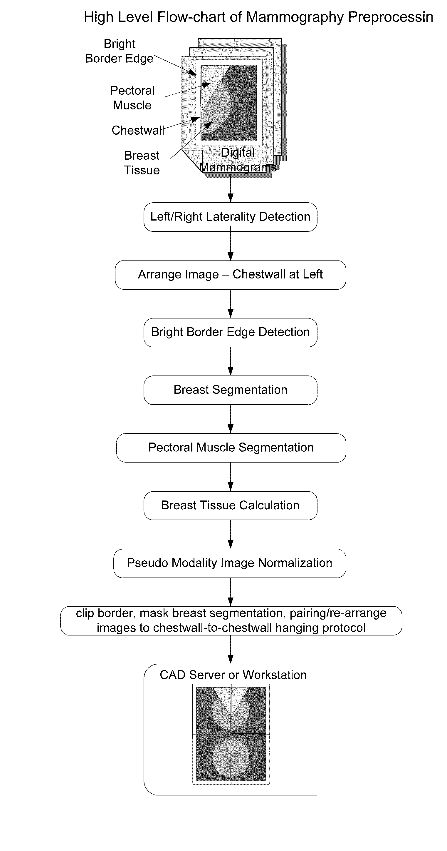
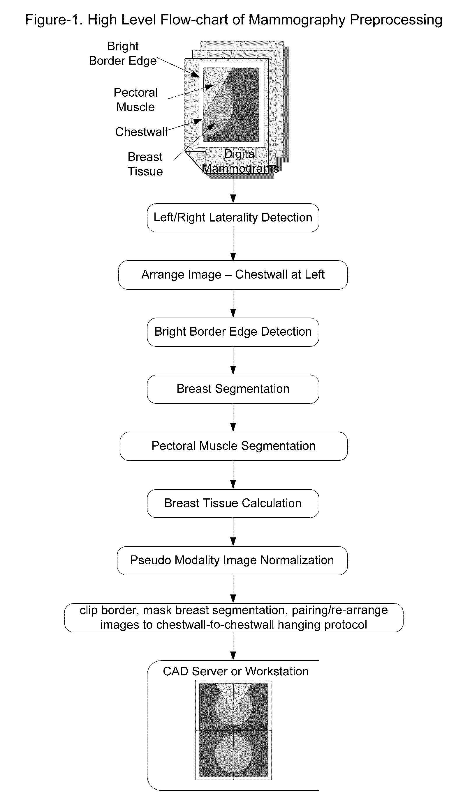
[0020] The mammography preprocessing steps, as shown in FIG. 1, include (1) detecting chestwall side, or left / right laterality of the mammogram image; (2) flipping the image along the vertical axial if the chestwall is not at left; (3) detecting bright or black gap between chestwall and the edge of the image; (4) segmenting breast tissue from the image background; (5) detecting the curved line separation between breast tissue and pectoral muscle; (6) use segmentation information to normalize the image to a consistent pseudo modality; (7) re-arranging the image so to produce mammography standard chestwall-to-chestwall hanging protocol.

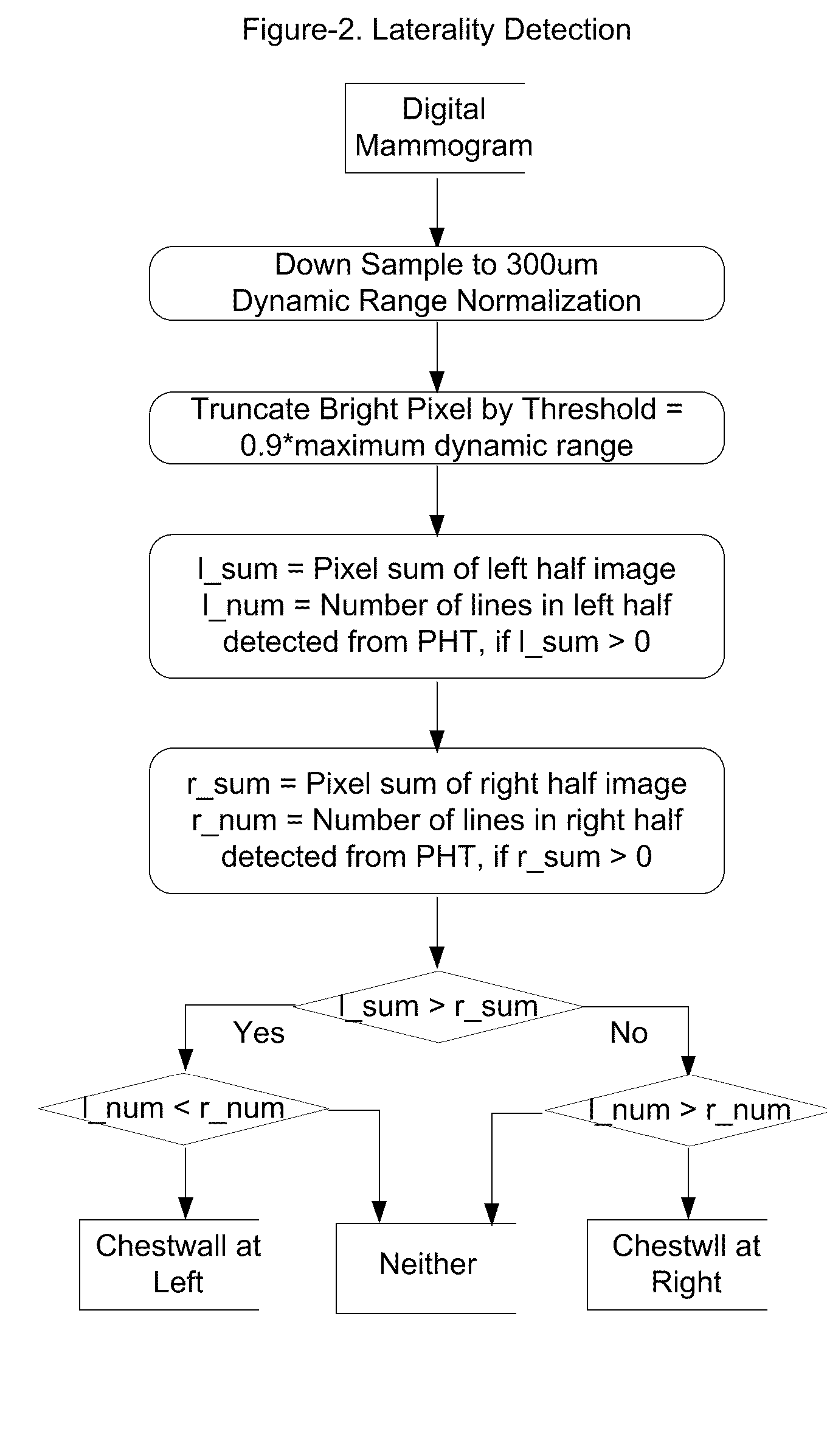
[0021] As shown in FIG. 2, the idea of the detection of the chestwall side for left or right laterality is based on the assumption that the breast tissue intensity is mostly distributed at the chestwall side and the low intensity of the air background at the opposite side.

[0022] As shown in FIG. 3, continuing from the down-sampled image, and flipping ...


PUM


Abstract

A method and apparatus are disclosed for an image preprocessing device that automatically detects chestwall laterality; removes border artifacts; and segments breast tissue and pectoral muscle from digital mammograms. The algorithms in the preprocessing device utilize the computer cache, a vertical Sobel filter and a probabilistic Hough transform to detect curved edges. The preprocessing result, along with a pseudo-modality normalized image, can be used as input to a CAD (computer-aided detection) server or to a mammography image review workstation. In the case of workstation input, the preprocessing results improve the protocol for chestwall-to-chestwall image hanging, and support optimal image contrast display of each segmented region.

Description

CROSS-REFERENCE TO RELATED APPLICATIONS Related U.S. Application Data [0001] Provisional application No. 60 / 923,188, filed on Apr. 13, 2007 U.S. Patent Documents [0002] U.S. Pat. No. 5,572,565 November 1996 Abdel-Mottaleb “Automatic segmentation, skinline and nipple detection in digital mammograms”[0003] U.S. Pat. No. 5,825,910 September 1998 Vafai “Automatic segmentation and skinline detection in digital mammograms”[0004] U.S. Pat. No. 6,035,056 March 2000 Karssemeijer “Method and apparatus for automatic muscle segmentation in digital mammograms”[0005] U.S. Pat. No. 6,091,841 July 2000 Roger et al. “Method and system for segmenting desired regions in digital mammograms”Other Publications [0006] N. Kiryati, Y. Eldar and A. M. Bruckstein, “A Probabilistic Hough Transform”, Pattern Recognition vol. 24, pp. 303-316, 1991STATEMENT REGARDING FEDERALLY SPONSORED RESEARCH OR DEVELOPMENT [0007] Not Applicable. BACKGROUND OF THE INVENTION [0008] The present invention relates generally to the...

Claims


Application Information

Patent Timeline
IPC IPC(8): G06K9/00
CPCG06T7/0083G06T2207/30068G06T2207/10116G06T7/12
Inventor ZHANG, HEIDIHEFFERNAN, PATRICK
Owner THREE PALM SOFTWARE
Why Eureka
Try Eureka