
Slice image display apparatus, method and recording-medium having stored therein program

Inactive Publication Date: 2009-10-15
FUJIFILM CORP
View PDF10 Cites 10 Cited by
  • Summary
  • Abstract
  • Description
  • Claims
  • Application Information

AI Technical Summary

Benefits of technology

[0010]In view of the foregoing circumstances, the inventor of the present invention has noted that when blood (nutrition) is being sent to a node through blood vessels, the probability that the node is malignant is high, and therefore, observation of the connection condition between the node and the blood vessels is essential to recognize the pathological condition of a patient. It is an object of the present invention to provide a slice image display apparatus and method that can more conveniently generate slice images based on a predetermined point in a hilum-of-lung area and a predetermined point in a shadow area.
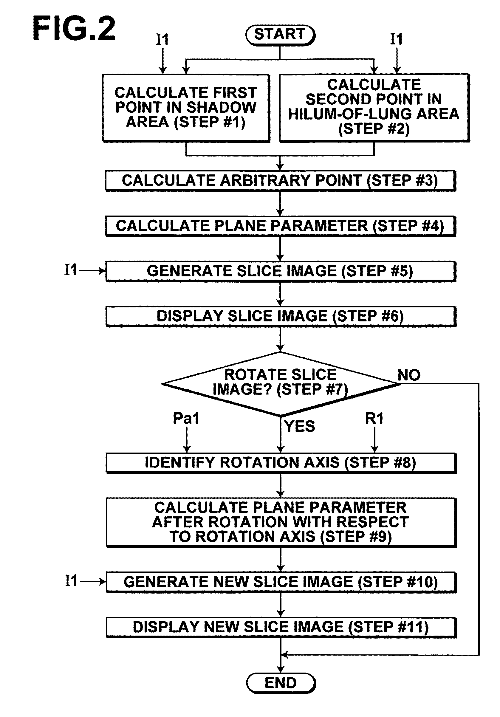
[0037]According to a slice image display apparatus and method of the present invention and a computer-readable recording medium having stored therein a program for the apparatus and method of the present invention, a shadow area in a lung-field area of a subject is detected from tomographic images on predetermined sectional planes, the tomographic images constituting a three-dimensional image representing the subject. Further, a hilum-of-lung area in the lung-field area of the subject is detected from the tomographic images. Further, a slice image that passes through a first point that is a predetermined point in the detected shadow area, a second point that is a predetermined point in the detected hilum-of-lung area, and an arbitrary point that has been set in advance is generated and displayed. Therefore, it is possible to easily generate and display a slice image that makes it possible to easily recognize blood vessels that radially extend from the hilum of lung and / or the course of a trachea. Further, a positional relationship with a lesion and / or a connection relationship with the lesion can be easily recognized.

Problems solved by technology

Further, the operations must be repeated, and that is troublesome.

Method used


Image

  • Slice image display apparatus, method and recording-medium having stored therein program
  • Slice image display apparatus, method and recording-medium having stored therein program
  • Slice image display apparatus, method and recording-medium having stored therein program

Examples


Embodiment Construction

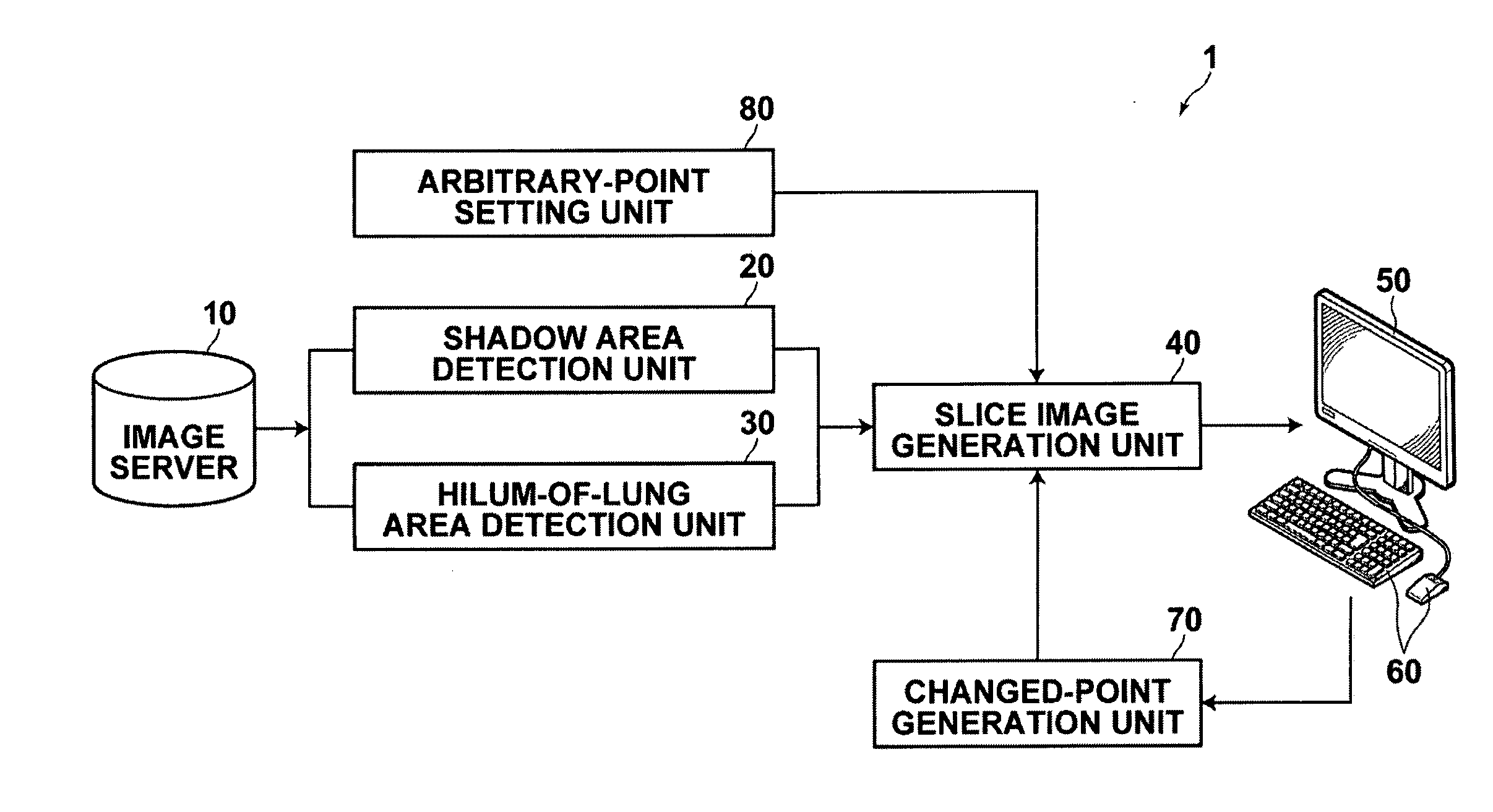
[0045]Hereinafter, an embodiment of the present invention will be described with reference to drawings. In this embodiment, a slice image display apparatus 1 of the present invention generates a slice image from a three-dimensional image and displays the generated slice image.

[0046]First, the slice image display apparatus 1 illustrated in FIG. 1 will be described.

[0047]The slice image display apparatus 1 illustrated in FIG. 1 includes an image server 10, a shadow area detection unit 20, a hilum-of-lung area detection unit 30, an arbitrary-point setting unit 80, a slice image generation unit 40 and a display unit 50. The image server 10 stores a three-dimensional image representing a subject, which is obtained by a CT apparatus or the like. The shadow area detection unit 20 detects a shadow area in a lung-field area of the subject from tomographic images on predetermined sectional planes of the three-dimensional image that is stored in the image server 10. The hilum-of-lung area dete...


PUM


Abstract

A shadow area in a lung-field area of a subject is detected from tomographic images constituting a three-dimensional image representing the subject. A hilum-of-lung area in the lung-field area of the subject is detected from the tomographic images. Further, a slice image that passes through a first point that is a predetermined point in the detected shadow area, a second point that is a predetermined point in the detected hilum-of-lung area, and an arbitrary point that has been set in advance is generated. The generated slice image is displayed.

Description

BACKGROUND OF THE INVENTION[0001]1. Field of the Invention[0002]The present invention relates to a slice image display apparatus and method for displaying a slice image based on voxel data that represents spatial distribution of an object as a three-dimensional image. Further, the present invention relates to a computer-readable recording medium having stored therein a program for the apparatus and method.[0003]2. Description of the Related Art[0004]In recent years, as CT (computed tomography) apparatuses and the like have been developed, it has become possible to obtain tomographic images that accurately represent anatomical structures of human bodies. Further, three-dimensional images have been reconstructed based on a plurality of tomographic images obtained by imaging a subject at different sectional positions (slice positions).[0005]In a method for displaying a three-dimensional medical image, the three-dimensional medical image is generally handled as a three-dimensional array...

Claims


Application Information

Patent Timeline
IPC IPC(8): H05G1/60G06K9/00
CPCG06T2219/008G06T19/00
Inventor MORIYA, YOSHIYUKI
Owner FUJIFILM CORP
Learn More