

External ear-placed non-invasive physiological sensor
a physiological sensor and external measurement technology, applied in the field of external measurement with an external physiological sensor, can solve the problems of inability to use tissue, location is impractical for pre-hospital or emergency use, and accuracy up to 120 seconds in the measurement made by the sensor, so as to reduce the latency of monitoring data, the effect of quick tracking to the saturation of the core arteries
Active Publication Date: 2009-11-05
RGT UNIV OF CALIFORNIA +1
View PDF4 Cites 357 Cited by
- Summary
- Abstract
- Description
- Claims
- Application Information
AI Technical Summary
Benefits of technology
[0004]Optical sensors are widely used across clinical settings, such as operating rooms, emergency rooms, post anesthesia care units, critical care units, outpatient surgery and physiological labs, to name a few. Studies have suggested that conditions producing peripheral vasoconstriction, such as decreased ambient temperature or hypoperfusion states, can cause delays of accuracy up to 120 seconds in measurements made by the sens
Problems solved by technology
Studies have suggested that conditions producing peripheral vasoconstriction, such as decreased ambient temperature or hypoperfusion states, can cause delays of accuracy up to 120 seconds in measurements made by t
Method used
View moreImage
Smart ImageViewing Examples

Examples
 PUM
 Abstract
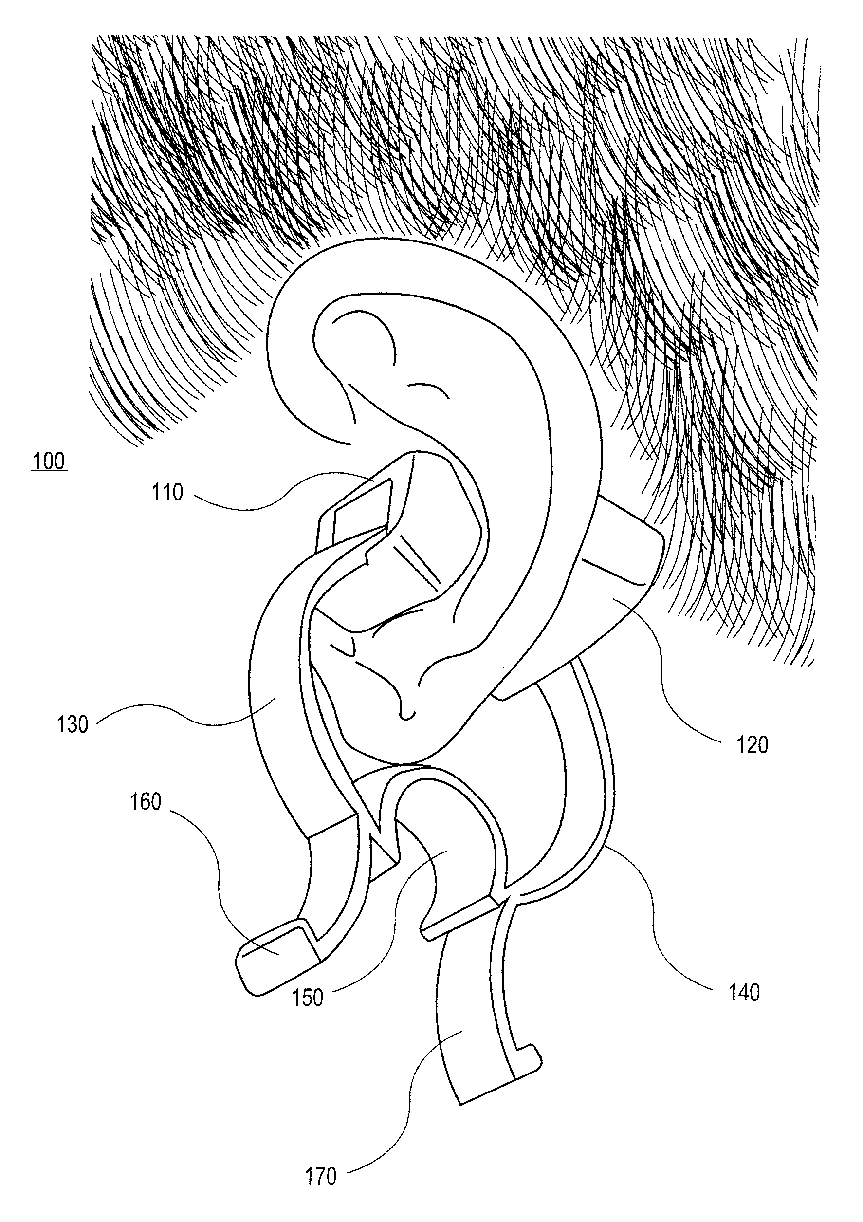
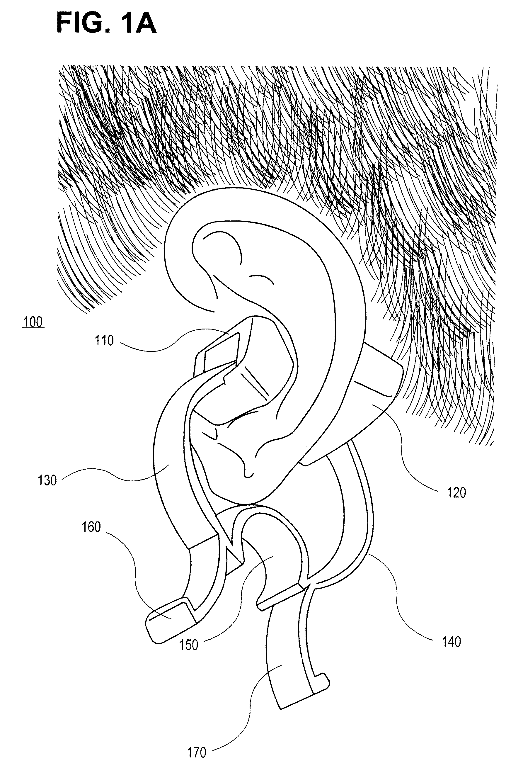
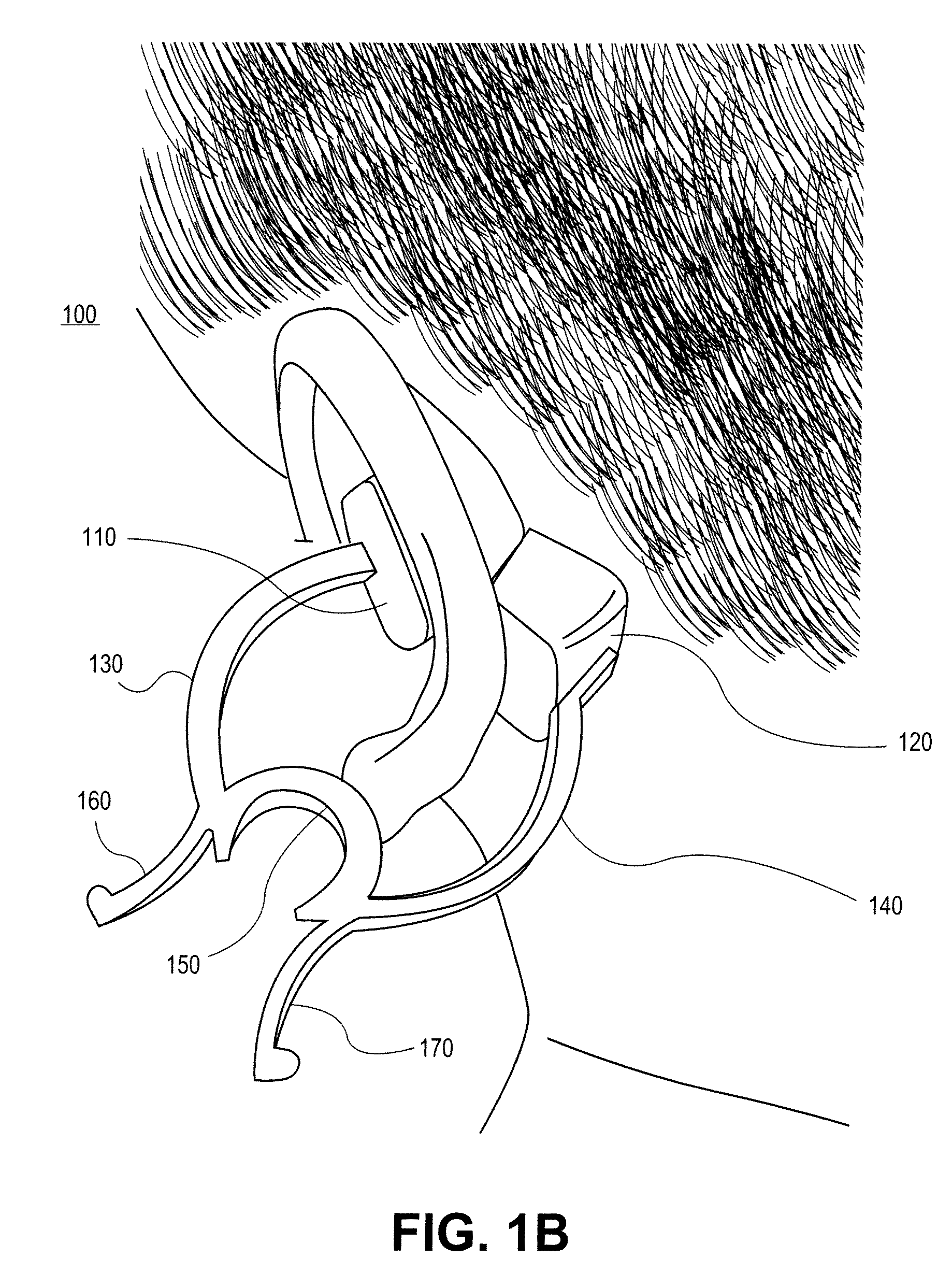
In one embodiment, a non-invasive physiological sensor assembly is capable of attachment to a tissue site of the ear comprising of cartilaginous structures of the ear, providing low latency of physiological measurements as well as a secure attachment.
Description
CROSS-REFERENCE TO RELATED APPLICATIONS[0001]The present application claims the benefit under 35 U.S.C. 119(c) to U.S. Provisional Application No. 61 / 050,085, filed May 2, 2008, titled, “EXTERNAL EAR-PLACED PULSE OXIMETRY PROBE” which is hereby incorporated by reference in its entirety, including specifically but not limited to the systems and methods relating to an external ear-placed non-invasive physiological sensor.FIELD OF THE DISCLOSURE[0002]The present disclosure generally relates to apparatus and methods for non-invasive physiological sensors, and more specifically to methods and apparatus for external measurement with an ear-placed non-invasive physiological sensor.BACKGROUND OF THE DISCLOSURE[0003]Non-invasive physiological sensors are applied to the body for monitoring or making measurements indicative of a patient's health. One application for a non-invasive physiological sensor is pulse oximetry, which provides a noninvasive procedure for measuring the oxygen status of ...
Claims
Login to View More Application Information
Patent Timeline
 IPC IPC(8): A61B5/1455
CPCA61B5/14546A61B5/6838A61B5/6815A61B5/14552A61B5/6817
InventorDAVIS, DANIEL
OwnerRGT UNIV OF CALIFORNIA