Method for interactively determining a bounding surface for segmenting a lesion in a medical image

a technology of medical image and bounding surface, applied in the field of digital imaging, can solve the problems of many voxels that could be easily ignored, unconstrained growth, and additional constraints

Inactive Publication Date: 2009-11-12
CARESTREAM HEALTH INC
View PDF5 Cites 25 Cited by
  • Summary
  • Abstract
  • Description
  • Claims
  • Application Information

AI Technical Summary

Problems solved by technology

In some cases, this simple stopping condition leads to unconstrained growth, as the signal values of diseased tissue and healthy tissue are similar.
This results in additional constraints, such as maximum distance away from the initial seed point.
Unfortunately, the approach results in many voxels that could be readily ignored, and the boundary of the box does not reflect anatomical shapes, which are generally smooth without sharp rectilinear corners.
It is known that a human has unexcelled capability to interpret visual information, even when the information is obscured or ambiguous.
However, identifying all the voxels within the diseased tissue is tedious and often accompli...

Method used


Image

  • Method for interactively determining a bounding surface for segmenting a lesion in a medical image
  • Method for interactively determining a bounding surface for segmenting a lesion in a medical image
  • Method for interactively determining a bounding surface for segmenting a lesion in a medical image

Examples


Embodiment Construction

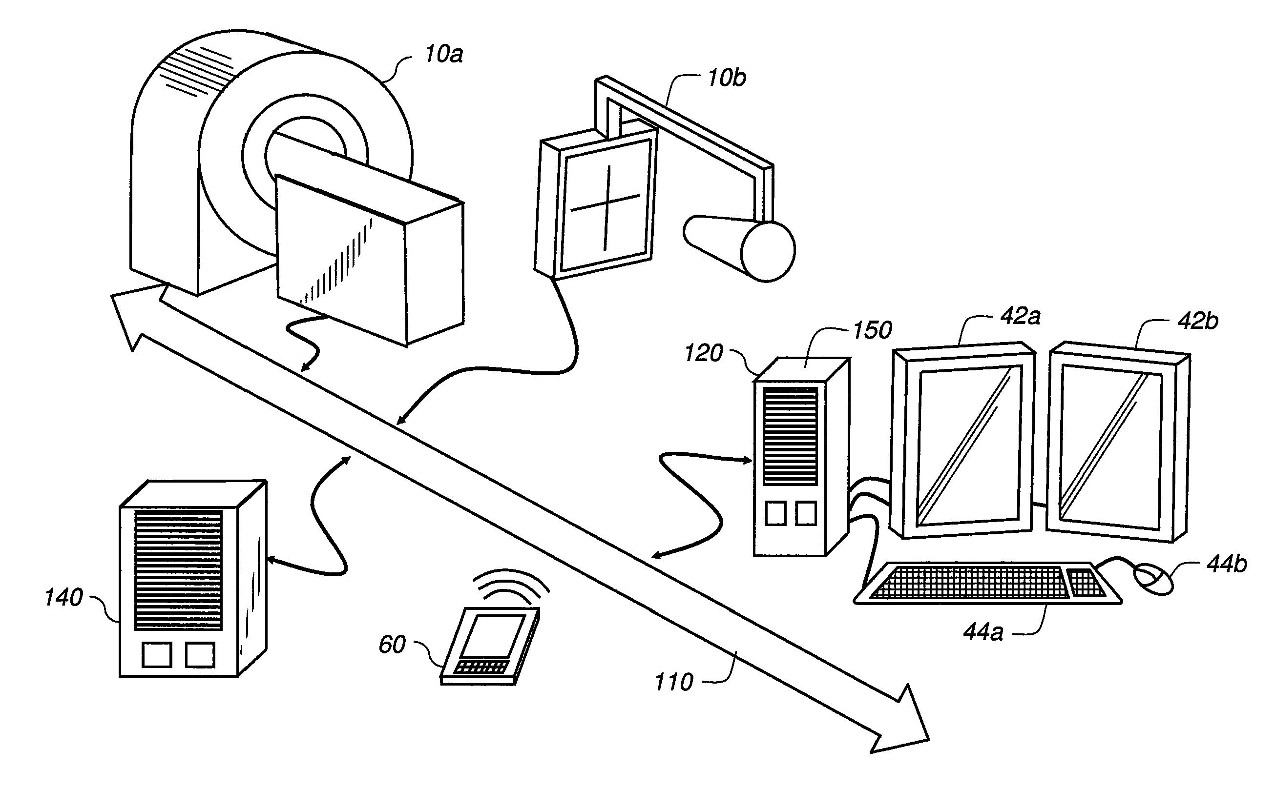
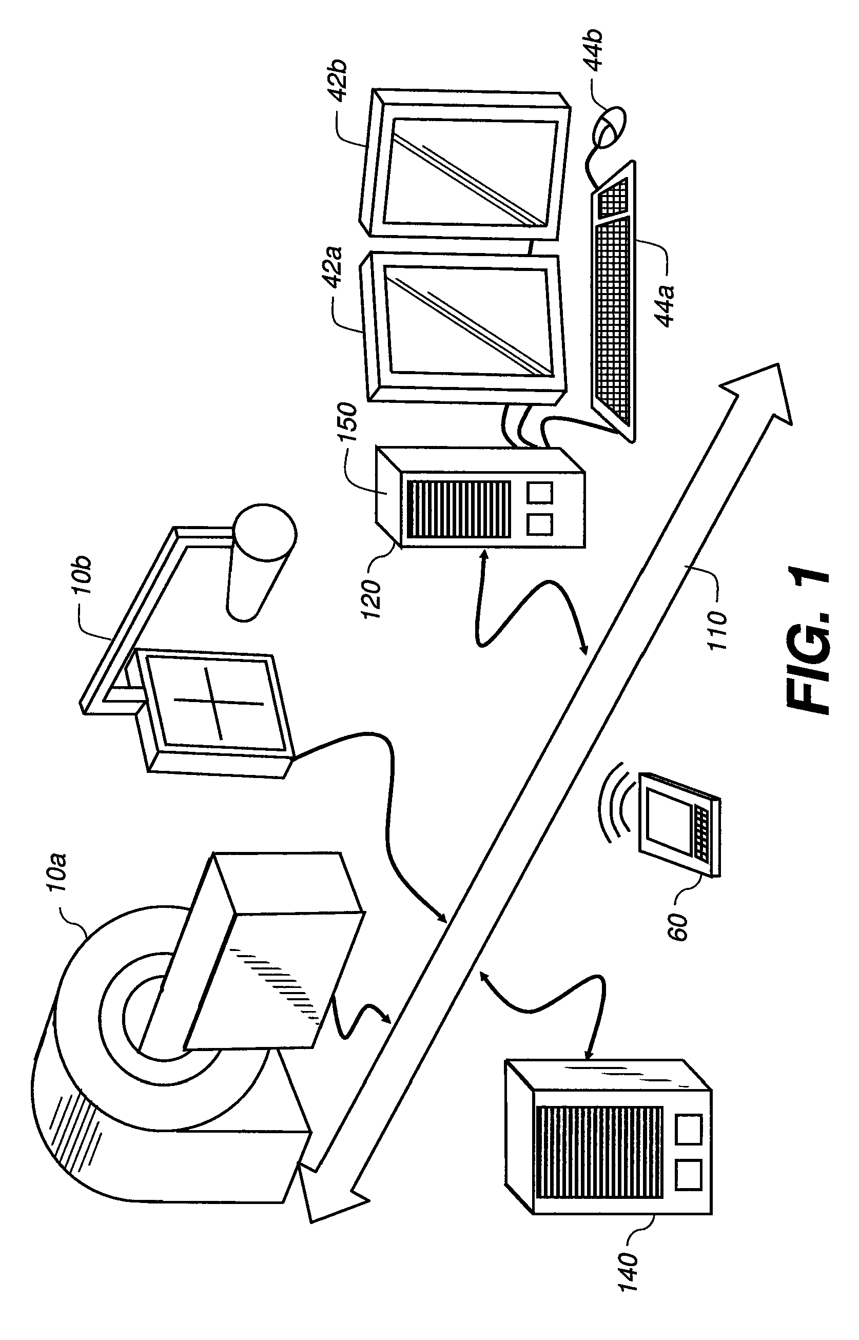
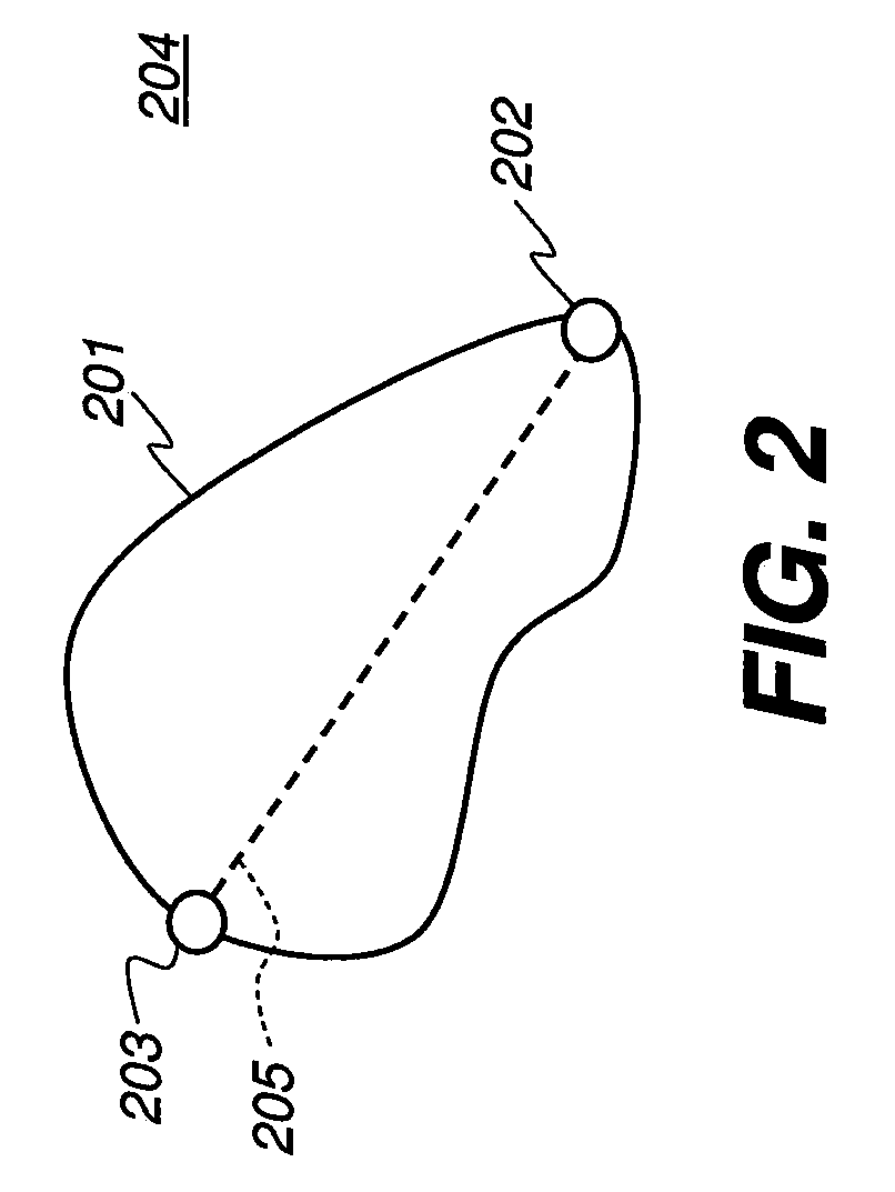
[0024]Exemplary embodiments of the present disclosure provide the operator of a PACS medical imaging diagnostic workstation the ability to segment anatomical regions of interest in digital medical images with a limited amount of input. The input addresses the knowledge of the user to identify the object of interest and to give some rudimentary control over the location, extent, orientation, and shape of the object. The volume of interest generated is compatible with anatomical objects. For example, in an embodiment of the present disclosure, the boundary of the volume of interest may not possess sharp corners or edges. Moreover, the test whether a voxel is interior or exterior to the volume is simple to implement.

[0025]Reference is made to commonly assigned application U.S. Ser. No. 61 / 050,723 (Docket No. 94765), entitled “STATISTICS COLLECTION FOR LESION SEGMENTATION”, provisionally filed on May 6, 2008, the entire disclosure of which is incorporated herein by reference.

[0026]Refer...


PUM


Abstract

A method of bounding an anatomical object of interest in a 3-dimensional volume image includes displaying an image of at least a portion of the object, selecting a plurality of points in the displayed image, at least a first and second point of the plurality of points spanning the object, forming a non-rectilinear surface bounding the plurality of points, identifying a seed point within the surface and extracting a plurality of statistical values corresponding to image voxels disposed proximate the seed point, and classifying image voxels within the surface into a first class and a second class based on the plurality of statistical values.

Description

CROSS REFERENCE TO RELATED APPLICATIONS[0001]Priority is claimed from U.S. Ser. No. 61 / 050,752, provisionally filed on May 6, 2008, entitled “METHOD FOR INTERACTIVELY DETERMINING A BOUNDING SURFACE FOR SEGMENTING A LEDION IN A MEDICAL IMAGE”, in the name of Edward B. Gindele et al., commonly assigned and incorporated herein by reference.FIELD OF THE INVENTION[0002]The present invention relates to the field of digital imaging, and more particularly to segmenting a lesion in a digital medical image.BACKGROUND OF THE INVENTION[0003]Image segmentation is a branch of digital image processing that performs the task of categorizing, or classifying, the elements of a digital image into one or more class types. For medical imaging applications, it is common that image segmentation is performed on the voxels (volume elements) of a 3-dimensional image data set with the classification types related to anatomical structure. One task is segmenting regions within the image data set that are consid...

Claims


Application Information

Patent Timeline
IPC IPC(8): G06K9/00
CPCG06T7/0081G06T2207/30004G06T2207/20156G06T2207/20104G06T7/11
Inventor GINDELE, EDWARD B.RAY, LAWRENCE A.SIMON, RICHARD A.
Owner CARESTREAM HEALTH INC
Why Eureka
Try Eureka