
System for three-dimensional medical instrument navigation

a three-dimensional navigation and medical instrument technology, applied in the field of road mapping procedures, can solve the problems of user's image providing limited depth perception, reducing desired depth cues, and reducing highlights and shadows

Inactive Publication Date: 2009-11-12
THE BOARD OF TRUSTEES OF THE LELAND STANFORD JUNIOR UNIV
View PDF15 Cites 44 Cited by
  • Summary
  • Abstract
  • Description
  • Claims
  • Application Information

AI Technical Summary

Problems solved by technology

Due to vessel self-occlusion and blending that can occur when overlaying two 2D images, however, desired depth cues, such as highlights and shadows are often diminished, leaving the user with an image that provides limited depth perception.

Method used


Image

  • System for three-dimensional medical instrument navigation
  • System for three-dimensional medical instrument navigation
  • System for three-dimensional medical instrument navigation

Examples


Embodiment Construction

Definitions

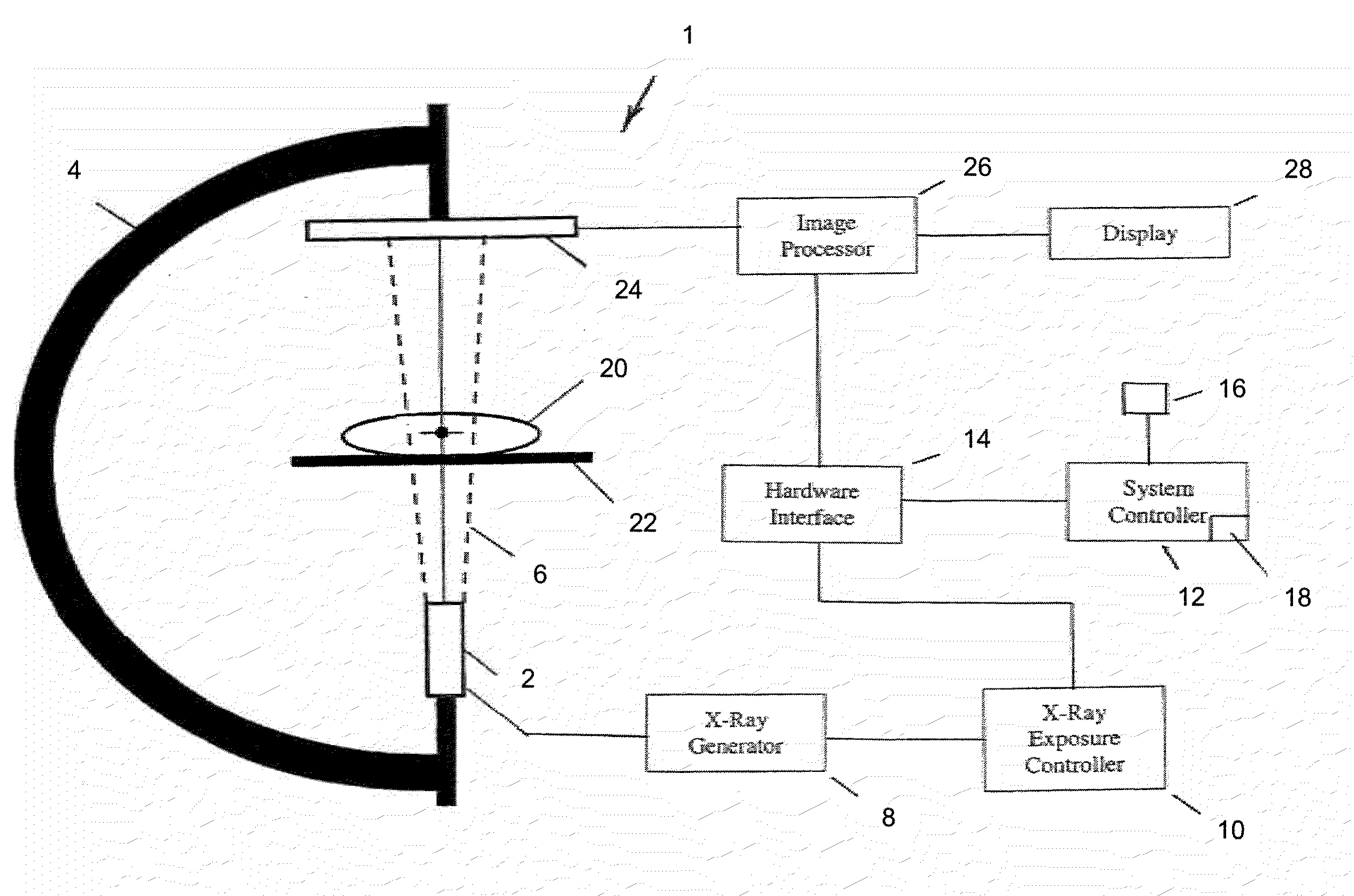
[0019]An angiogram uses a radiopaque substance (i.e., a contrast agent) to make blood vessels visible under x-ray imaging. A roadmapping mask is a digitally subtracted angiogram generated by computer processes which compare an x-ray image of a region of the body before and after a contrast agent has been injected arterially into the body. A fluoroscopic image is an x-ray image showing internal tissues of a region of the body. A live fluoroscopic image is an x-ray image showing live movement of internal tissues of a region of the body. A superimposed image is an image in which an original or adjusted roadmapping mask is combined with a live fluoroscopic image. “Combining” a roadmap mask with live fluoroscopy is achieved by digitally subtracting the adjusted mask in real time from the live fluoroscopic image. Since the mask contains a representation of the contrast media (i.e., the blood vessels) and the live fluoroscopic image does not, the contrast media shows up as white...


PUM


Abstract

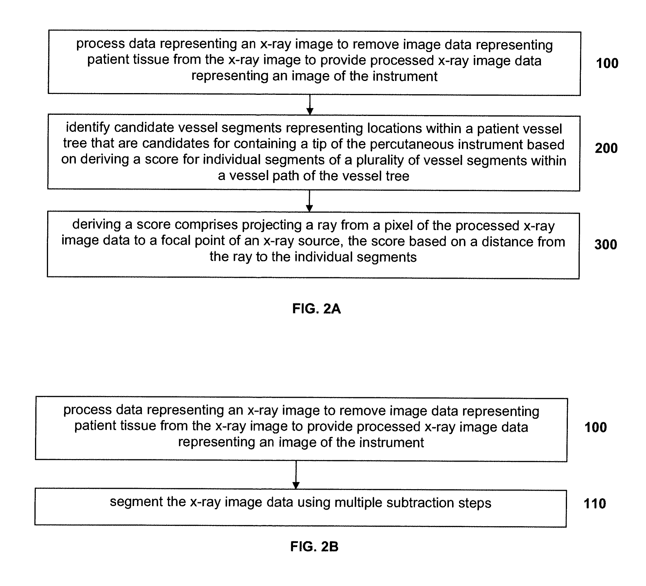
A system and method are disclosed for reconstructing an instrument in 3 dimensions for use during interventional medical procedures to provide enhanced instrument visualization with respect to a patient's vasculature. A patient vessel tree volume is co-registered with a live fluoroscopic image of a percutaneously-inserted instrument, such as a guidewire. The fluoroscopic image is segmented to eliminate images of surrounding tissue and to retain the guidewire image. The black pixels of the image are backprojected to the focal point of the x-ray source, through the co-registered vessel tree. The vessel tree is divided into segments that are scored based on proximity to the backprojected black pixels. Candidate instrument-containing vessel paths are identified based on the scores of the segments, and errant candidate vessel paths are eliminated to produce a refined list of candidate paths. Thresholding and visualization are performed to further refine the candidate vessel paths. 3D curve fitting is used to reconstruct an image of the instrument along with a 3D rendering of the final vessel path.

Description

CROSS-REFERENCE TO RELATED APPLICATIONS[0001]This is a non-provisional application of pending U.S. provisional patent application Ser. No. 61 / 052,312 filed May 12, 2008 by Markus Kukuk et al.FIELD OF THE DISCLOSURE[0002]The disclosure is related to roadmapping procedures in general, and more particularly to procedures for enhanced visualization of guidewire placement within patient blood vessels.BACKGROUND[0003]The process known as road-mapping is used in the selective catheterization of vessels in the framework of an interventional treatment. In these minimally invasive angiographic interventions, a 3-dimensional (3D) image of a patient's vasculature is obtained for use as a “road-map,” to assist a physician to efficiently guide or navigate instruments such as catheters or guide-wires through the patient's vasculature. Intravascular procedures are often performed using a C-arm x-ray imaging system which includes an adjustable x-ray source and an x-ray detector.[0004]Image based nav...

Claims


Application Information

Patent Timeline
Patent Type & Authority Applications(United States)
IPC IPC(8): G06K9/00H05G1/64
CPCG06T7/0044G06T2207/30101G06T2207/20116G06T2207/10121G06T7/74
Inventor KUKUK, MARKUSNAPEL, SANDY
Owner THE BOARD OF TRUSTEES OF THE LELAND STANFORD JUNIOR UNIV
Learn More