
Fast mapping of volumetric density data onto a two-dimensional screen

a volumetric density and two-dimensional screen technology, applied in the field of image-guided surgery, can solve the problems of avoiding the use of fiducials altogether, limiting the number of times and duration of fluoroscopic imaging, and virtually impossible to retain fiducials between scanning and surgical operations

Inactive Publication Date: 2005-06-14
STRYKER EURO OPERATIONS HLDG LLC
View PDF15 Cites 73 Cited by
  • Summary
  • Abstract
  • Description
  • Claims
  • Application Information

AI Technical Summary

Benefits of technology

[0014]A computational device in the system is operatively connected to the data file and display screen for (i) defining a plurality of rays extending between a selected source point and each of a plurality of points in a two-dimensional XY array, where (ia) the array points correspond to the array pixels in the display screen, and (ib) at least some of the rays pass through a plurality of scan-date voxels in the patient target region, (ii) for each ray, distributing the bit positions of the M-bit voxel density values among the multiple N-bit registers of the associated pixel, such that one or more bit positions of each M-bit value is assigned to a selected register of the associated pixel, (iii) for each ray, summing the M-bit values of the voxels along that ray by (iiia) individually summing the one or more bit-position values in each associated pixel register, and (iiib) determining the sum of the M-bit values along the associated ray from the values in the individual registers, and (iiic) plurality of passes over the previous step to further reduce a chance of register overflow and (iv) applying the summed tissue-density values along each ray to the associated pixel in the display screen.

Problems solved by technology

For other surgical regions, e.g., the spine, it is virtually impossible to retain fiducials at fixed patient coordinates between the scanning and surgical operations.
Ideally, the surgeon would want to forego the use of fiducials altogether, since their placement involves additional inconvenience to the patient and surgeon.
Unfortunately, the surgeon will be limited in the number or times and duration of fluoroscopic imaging, due to the need to limit the amount of x-ray exposure to both the patient and medical personnel.

Method used


Image

  • Fast mapping of volumetric density data onto a two-dimensional screen
  • Fast mapping of volumetric density data onto a two-dimensional screen
  • Fast mapping of volumetric density data onto a two-dimensional screen

Examples


Embodiment Construction

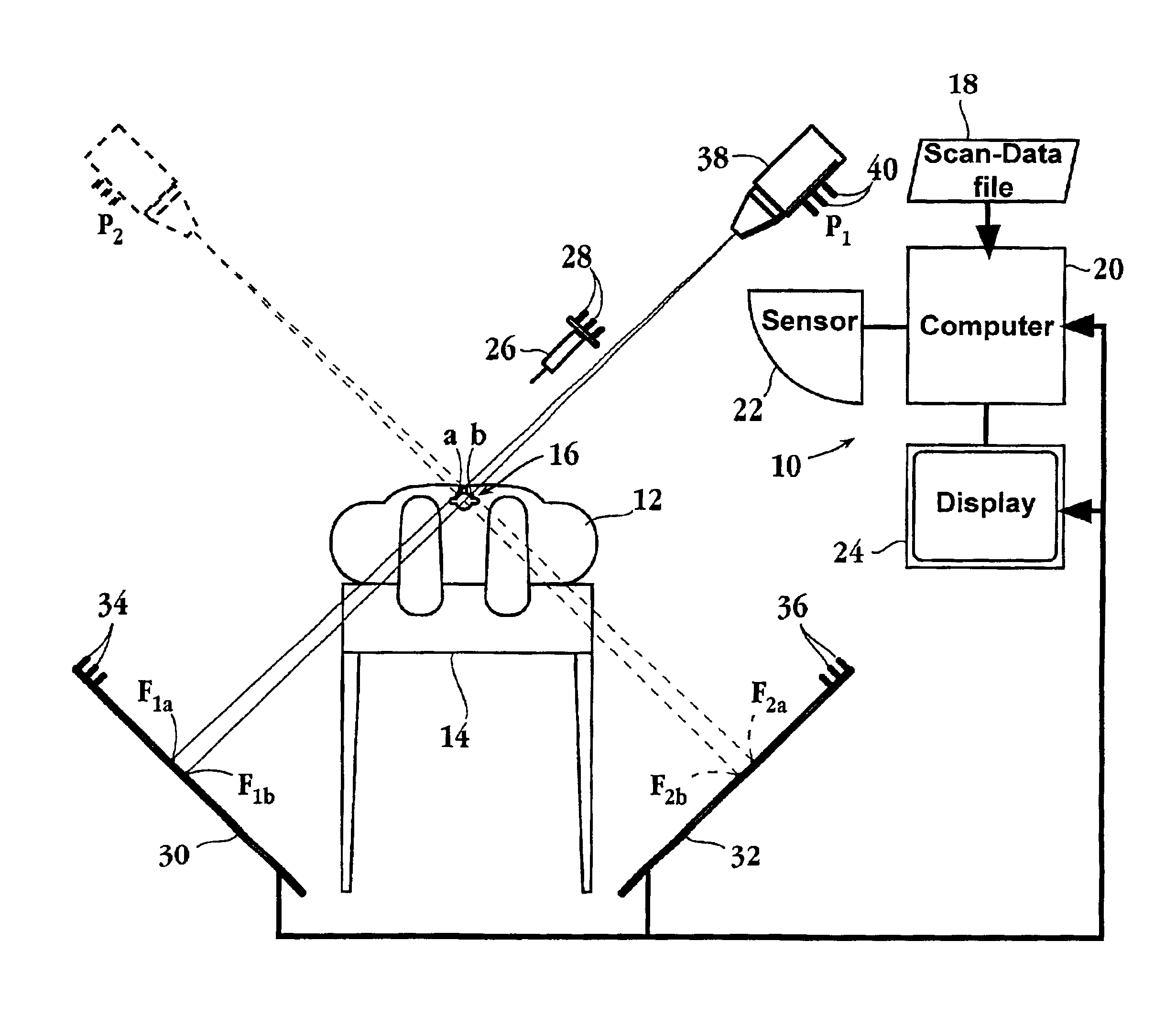
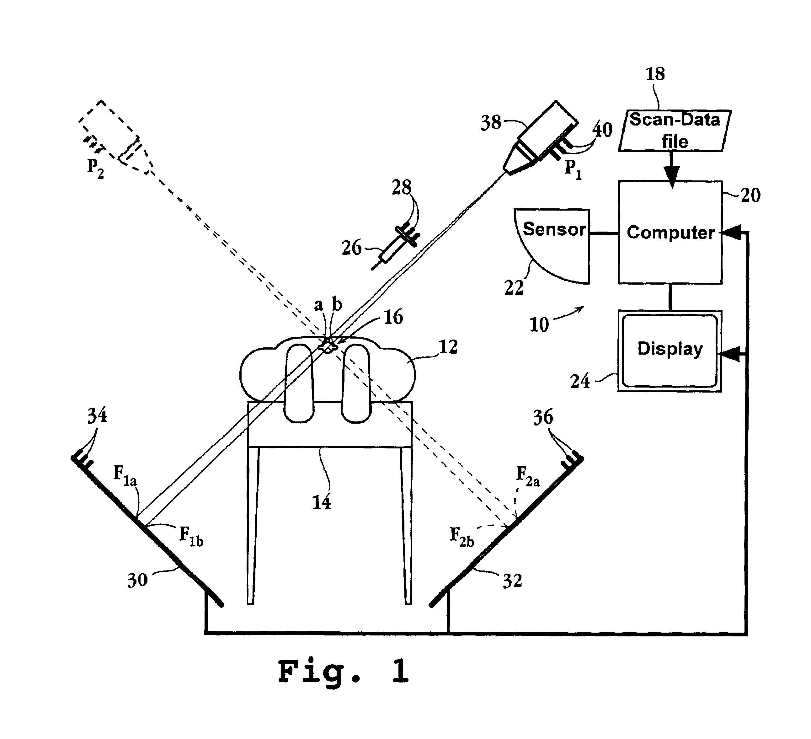
[0026]The present invention is designed to enhance several aspects of image-guided surgery. In each case, the invention depends on or uses pre-operative volumetric scan data of a patient target region. Typically, the scan data acquired are computerized tomography (CT), magnetic resonance imaging (MRI), or ultrasound scan data, obtained according to well-known methods. The scan data are conventionally stored in digitized form in a scan-data file, for later use in the invention. For purposes of the present invention, the scan data have three important characteristics.

[0027]First, the target region (volume) that is scanned is divided into a three-dimensional voxel space, where each voxel (small volume element) typically represents an x-y element in the “plane” of scans taken along the z direction. (Voxels defined by other types of coordinate systems, e.g., polar coordinates, may also be employed). Second, each voxel is associated with an M-bit density value that characterizes the scan ...


PUM


Abstract

A method and system for constructing from volumetric CT or MRI scan data of a patient region, a virtual two-dimensional fluoroscopic image of the patient region as seen from a selected point in space are disclosed. In practicing the method, a plurality of rays are constructed between a selected view point and each of a plurality of points in an XY array, where at least some of the rays pass through the patient target region, the points in the XY array correspond to the XY array of pixels in a display screen, and each pixel in the display screen includes multiple N-bit registers for receiving digital-scale values for each of multiple colors. For each pixel element, a sum of all M-bit density values associated with voxels along a ray extending from the selected point to the associated pixel element is calculated. The summing is carried out by distributing the M bits in the voxel density values among the multiple N-bit registers of that pixel, such that such that one or more bit positions of each M-bit value is assigned to a selected register. The image constructed of gray-scale values representing the summed M-bit density values at each pixel in the screen is displayed to the user.

Description

[0001]This application claims priority to U.S. Provisional Patent Application Ser. No. 60 / 230,631 filed Sep. 7, 2000, which is hereby incorporated in its entirety by reference.FIELD OF THE INVENTION[0002]This invention relates to image-guided surgery, and in particular, to registration and fluoroscopic imaging methods.BACKGROUND OF THE INVENTION[0003]In a typical image-guided surgical procedure, pre-operative scan data, e.g., CT or MRI scan data, of a patient region is obtained. This is done conventionally by immobilizing a patient during a scan with a CT or MRI imaging machine. The patient is then moved to a surgical setting for treatment of the target region. During the surgical procedure, the scan data is used to reconstruct hidden or subsurface images of the patient target region, to guide or assist the surgeon in the surgical procedure. For example, the scan data may be used to reconstruct a subsurface image of the target region as seen from the position and orientation of a su...

Claims


Application Information

Patent Timeline
IPC IPC(8): A61B19/00
CPCA61B19/52A61B19/5244A61B2019/5291A61B2019/5289A61B2019/5255A61B34/20A61B90/36A61B2034/2055A61B2090/364A61B2090/365
Inventor GRZESZCZUK, ROBERT
Owner STRYKER EURO OPERATIONS HLDG LLC
Learn More