Apparatus and method for tracking feature's position in human body

Active Publication Date: 2009-10-15
ARINETA
View PDF3 Cites 76 Cited by
  • Summary
  • Abstract
  • Description
  • Claims
  • Application Information

AI Technical Summary

Benefits of technology

[0002]The invention relates to tracking position of organs, lesions or devices in the human body, and more specifically it

Problems solved by technology

However, conventional fluoroscopy or angiography provide 2D projection images and do not disclose the complete 3D structure underneath.
However, CT scanners typically require minutes to scan a whole organ, reconstruct the slice images and process the data to 3D presentation.
Therefore, CT scanners cannot typically be used for real time monitoring of position in three dimensions during in

Method used


Image

  • Apparatus and method for tracking feature's position in human body
  • Apparatus and method for tracking feature's position in human body
  • Apparatus and method for tracking feature's position in human body

Examples


Embodiment Construction

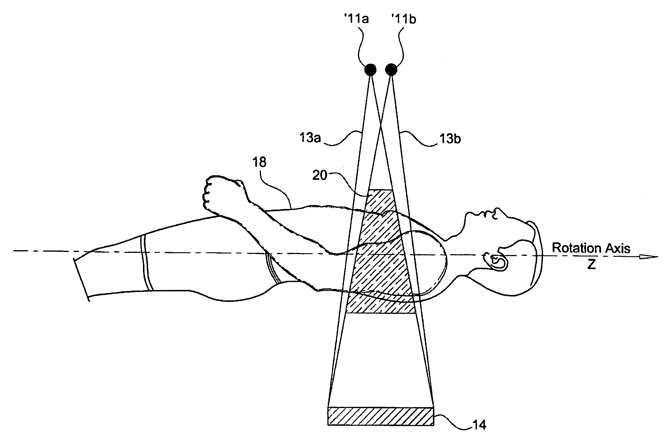
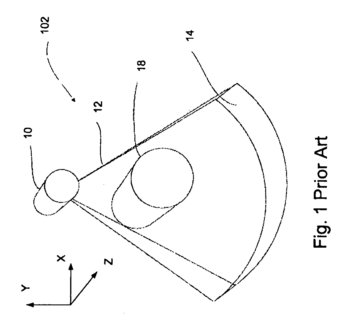
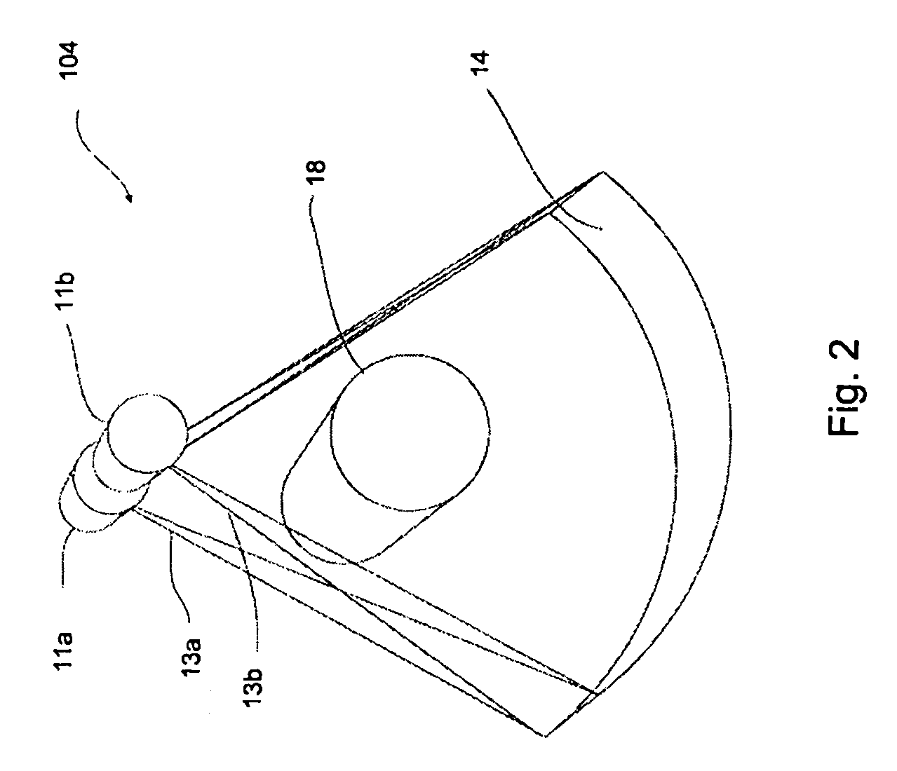
[0070]The invention relates to tracking position of organs, lesions or devices in the human body, and more specifically it relates to application of Computed Tomography (CT) and Stereoscopic Fluoroscopy imaging techniques during interventional procedures.

[0071]Before explaining the embodiments of the invention in detail, it is to be understood that the invention is not limited in its application to the details of construction and the arrangement of the components set forth in the following description or illustrated in the drawings. The invention is capable of other embodiments or of being practiced or carried out in various ways. Also, it is to be understood that the phraseology and terminology employed herein is for the purpose of description and should not be regarded as limiting.

[0072]In discussion of the various figures described herein below, like numbers refer to like parts. The drawings are generally not to scale.

[0073]For clarity, non-essential elements were omitted from so...


PUM


Abstract

A CT scanner for scanning a subject is provided, the scanner comprising: a gantry capable of rotating about a scanned subject; at least two cone beam X-Ray sources displaced from each other mounted on said gantry; at least one 2D detector array mounted on said gantry, said detector is capable of receiving radiation emitted by said at least two X-Ray sources and attenuated by the subject to be scanned; a first image processor capable of generating and displaying CT images of a volume within the subject; a second image processor capable of generating projection X-Ray images of said volume, wherein the images are responsive to X-Ray separately emitted by each of said at least two cone beam X-Ray sources; and a third image processor capable of generating and displaying fluoroscopic images composed of said projection X-Ray images, wherein said fluoroscopic images are spatially registered to said CT images.

Description

CROSS-REFERENCE TO RELATED APPLICATION[0001]This application claims the benefit of U.S. Provisional Patent Application Ser. No. 61 / 123,584, filed Apr. 10, 2008, the entire contents of which are incorporated by reference, as if fully set forth herein.FIELD OF THE INVENTION[0002]The invention relates to tracking position of organs, lesions or devices in the human body, and more specifically it to application of Computed Tomography (CT) and Stereoscopic Fluoroscopy imaging techniques during interventional procedures.BACKGROUND OF THE INVENTION[0003]Medical procedures that require tracking of the position of a lesion or device in the human body are very common. A lesion is referred to herein as a tumor, diseased artery or any other diseased or target tissue. For example, in percutaneous coronary intervention (PCI) procedures a balloon is inserted through a catheter from an artery in the groin to a narrowed section in a coronary artery. The balloon is then inflated, compressing the plaqu...

Claims


Application Information

Patent Timeline
IPC IPC(8): A61B6/00
CPCA61B6/022A61B6/032A61B6/4014A61B6/4028A61B6/504A61B6/541A61B6/481
Inventor DAFNI, EHUDBRADA, RAFAEL SHMUEL
Owner ARINETA
Why Eureka
Try Eureka