Method for Detecting Contours in Images of Biological Cells

Inactive Publication Date: 2009-12-03
EVOTEC TECH GMBH
View PDF3 Cites 20 Cited by
  • Summary
  • Abstract
  • Description
  • Claims
  • Application Information

AI Technical Summary

Benefits of technology

[0032]However, promising results are also obtained in diffuse regions of cytoplasm that, overall, even seem to be convex. In this case, it can be demonstrated that a closer clustering of the axes and an associated flattening of the splines also yield

Problems solved by technology

The problem of segmentation is complex since cells have a very heterogeneous morphology that is difficult to model.
Further, difficulties are caused by occlusion as well as by cases of neighbor

Method used


Image

  • Method for Detecting Contours in Images of Biological Cells
  • Method for Detecting Contours in Images of Biological Cells
  • Method for Detecting Contours in Images of Biological Cells

Examples


embodiment 1

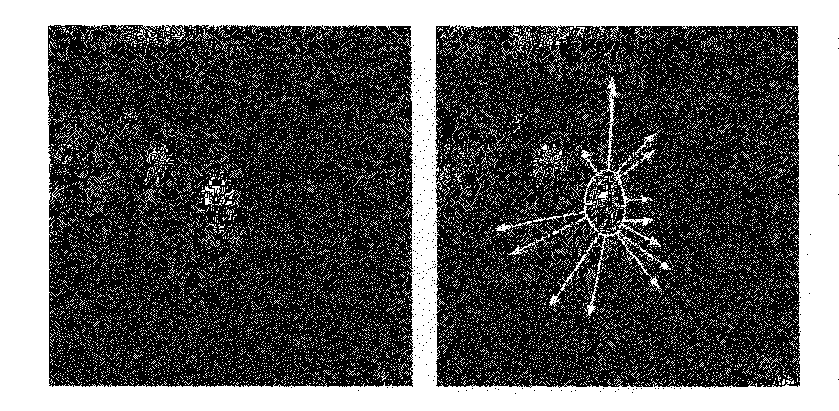
[0052]The model of an adherent cell presented herein puts emphasis on the calculation of the membrane, which, die to the data being present in the second dimension, means a line. At the same time, a simple inner structure for the construction of a cell is proposed.

[0053]Morphology of the Cell

[0054]The shape of a cell substantially results from the cytoskeleton, a combination of fibers internal to the cell, on the one hand, and the membrane, which is a bordering surface enclosing the cell, on the other hand. The skeleton is made from a framework of fibers connected at discrete points. Terminal points of the fibers penetrate the membrane and, by means of receptors, form a connection to the surrounding tissue or to the substrate. As used in the present embodiment, the terms inner and outer adhesion points refer to the adhesion points among the fibers or to the connection points of the fibers and materials surrounding the cell. Fibers having an outer adhesion point may often be consider...

embodiment 2

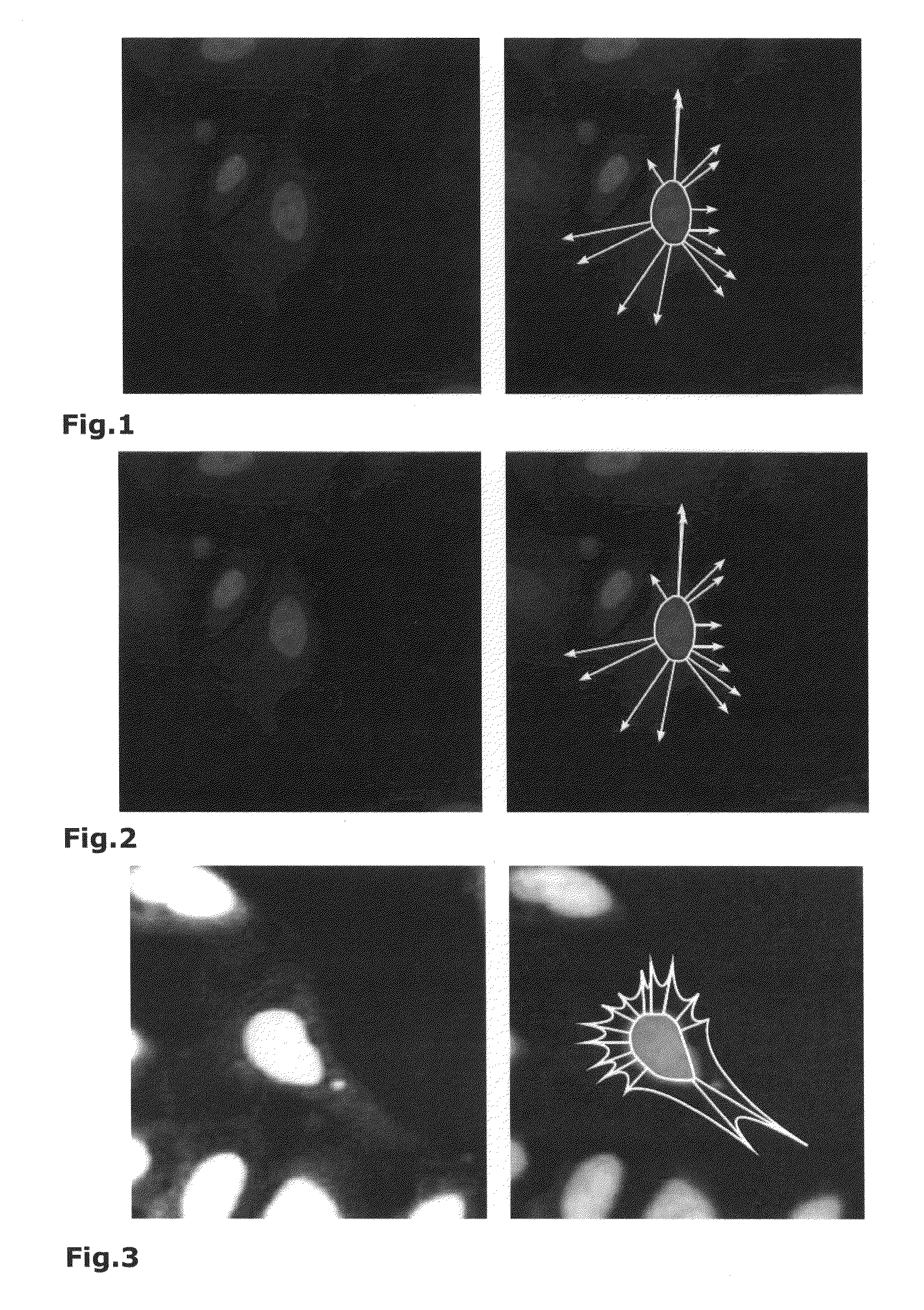
[0084]In less dense monolayers cells grow with a relatively high degree of freedom so that the cytoplasm with the surrounding membrane takes an irregular shape. The nucleus of a cell is then often offset from the centre and the tensions between outer adhesion points and the inside of the cell can no longer be described as a circular nucleus-related bundle of vectors.

[0085]A model that would be realistic in this sense should thus also include inner adhesion points besides the outer adhesion points.

[0086]Within a cell, the adhesion points are interconnected by actine fibers (Wang, Ning; Naruse, Keiji; Stamenovic, Dimitrije; Fredberg, Jeffrey J.; Mijailovich, Srboljub M.; Tolic-Norrelykke, Iva M.; Polte, Thomas; Mannix, Robert; Ingber, Donald E.: Mechanical behaviour in living cells consistent with the tensegrity model. In: Proc Natl Acad Sci USA (2001), July, No. 14, p. 7765-7770); they form the terminal points of those fibers. The connections are subject to mechanical forces (Wang, N...

embodiment 4

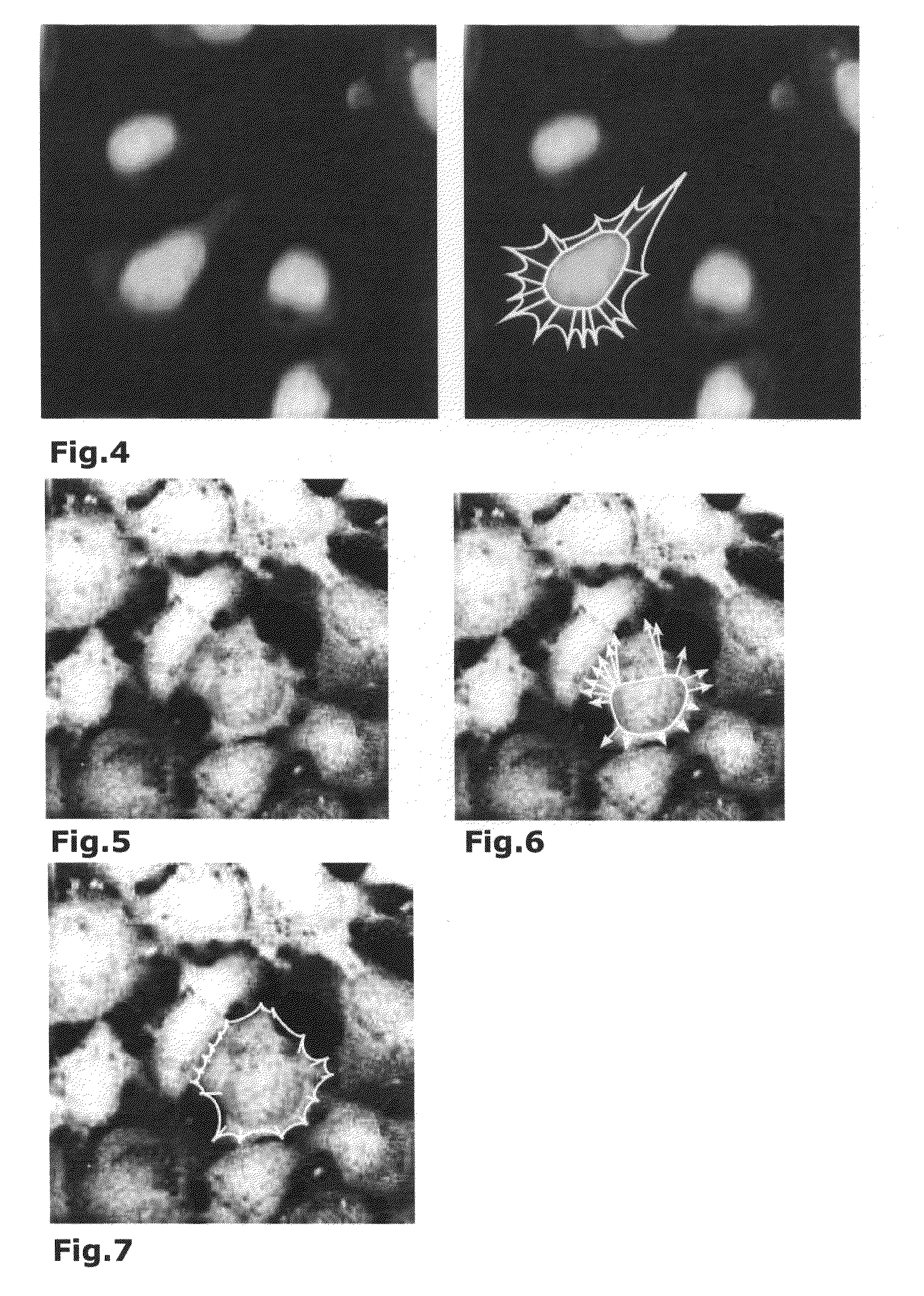
[0139]In a preferred embodiment of the present method an analysis was applied to further cell types. Concretely, those also were cells in cancerous bone s compared to the preceding Figures. In the cell tissue, the larger distances cause a higher degree of freedom of the formation of the cell contour of an individual cell. The above described landmarks were again used in the parametrization of splines by means of direction vectors (axes of growth). o longer be sorted in the order of their positions on the membrane contour; the present example further demonstrates that for cells with a spacious extension of the cytoplasm, a position of the axes of growth that is more independent from the nucleus—that is, a local orientation—is advantageous and can be determined with the method forming the basis of the disclosure.

[0140]thogonal to each other are determined. Thereafter, the set of landmarks was divided into two subsets A and B such that A included the marks situated in the first of the ...


PUM


Abstract

A method for identifying membrane contours in images of biological cells is described and comprises the following steps:
  • detection of a substructure of a biological cell, where the substructure serves to localize the biological cell in the image,
  • detection of a plurality of landmarks taking account of the spatial position of the substructure,
  • determination of line segments between pairs of spatially adjacent landmarks,
and combining the line segments to a membrane contour.
In particular, physical/biological information concerning cell membrane stabilization is used as a basis for determining the line segments.

Description

BACKGROUND[0001]1. Field of the Disclosure[0002]Automatic segmentation of cells in microscope images is one of the most important objects of image analysis in the domain of biology and pharmaceutical research.[0003]2. Discussion of the Background Art[0004]To address biological problems, structures of cells, e.g. receptors or parts of cytoskeletons, are marked with fluorescent dyes. In reaction to external stimuli these structures may react with a re-arrangement. A quantification of such observations requires the segmentation of individual cells within an image. For high-throughput applications, it is of interest to obtain fully automatic methods.[0005]The problem of segmentation is complex since cells have a very heterogeneous morphology that is difficult to model. Further, difficulties are caused by occlusion as well as by cases of neighboring cells grown together, whose interface is hard to associate clearly.[0006]The scientific field of cell recognition in digital images has bred...

Claims


Application Information

Patent Timeline
IPC IPC(8): G06K9/00
CPCG01N15/1475
Inventor JETZEK, FRITZ
Owner EVOTEC TECH GMBH
Why Eureka
Try Eureka