Medical imaging apparatus

a technology of medical imaging and equipment, applied in the field of medical imaging equipment, can solve the problems of inability to perform the fundus imaging that is originally to be performed at a proper timing, the operation may not recognize in which imaging mode the current imaging is performed, and the proper timing of the fluorescence imaging may be missed, so as to improve the efficiency of examination and increase the opportunity for concurrent fundus imaging

Inactive Publication Date: 2009-12-31
CANON KK
View PDF8 Cites 0 Cited by
  • Summary
  • Abstract
  • Description
  • Claims
  • Application Information

AI Technical Summary

Benefits of technology

[0016]The present invention is directed to a medical imaging apparatus capable of impr

Problems solved by technology

However, when fluorescence imaging and color imaging of a subject are concurrently performed, an operator may not recognize in which imaging mode the current imaging is performed.
However, when the fundus imaging is excessively inserted in the fundus imaging of the subject, the fundus imaging that is originally to be performed cannot be performed at a proper timing.
Even when the changing of the subjects is smoothly performed, the proper timing of the fluorescence imaging m

Method used


Image

  • Medical imaging apparatus
  • Medical imaging apparatus
  • Medical imaging apparatus

Examples


Embodiment Construction

[0040]Various exemplary embodiments, features, and aspects of the invention will be described in detail below with reference to the drawings.

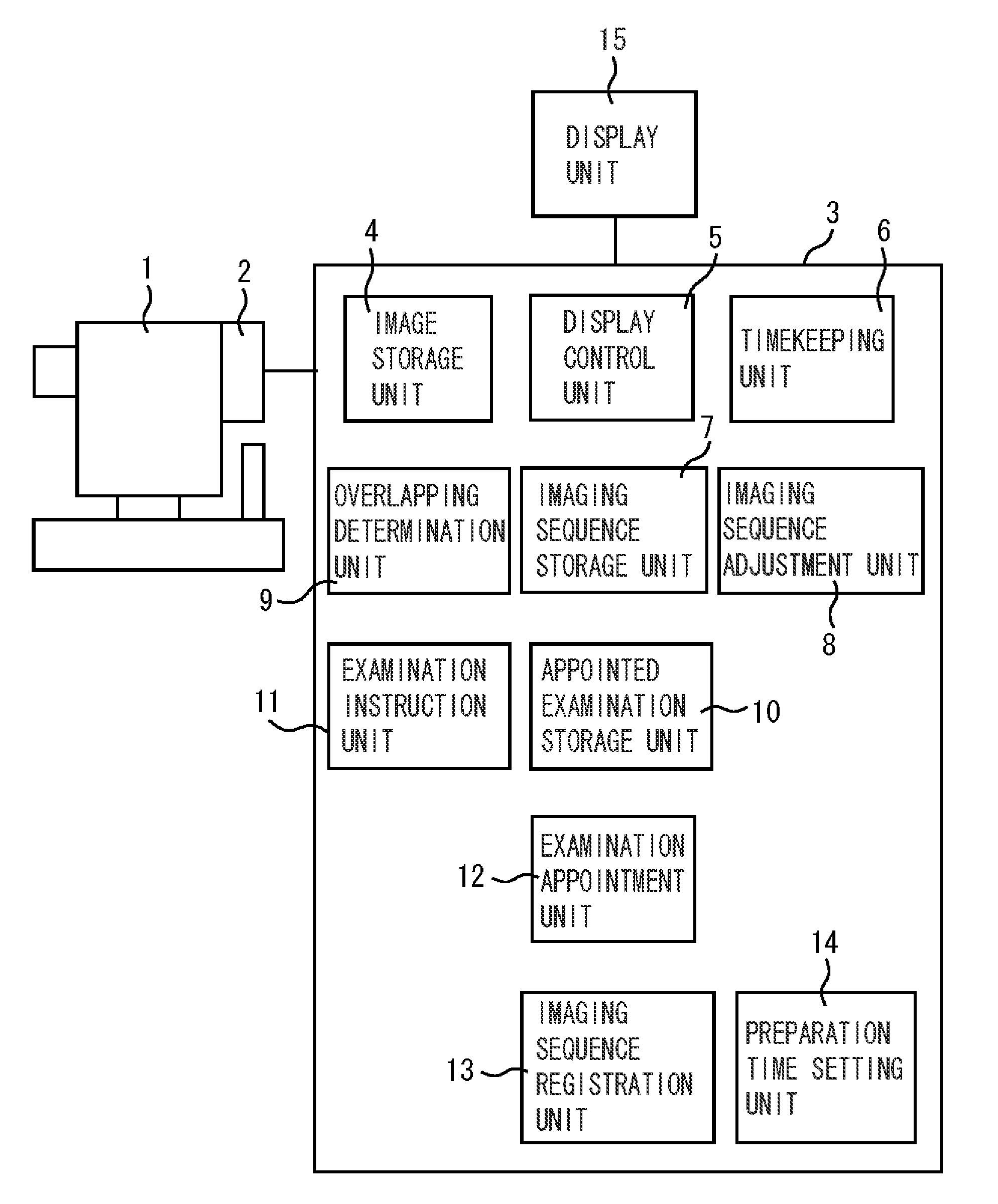
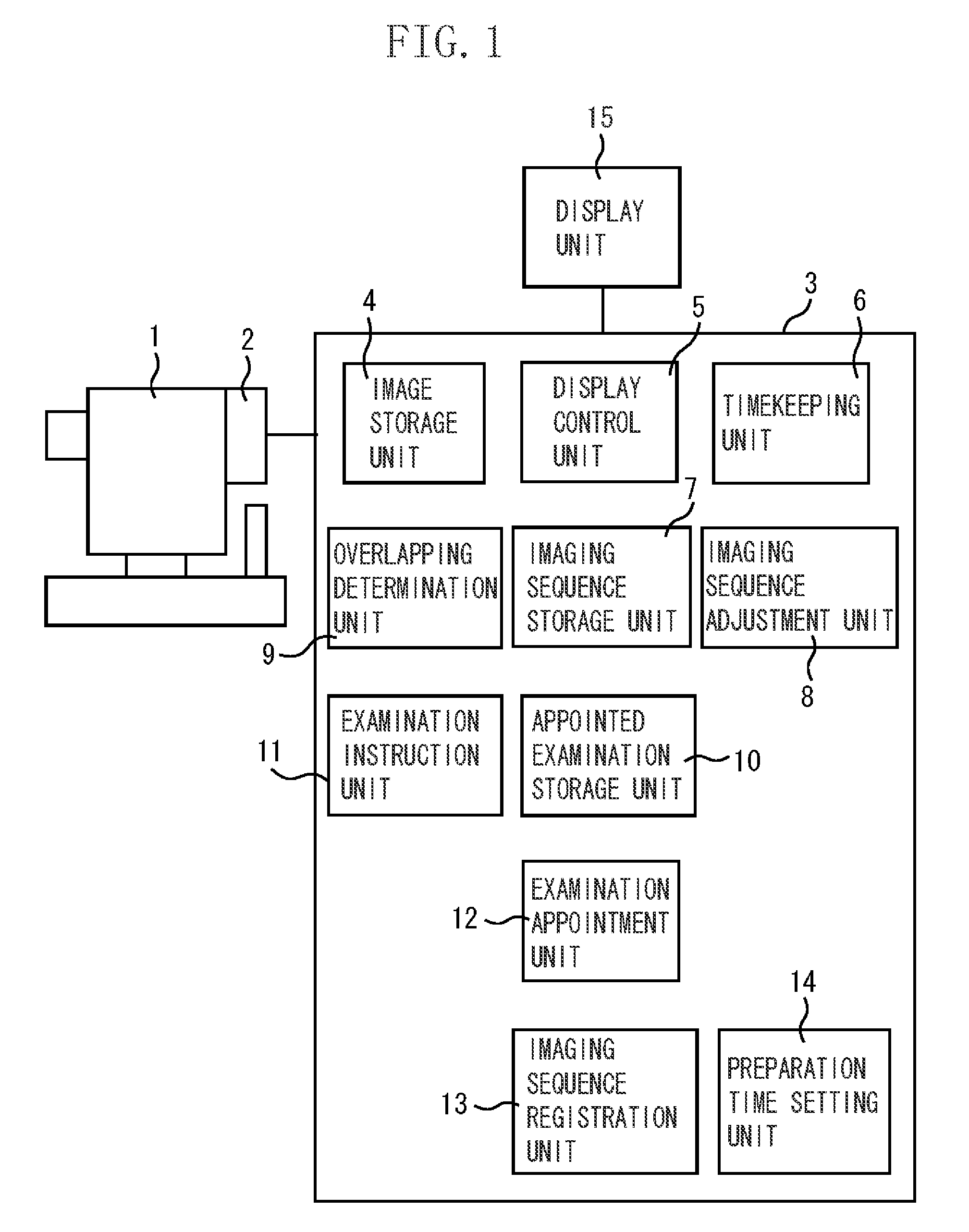
[0041]FIG. 1 illustrates a configuration of a fundus imaging apparatus according to a first exemplary embodiment, which serves as a medical imaging apparatus.

[0042]A digital camera 2 including an image sensor that captures an image of a fundus of a subject's eye is attached to a fundus camera 1. An image captured by the digital camera 2 is output to an information processing apparatus 3.

[0043]The information processing apparatus 3 includes an image storage unit 4, a display control unit 5, a timer unit 6, an imaging sequence storage unit 7, an imaging sequence adjustment unit 8, an overlapping determination unit 9, an appointed examination storage unit 10, an examination instruction unit 11, an examination appointment unit 12, an imaging sequence registration unit 13, and a preparation time setting unit 14. A display unit 15 is connected to the...


PUM


Abstract

A medical imaging apparatus capable of concurrently performing a plurality of imaging operations including an imaging sequence setting unit configured to set a sequence for each of the plurality of imaging operations, an overlapping determination unit configured to determine temporal overlapping between an imaging sequence of an imaging operation that is currently performed and an imaging sequence for an imaging operation that is to be started, and an imaging sequence adjustment unit configured to adjust, based on a result of determination made by the overlapping determination unit, the imaging sequence for the imaging operation that is to be started.

Description

BACKGROUND OF THE INVENTION[0001]1. Field of the Invention[0002]The present invention relates to a medical imaging apparatus, such as a fundus camera to be used for a group examination, at an ophthalmologist's office, and the like.[0003]2. Description of the Related Art[0004]In these days, fundus imaging using a fundus camera has been widely used for screening in group examinations, and diagnosis for ophthalmological disease. In recent years, a method for recording a fundus image as digital data has been widely used. Imaged data is recorded in portable type recording media and hard disk drives built in personal computers (PCs).[0005]In order to improve efficiency of fundus imaging, fundus cameras capable of concurrently performing a plurality of fundus imaging operations have become widely used. Particularly, when fluorescence imaging using a fluorescence agent is performed in an ophthalmologist's office, in many cases, when several minutes or several tens of minutes have passed aft...

Claims


Application Information

Patent Timeline
IPC IPC(8): A61B3/14A61B3/12
CPCA61B3/12
Inventor UCHIDA, HIROKI
Owner CANON KK
Why Eureka
Try Eureka