Automatic Cell Migration and Proliferation Analysis

a cell migration and proliferation analysis technology, applied in the field of biological and cellular image processing, can solve the problems of piecewise segmentation failing to capture the object, the method of kachouie and fieguth (2005) is not robust in the presence of noise, and the method is not fully automati

Active Publication Date: 2010-04-01
KARAM LINA JAMIL +1
View PDF8 Cites 39 Cited by
  • Summary
  • Abstract
  • Description
  • Claims
  • Application Information

AI Technical Summary

Problems solved by technology

Cell segmentation is one of the many challenging tasks in cellular image processing.
But the method of Kachouie and Fieguth (2005) is not robust in the presence of noise.
Moreover, this method is not fully automatic as it requires the knowledge of the cell cluster location relative to the initial boundary of the evolving level-set function.
In some cases, these mean values are very close to each other, which causes the piecewise segmentation to fail in capturing the object (ROI).
However, this method is only applicable to high resolution images, and therefore has limited usefulness in cellular imaging.
However, this method is also only applicable to high resolution images, and the samples must be prepared prior to the analysis of the image.

Method used


Image

  • Automatic Cell Migration and Proliferation Analysis
  • Automatic Cell Migration and Proliferation Analysis
  • Automatic Cell Migration and Proliferation Analysis

Examples


example 1

Cell Evolution Analysis (CEA) Scheme

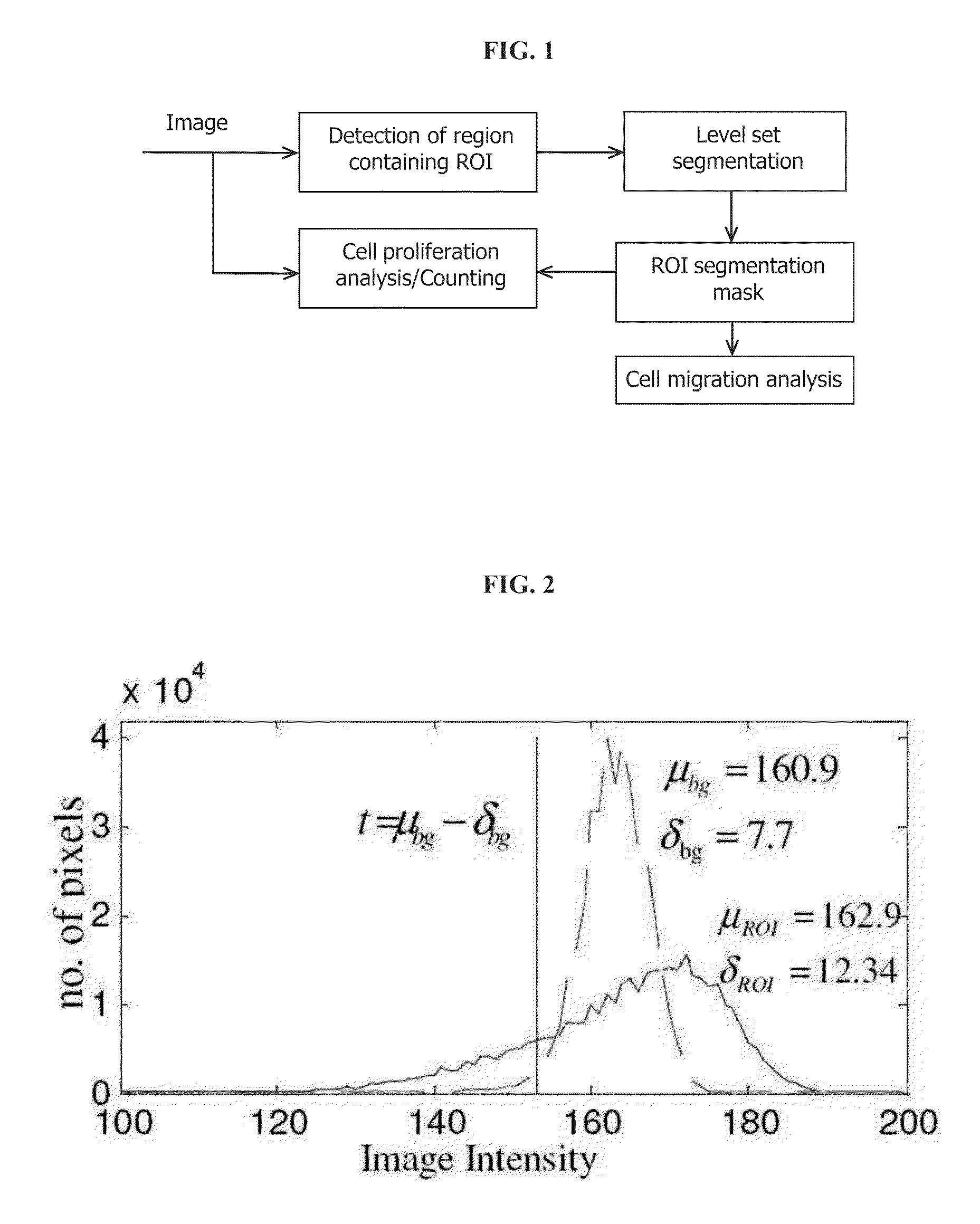
[0042]The cell evolution analysis (CEA) scheme of one embodiment consists of the following two main parts (FIG. 1):

[0043]1—Cell migration analysis—achieved by determining and segmenting the region where the cells are concentrated, and then tracking the evolution of the contour of that region over time. This cell cluster region is denoted as the region-of-interest (ROI).

[0044]2—Cell proliferation analysis—achieved by segmenting and counting the cells inside the ROI at different time points.

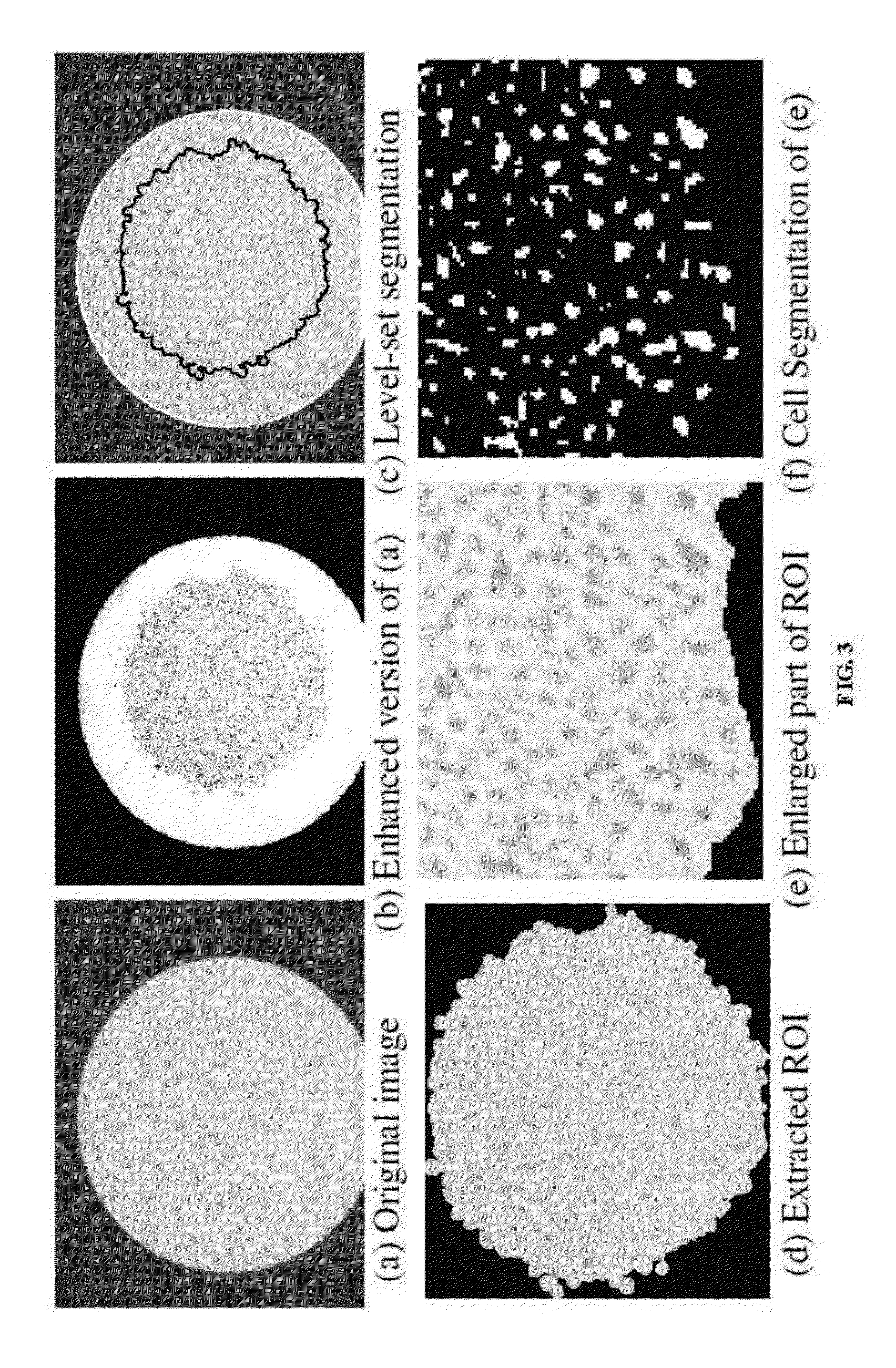
[0045]As shown in FIGS. 3A and 3B, the considered illustrative bladder cancer cell images consist, each, of three different regions: 1) the ROI, 2) the region between the ROI and the outer circle (bright area), and 3) the region outside the outer circle (dark area). An automatic segmentation method needs to be devised to distinguish between these three regions. For the level-set based segmentation methods, two level-set functions are needed for the considered ima...

example 2

Cell Migration Analysis Using a Structure Tensor Feature Space

A. Cell Migration Analysis Scheme

[0061]The overall migration analysis is done by segmenting and tracking the ROI (Cell cluster) area at different time instances. Given two extracted ROI areas, At1 and At2, (t2>t1), the overall cell migration is given by (At2−At1) / (t2−t1).

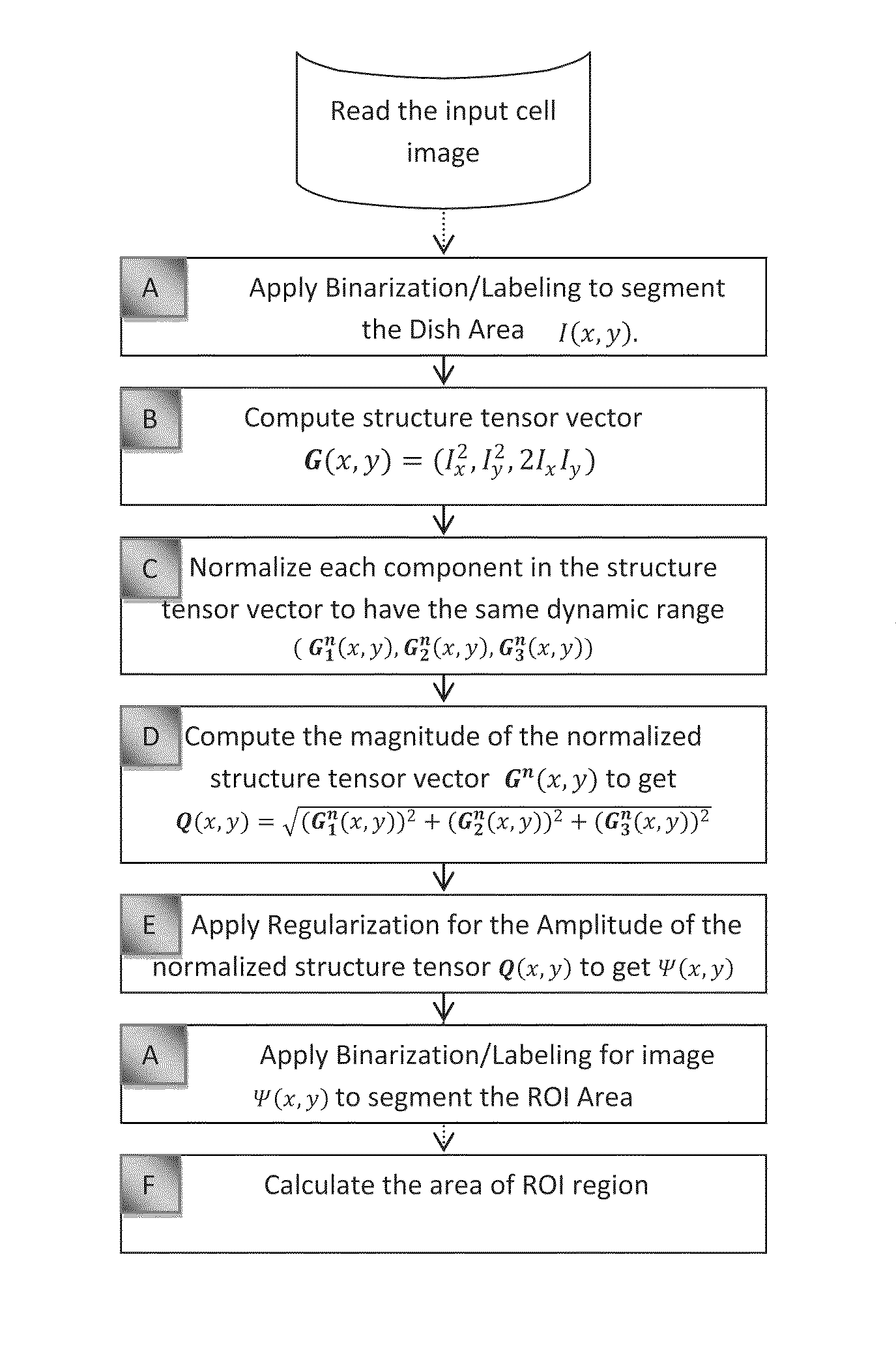
[0062]The procedure starts by extracting the outer-circle using piecewise level-set segmentation (Vese and Chan, 2002) with one level-set function or using a histogram-based thresholding procedure. Then, the ROI is extracted by using an unsupervised texture segmentation procedure. This procedure consists of computing structure tensor features. These features can either be, for example, the components of a structure tensor vector that is computed at each pixel in the image, or can the amplitude of the structure tensor vector at each image pixel. The obtained structure tensor based image is then reguralized. This regularization can be performed for example ...


PUM


Abstract

The disclosed methods and apparatus provide for the automatic segmentation and analysis of the overall migration rate and proliferation rate of cells that may be used with any resolution image and without the need to prepare the sample or the image before the image is analyzed. In particular embodiments, the method or apparatus of analyzing a cell image comprise performing a structure tensor analysis and / or regularization and / or a histogram-based analysis and / or a level-set analysis to classify pixels in the image into a region of interest (ROI), corresponding to cell clusters, and a non-significant region. Methods and apparatus for cell migration analysis comprise means for computing the areas of the segmented ROIs for a set of images. Methods and apparatus for cell proliferation analysis comprise means for counting the number of cells within the segmented ROIs for a set of images.

Description

CROSS-REFERENCE TO RELATED APPLICATION[0001]This application claims the benefit of provisional patent application Ser. No. 61 / 042,533 filed on 2008 Apr. 4 by the present inventors.BACKGROUND OF THE INVENTION[0002]I. Field of the Invention[0003]The present invention relates generally to the fields of biology and cellular image processing. In certain aspects, the invention is related to methods for cell segmentation and cellular image processing.[0004]II. Description of Related Art[0005]Studying the migration and proliferation behavior of cells contributes to the understanding of biological processes and disease pathologies such as cancer, angiogenesis, vascular stenosis and arthritis. To this end, cellular imaging coupled with image processing and segmentation techniques, has been used to provide an automatic and improved analysis of the cellular behavior.[0006]Cell segmentation is one of the many challenging tasks in cellular image processing. Several methods have been proposed and ...

Claims


Application Information

Patent Timeline
Patent Type & Authority Applications(United States)
IPC IPC(8): G06K9/48G06T7/00G06T7/60
CPCG06K9/00134G06K9/0014G06T7/0012G06T2207/30024G06T7/401G06T2207/20104G06T2207/20161G06T7/0081G06T7/11G06T7/41G06V20/693G06V20/695
Inventor KARAM, LINA JAMILSAID, ASAAD
Owner KARAM LINA JAMIL
Why Eureka
Try Eureka