Puncture Treatment Supporting Apparatus

a technology for supporting equipment and puncture, which is applied in the field of puncture treatment supporting equipment, can solve the problems of difficult to perform puncture operation,

Inactive Publication Date: 2010-05-27
HITACHI MEDICAL CORP
View PDF8 Cites 57 Cited by
  • Summary
  • Abstract
  • Description
  • Claims
  • Application Information

AI Technical Summary

Problems solved by technology

While the operator develops a puncture plan in advance by imaging the position of a tumor in the body of an object, it is difficult to

Method used


Image

  • Puncture Treatment Supporting Apparatus
  • Puncture Treatment Supporting Apparatus
  • Puncture Treatment Supporting Apparatus

Examples


Embodiment Construction

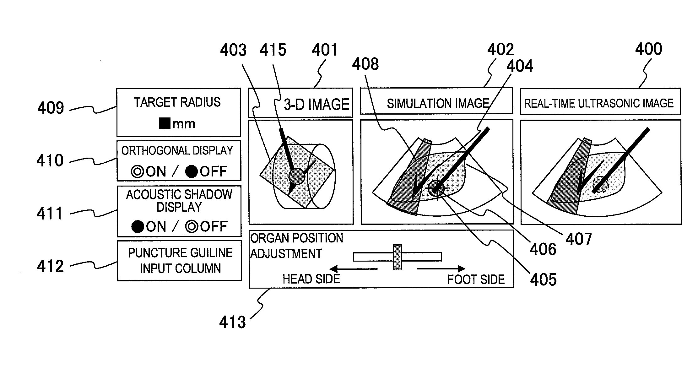
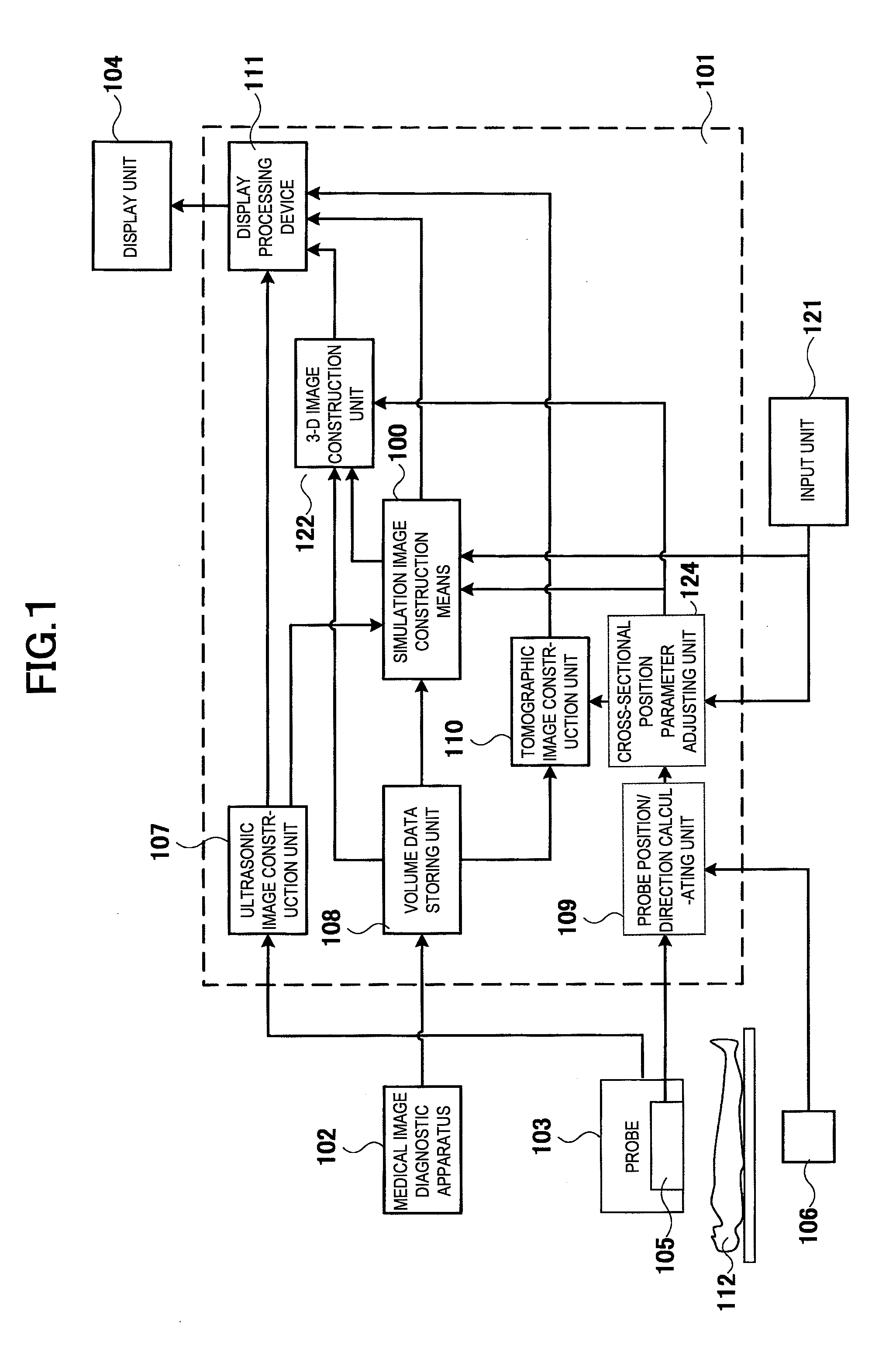
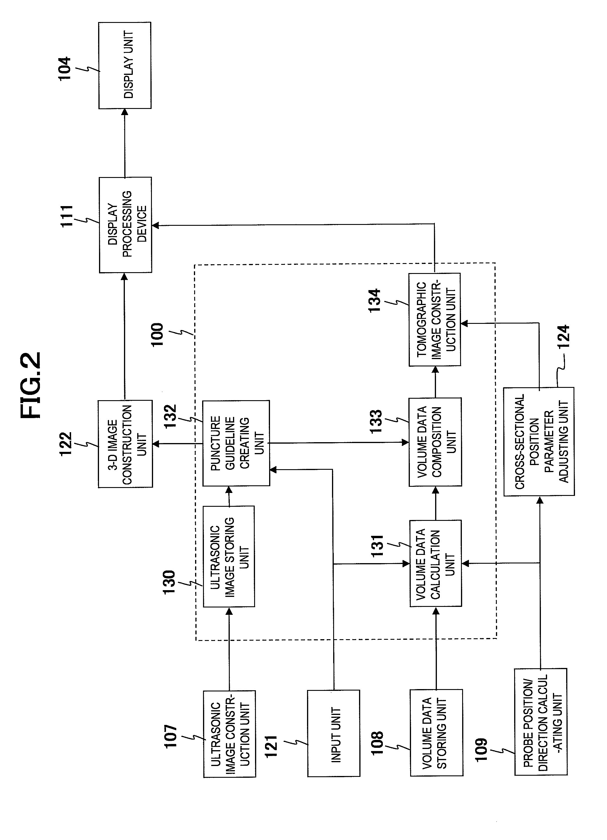
[0032]Hereinafter, the system configuration of the puncture treatment supporting apparatus related to the present invention will be described using FIG. 1.

[0033]The puncture treatment supporting apparatus comprises:

[0034]a medical image diagnostic apparatus 102 such as an X-ray CT apparatus or an MRI apparatus;

[0035]a probe 103 for transmitting / receiving ultrasonic waves to / from an object 112;

[0036]a probe position sensor 105 formed together with the probe 103;

[0037]a source 106 placed in the vicinity of the object 112, and

[0038]for detecting the movement of the probe position sensor 105 by a magnetic field, etc.;

[0039]an image processing device 101 for imaging the image data obtained from the medical image diagnostic apparatus 102 or the probe 103; and

[0040]a display unit 104 for displaying the image processed in the image processing device 101.

[0041]Further, a central control device (not shown in the diagram) is provided in the image processing device 101, and controls the respect...


PUM


Abstract

[Problems] To provide a puncture treatment supporting apparatus enabling puncture treatment by previously performing puncture simulation and enabling its treatment evaluation.
[Means for Solving Problems] A puncture treatment supporting apparatus comprises an ultrasonic probe, ultrasonic image construction means for constructing an ultrasonic image, volume data storing means for storing volume data on a medical image diagnostic apparatus, probe position/direction detecting means for detecting the position and direction of the ultrasonic probe, tomographic image construction means for constructing tomographic image having the same cross section as the ultrasonic image by using the information on the position and direction of the ultrasonic probe, display means for displaying these images, and puncture means for inserting a puncture needle.
The apparatus, further comprises simulation image constructing means for constructing a simulation image by adding a puncture guideline indicating the puncture position and direction of the puncture needle to the tomographic image, and the display means displays the simulation image along with the ultrasonic image.

Description

TECHNICAL FIELD[0001]The present invention relates to a puncture treatment supporting apparatus for drawing up a puncture operation plan by simulation and displaying the puncture operation proceedings.BACKGROUND ART[0002]Ultrasonic diagnostic apparatuses obtain a tomographic image of soft tissues in a living body using ultrasonic pulse reflection method. Ultrasonic diagnostic apparatuses are compact in size and inexpensive compared to other medial image diagnostic apparatuses, highly safe since there is no exposure to radiation such as X-rays, and has features such as being capable of imaging blood flow, thus are widely used, for example, in digestive organs departments, urology departments, and obstetrics and gynecology departments.[0003]The ultrasonic diagnostic apparatus is used for inserting a puncture needle into an object while observing an ultrasonic image, obtaining a part of tumor cells for a sample using the puncture needle or cauterizing the tumor using an RF coil provide...

Claims


Application Information

Patent Timeline
IPC IPC(8): A61B8/14
CPCA61B8/0833A61B19/201A61B19/50G06T2219/028A61B2017/3413G06T19/00A61B19/52A61B90/36A61B90/11A61B34/10
Inventor ARAI, OSAMUIWASAKI, TAKAO
Owner HITACHI MEDICAL CORP
Why Eureka
Try Eureka