Porcine biliary tract imaging

a biliary tract and imaging technology, applied in the field of near infrared spectroscopy, can solve the problems of bile duct injury (bdi), no technology has been introduced that enables surgeons, and images are not clear, so as to facilitate tissue identification and facilitate the characterization of one or more tissues

Inactive Publication Date: 2010-06-24
BOARD OF RGT THE UNIV OF TEXAS SYST
View PDF4 Cites 19 Cited by
  • Summary
  • Abstract
  • Description
  • Claims
  • Application Information

AI Technical Summary

Benefits of technology

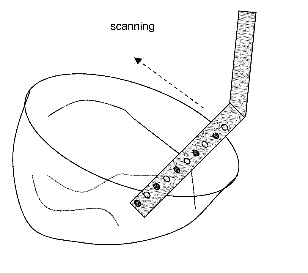
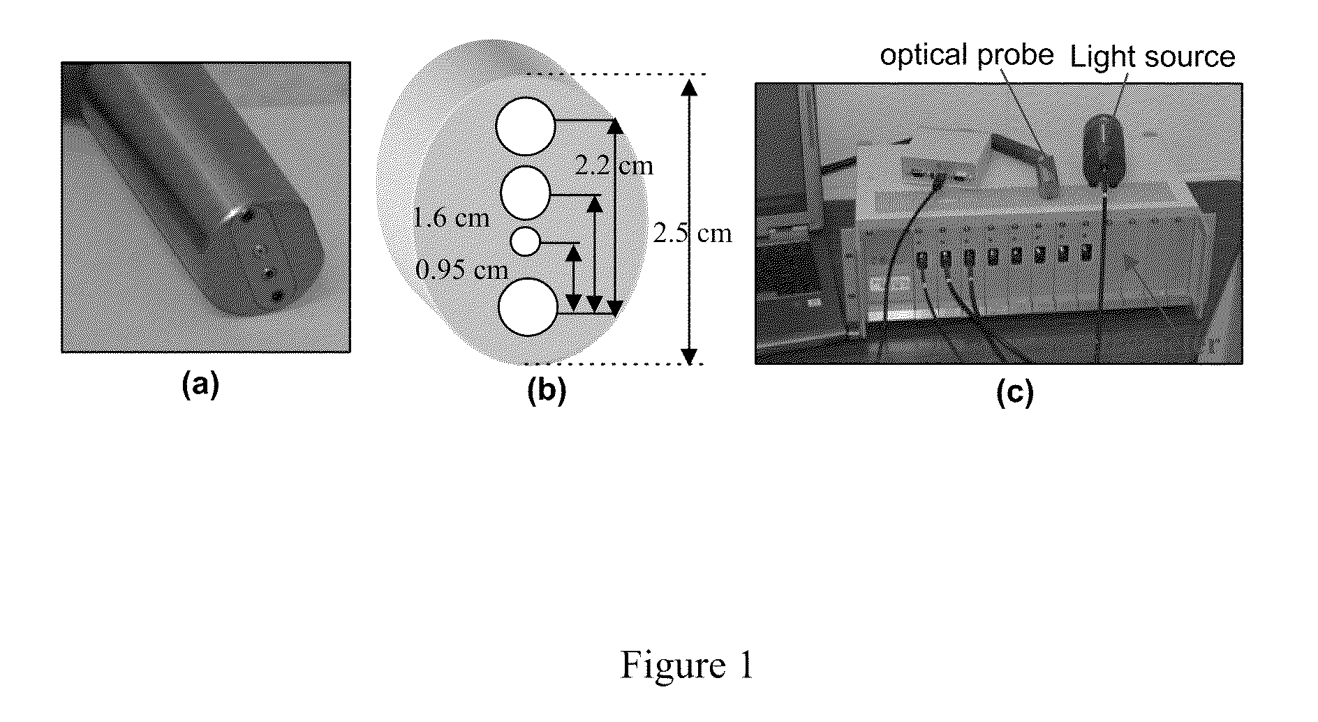
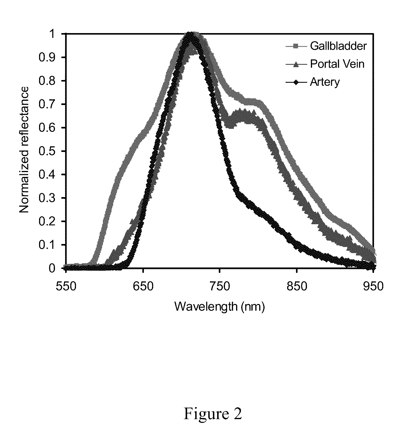
[0011]In one aspect, the present invention is an apparatus to visualizes one or more tissues in vivo. The apparatus has an electromagnetic radiation source capable of producing a continuous wave broadband light, at least one optical probe connected to the electromagnetic radiation source, a multi-channel CCD array spectrometer detector connected to the optical probe capable of collecting near infrared wavelength emissions, at least one computer connected to the multi-channel CCD array spectrometer detector. The computer includes one or more image evaluation algorithms, and at least one display connected to the computer to generate images from the one or more tissues and display results from the image evaluation algorithms. The optical probe is typically a fiber optical bundle having one or more non-bifurcated channels for light delivery fibers and one or more bifurcated channels to collect the near infrared wavelength emissions. The near infrared wavelengths emissions used has wavelength between about 550 nm and about 900 nm. In one aspect, the present invention examines tissues such as biliary tree of an animal or human. In another aspect, the present inventions uses one or more image evaluation algorithms such as a Radial Basis Function algorithm to facilitate the characterization of the one or more tissue based on observed reflectance spectra and to distinguish between tissue structures, a Minimal Distance Method algorithm to classify the one or more tissues, a two-layer diffusion model algorithm to localize heterogeneities, and / or a linearized image reconstruction algorithm to obtain ultrasound-like, two-dimensional images.
[0012]Yet in another aspect, the present invention demonstrates methods of imaging one or more tissues (e.g. a biliary tree) in an animal or a human subject during a surgical operation. The method typically includes directing a continuous wave broadband light towards the one or more tissues using at least one optical probe, collecting near infrared wavelength emissions from the one or more tissues using the optical probe, analyzing the near infrared wavelength emissions using one or more image evaluation algorithms, and / or reconstructing a two-dimensional or three-dimensional optical map of the one or more tissues based on results derived from the one or more image evaluation algorithms. The tissue examined using the present invention can use a Radial Basis Function algorithm to facilitate tissue identification based on observed reflectance spectra and to distinguish between tissue structures or use a Minimal Distance Method algorithm to classify the tissues. In one aspect, the Minimal Distance Method uses the equation:
[0013]In some aspects, the one or more image evaluation algorithms also includes a two-layer diffusion model to localize heterogeneities, or a linearized image reconstruction algorithm to obtain ultrasound-like, two-dimensional images.

Problems solved by technology

The most important complication of the operation is bile duct injury (BDI).
As of yet no technology has been introduced that enables surgeons to visualize the bile ducts while operating.
Routine intraoperative ultrasonography has been proposed for delineating biliary anatomy; however, the images are not very clear and identifying the relevant anatomy is challenging.
Even in relatively small trials of routine intraoperative ultrasonography, major bile duct injuries do occur, demonstrating that the technique currently in use falls short as an injury-prevention strategy.

Method used


Image

  • Porcine biliary tract imaging
  • Porcine biliary tract imaging
  • Porcine biliary tract imaging

Examples


Embodiment Construction

[0065]While the making and using of various embodiments of the present invention are discussed in detail below, it should be appreciated that the present invention provides many applicable inventive concepts that can be embodied in a wide variety of specific contexts. The specific embodiments discussed herein are merely illustrative of specific ways to make and use the invention and do not delimit the scope of the invention.

[0066]To facilitate the understanding of this invention, a number of terms are defined below. Terms defined herein have meanings as commonly understood by a person of ordinary skill in the areas relevant to the present invention. Terms such as “a”, “an” and “the” are not intended to refer to only a singular entity, but include the general class of which a specific example may be used for illustration. The terminology herein is used to describe specific embodiments of the invention, but their usage does not delimit the invention, except as outlined in the claims.

[...


PUM


Abstract

The present invention includes apparatus and method to prevent surgical injury. The invention incorporates near infrared (NIR) spectroscopy, which capitalizes on near infrared light's ability to penetrate deeply into tissues and spectroscopic capability to discern tissue's chemical properties. The present invention further characterized the NIR optical properties of bile containing structures as a clinically useful probe.

Description

BACKGROUND OF THE INVENTION[0001]Without limiting the scope of the invention, its background is described in connection with near infrared (NIR) spectroscopy, more particularly, apparatus and methods to prevent biliary tract injury during a surgery using in-vivo near infrared spectroscopy.[0002]Approximately 400,000 cholecystectomies are performed annually in the United States. The most important complication of the operation is bile duct injury (BDI). Injury prevention relies mostly on an individual surgeon's skill. As of yet no technology has been introduced that enables surgeons to visualize the bile ducts while operating. Routine intraoperative ultrasonography has been proposed for delineating biliary anatomy; however, the images are not very clear and identifying the relevant anatomy is challenging. Even in relatively small trials of routine intraoperative ultrasonography, major bile duct injuries do occur, demonstrating that the technique currently in use falls short as an inj...

Claims


Application Information

Patent Timeline
Patent Type & Authority Applications(United States)
IPC IPC(8): A61B6/00
CPCA61B5/0059G01N21/359G01N21/4795
Inventor LIU, HANLILIVINGSTON, EDWARDWANG, BO PING
Owner BOARD OF RGT THE UNIV OF TEXAS SYST
Why Eureka
Try Eureka