Visualization of voxel data

a voxel data and visualization technology, applied in the field of medical images, can solve the problems of difficult for clinical users to efficiently extract information from data, inefficient analysis, and insufficient visualization of tissue within solid organs, and achieve the effect of improving correlation to anatomy and being easy to compar

Active Publication Date: 2010-08-12
KONINKLIJKE PHILIPS ELECTRONICS NV
View PDF6 Cites 33 Cited by
  • Summary
  • Abstract
  • Description
  • Claims
  • Application Information

AI Technical Summary

Benefits of technology

[0020]An effect of maintaining the direction extending along the inter-surface distance is that in connection with visualization of walled body parts, such as the left ventricle of the heart or other hollow organs or body parts, the transmurality is preserved. Transmurality information is lost in the bull's-eye plot. It is an advantage of the present invention that transmurality information is directly available for the property value, and moreover is available together with the continuous volumetric distribution of the property value. A more efficient quantitative analysis is thereby available to the clinician, facilitating a more efficient diagnosis process for the clinician.
[0021]In an advantageous embodiment, the mapping between the reference shape and the target shape is such that distinct values in the reference shape is mapped to distinct values in the target shape. In general may the mapping be injective or non-injective mapping. Non-injective mapping may be applied in the situation where the inner reference surface is in the form of a point. Typically, however, injective mapping is applied.
[0022]Typically, the mapping is from the outer surface to the inner surface, but in general, the mapping can be from the inner surface to the outer surface as well.
[0023]In an advantageous embodiment, one or more reference structures are identified in the one or more sets of voxel data and mapped onto the target shape, enabling a visualization of the target shape with the one or more reference structures overlayed or superimposed. While the target shape is correlated to the anatomy by its continuous volumetric nature and conservation of the inter-surface distance, it is still an abstraction of the anatomy. By mapping reference structures onto the target shape, an improved correlation to the anatomy may be provided, and moreover, structures which are related to a property value under investigation can directly be corrected to the distribution geometry of the property value. The presence of localized reference structures renders the target shape patient specific, which may be important in connection with the diagnosis.
[0024]In advantageous embodiments, a 3D spatial rendering of the set of voxel data is provided and visualized in combination with a visualization of the target shape. Optionally, a region may be selectable on the 3D spatial rendering or on a rendering of the target shape, and wherein by selecting a region on one of the 3D spatial rendering or on a rendering of the target shape, the corresponding region is marked on the other of the 3D spatial rendering or on rendering of the target shape. It is advantageous to provide a combined interactive visualization of the anatomy and the quantitative analysis data of the target shape. A comprehensive visualization may thereby be provided. By providing a combined visualization of the target shape and a 3D spatial rendering of the set of voxel data, and by interactive linking the two objects, it is made possible for the clinician to navigate through both anatomical aspects and schematic analysis aspects in a comprehensive way.
[0025]In connection with the visualization of the target shape alone or the combined visualization of the target shape and the 3D spatial rendering, various cut planes at various locations and orientations may be made. The user thereby has the flexibility to inspect the anatomy and the one or more property values from any viewpoint and to make a comparison with the existing visualization techniques.

Problems solved by technology

A limitation of this technique is that it does not adequately visualize the tissue within solid organs; it is optimized for visualization of surfaces and boundaries.
However, even though quantitative analysis is related to the anatomy via the bull's-eye plot, the bull's-eye plot is an abstraction that does not directly reflect the anatomy of the left ventricle.
In general, despite of the availability of detailed 3D images of the body part under investigation it is still challenging for the clinical user to efficiently extract information from the data.
This leads to inefficient analysis and decreases the reproducibility of the diagnostic work flow.

Method used


Image

  • Visualization of voxel data
  • Visualization of voxel data
  • Visualization of voxel data

Examples


embodiment

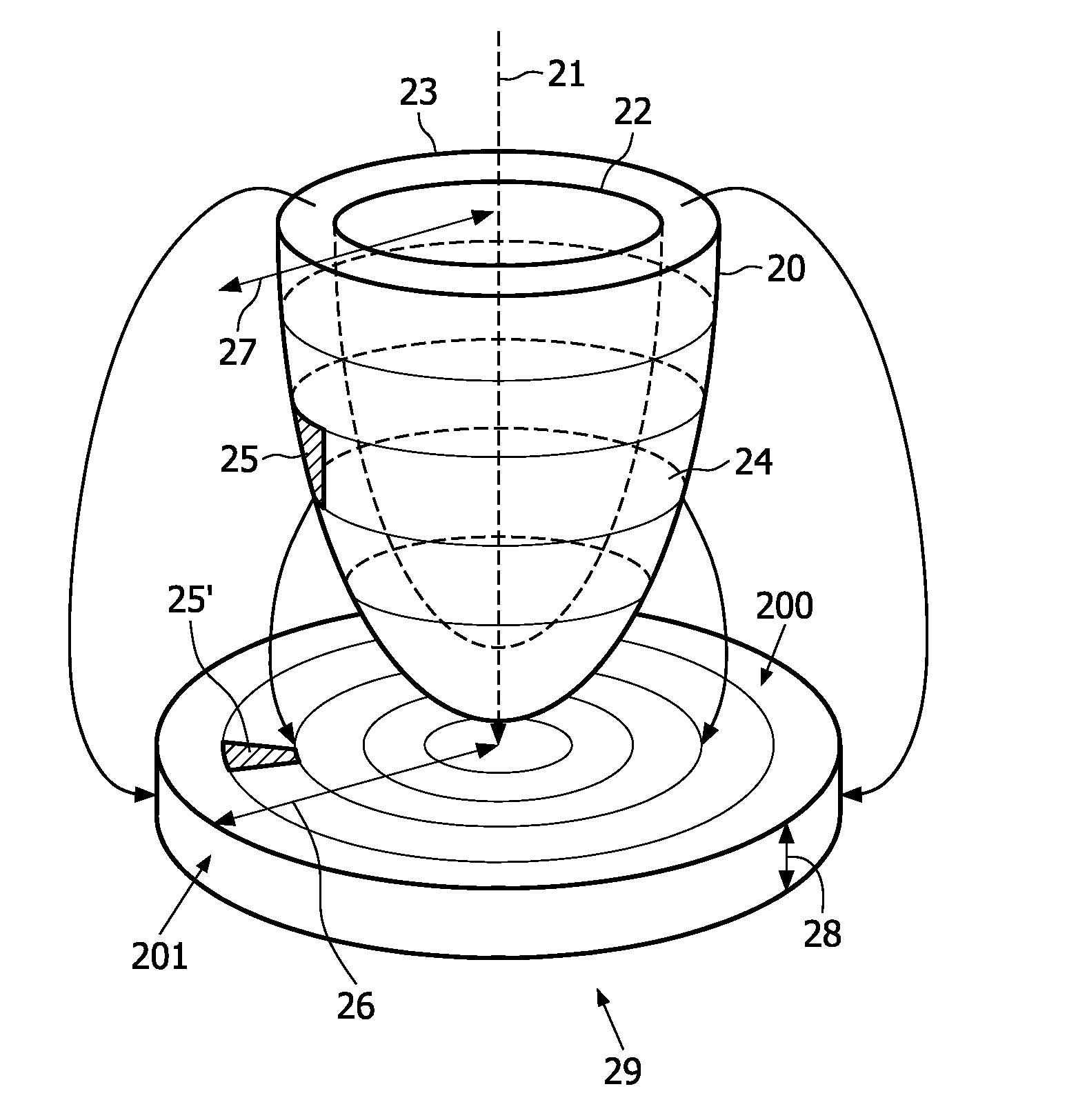
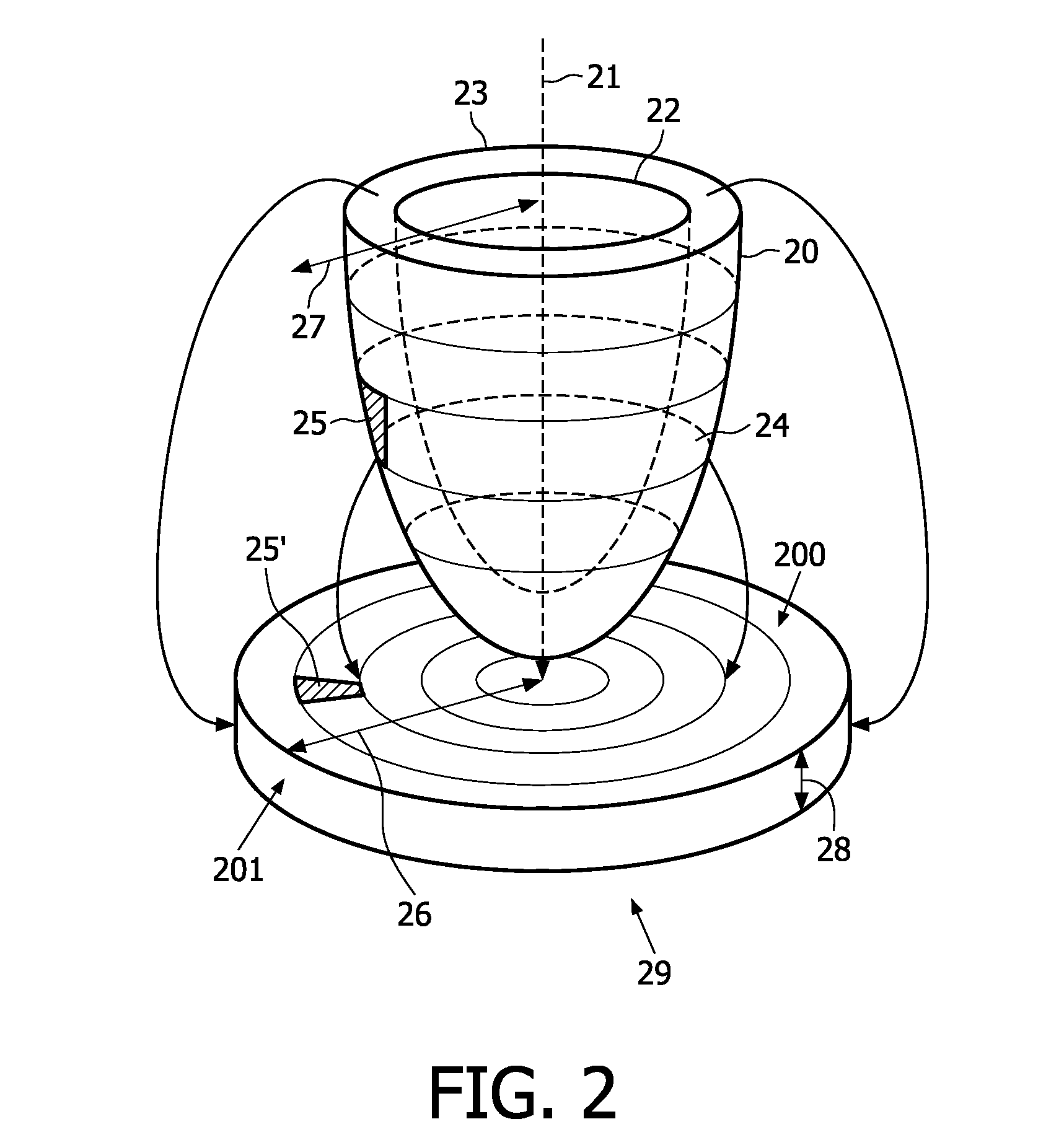
Implementation of the Volumetric Bull's Eye Plot

[0075]The VBEP may be seen as an unfolding of the left ventricle along the long axis followed by a reformatting into a cylinder. The unfolding is based on a parametrization of both the cylinder and the left ventricular myocardium. The parameterization of the cylinder is illustrated in FIG. 10. The long 100 and the short 101 axes of the reference shape may be extracted from a geometric heart model, which may be used to represent the voxel data of the whole heart scan. However, these axes may also be extracted by other means. For example, the axes can be extracted from the orientation information of the late enhancement scan. The endocardium and the epicardium are segmented as a pair of contours on each slice of the late enhancement scan. Each contour is represented by a uniformly sampled piecewise cubic Bèzier spline. In this context, uniformly sampled is to be understood as that the length of any segment of a spline s between s(t) and ...


PUM


Abstract

The invention relates to visualization of medical images, and in embodiments to the visualization of the left ventricle of the human heart or other organs. A method of visualizing one or more sets of voxel data is disclosed. The method comprising: providing one or more sets of voxel data, providing and segmenting the voxel data in accordance with a segment model. The segmented voxel data is reformatted to fit a reference shape (20) being defined by at least an inner (22) reference surface and an outer (23) reference surface. The reformatted voxel data is mapped to a target shape being defined by at least a first (29) target surface and a second (200) target surface. The target shape is moreover visualized. The mapping of the reformatted voxel data to a target shape is a mapping of one or more property values from the inner reference surface to the first target surface, and from the outer reference surface to the second target surface, and where a direction (26, 27) extending along the inter-surface distance of the reference shape is maintained in the target shape.

Description

FIELD OF THE INVENTION[0001]The invention relates to visualization of medical images, and in particular to the area of visualization of anatomy and associated quantitative analysis data derived from the medical images.BACKGROUND OF THE INVENTION[0002]Medical imaging systems such as magnetic resonance imaging (MRI) are capable of producing exact cross-sectional image data that express the physical properties related to the human or animal body. Reconstruction of three-dimensional (3D) images using collections of parallel 2D images representing cross-sectional data has been applied in the medical field for some time.[0003]Various rendering techniques are applied for 3D display of medical images. One such technique is surface rendering. A limitation of this technique is that it does not adequately visualize the tissue within solid organs; it is optimized for visualization of surfaces and boundaries.[0004]Another rendering technique is volume-rendering. Instead of overlaying surfaces us...

Claims


Application Information

Patent Timeline
Patent Type & Authority Applications(United States)
IPC IPC(8): G06T17/00G06K9/00G06T15/08
CPCG06T15/08
Inventor BREEUWER, MARCELOLIVAN BESCOS, JAVIERTERMEER, MAURICE ALAIN
Owner KONINKLIJKE PHILIPS ELECTRONICS NV
Why Eureka
Try Eureka