
Corneal endothelial tissue inserter

a technology of endothelial tissue and inserter, which is applied in the field of medical devices, can solve the problems of eye tissue lacking irrigation system, and achieve the effect of avoiding inadvertent adhesion to eye tissue and low manufacturing cos

Active Publication Date: 2010-10-28
FISCHER SURGICAL
View PDF4 Cites 11 Cited by
  • Summary
  • Abstract
  • Description
  • Claims
  • Application Information

AI Technical Summary

Benefits of technology

[0014]One object of the corneal endothelial tissue inserter is to provide an inserter that can readily collect, transport, and deliver endothelial tissue without folding.
[0016]Another object of the corneal endothelial tissue inserter is to provide such an inserter that readily provides complete control of the device by the surgeon before, during, and after usage.
[0018]Another object of the corneal endothelial tissue inserter is to provide such an inserter that has a tip arrangement that avoids inadvertent adherence to eye tissue during usage.
[0019]Another object of the corneal endothelial tissue inserter is to provide such an inserter that has a low cost of manufacturing so the surgeons and hospitals can readily purchase the inserter through existing medical supply outlets.

Problems solved by technology

This apparatus though lacks an irrigation system and folding of the cradle without an overlap.

Method used


Image

  • Corneal endothelial tissue inserter
  • Corneal endothelial tissue inserter
  • Corneal endothelial tissue inserter

Examples


Embodiment Construction

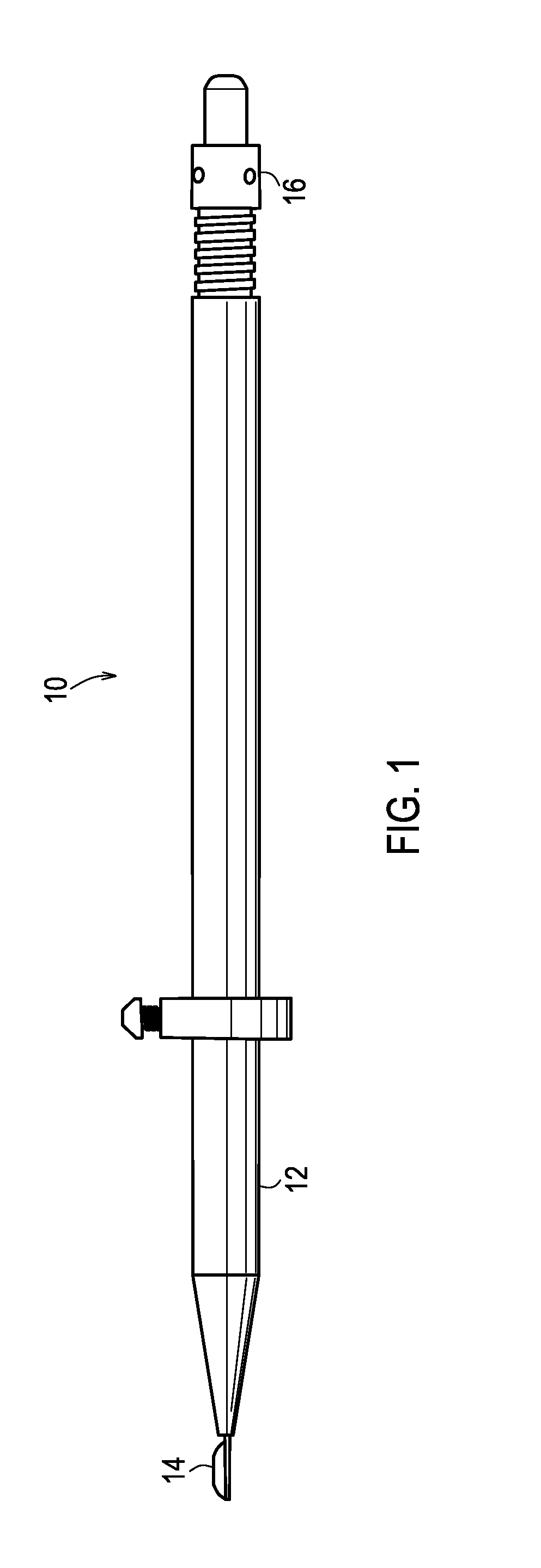
[0042]The present invention overcomes the prior art limitations by providing an instrument that retrieves and delivers endothelial tissue without damage to the tissue and recipient eye. Referring now to FIG. 1, it shows an alternate embodiment in a side view of an instrument 10. This instrument has a hollow rigid tube, like a barrel or handle 12 with a disposable tissue holder or transfer chamber 14 at a distal end of the instrument. The holder 14, or platform, advances forward or retracts rearward into the barrel 12 by pushing or pulling a tab 16 at a proximal end of the barrel 12. Referring to FIG. 2, it shows an alternate embodiment in a top view of the instrument 10. The instrument also includes a flow regulator ring 20 that also moves forward or rearward to advance or retract the tissue holder or platform 14. Next, FIG. 3 shows a side sectional view of the alternate embodiment of instrument 10 with the holder 14 retracted completely into barrel 12.

[0043]Turning to FIG. 4, the a...


PUM


Abstract

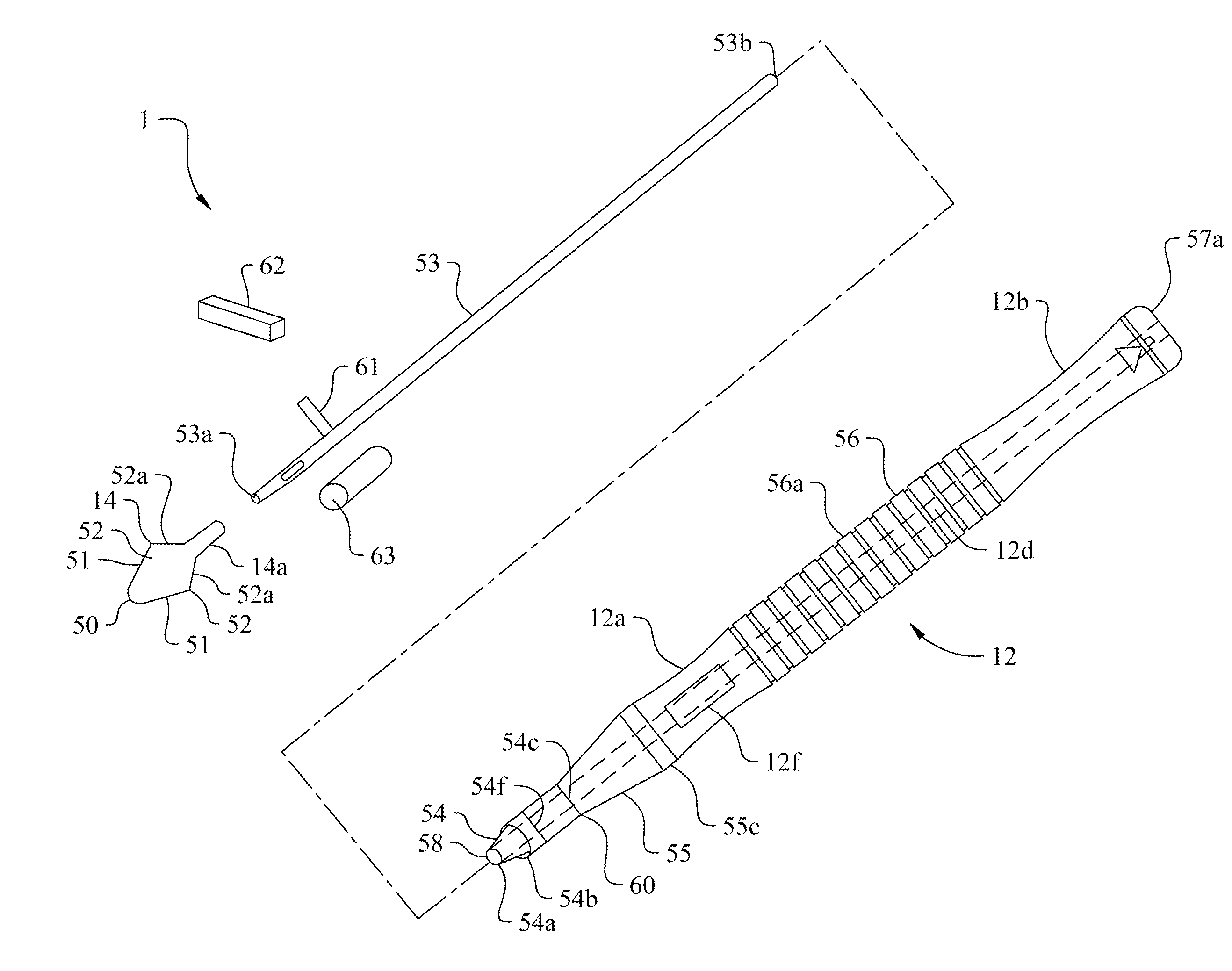
A device transfers donor endothelial cells to the cornea of a recipient without injuring the cells or the cornea during the transfer process. The device transfers living endothelial cells from a donor to a recipient with minimal or no trauma. The device includes a handle, an irrigation tube lengthwise within the handle, a button upon the handle concentric with the irrigation tube, a tapered nose opposite the button, a forward tube extending outwardly from the nose, and a platform joined to the irrigation tube. The forward tube has an elliptical cross section and two beveled features. The platform has two wings that curl gently around donor tissue upon retracting the platform into the forward tube. The handle has a groove for a stem of a knob that surgeon presses and pulls to move the platform.

Description

CROSS-REFERENCE TO RELATED APPLICATION[0001]This non-provisional application is a continuation in part of the pending non-provisional application for patent having the U.S. Ser. No. 12 / 027,822 filed on Feb. 7, 2008 which claims priority to the expired provisional application having the U.S. Ser. No. 60 / 889,236 filed on Feb. 9, 2007 and both are owned by the same assignee, and both disclosures are incorporated by reference.BACKGROUND OF THE INVENTION[0002]The corneal endothelial tissue inserter relates to medical devices that work with eye tissue and more specifically to a device that retracts an extremely thin layer of donor endothelial eye tissue and then places this tissue within the eye of a recipient.[0003]People have eyes to see. An eye has various parts that receive and focus light upon a retina that converts the light into electrical signals transmitted to a is person's brain for interpretation. In the vicinity of a person, light encounters a cornea as the first part of the e...

Claims


Application Information

Patent Timeline
IPC IPC(8): A61F9/00
CPCA61B17/0231A61F9/0017A61F2/142A61B2017/00969A61F2/148
Inventor NEUSIDL, WILLIAM B.FISCHER, CRAIG L.UTT, GREG GORDON
Owner FISCHER SURGICAL
Learn More