Biocompatible scaffold for ligament or tendon repair

a biocompatible, ligament or tendon technology, applied in the field of biocompatible tissue implant devices, can solve the problems of surgical intervention, articular cartilage tissue injury will not heal spontaneously, and the cartilage and underlying bone of the joint will gradually be destroyed

Inactive Publication Date: 2011-01-13
DEPUY MITEK INC
View PDF99 Cites 56 Cited by
  • Summary
  • Abstract
  • Description
  • Claims
  • Application Information

AI Technical Summary

Problems solved by technology

Damaged articular cartilage can severely restrict joint function, cause debilitating pain and may result in long term chronic diseases such as osteoarthritis, which gradually destroys the cartilage and underlying bone of the joint.
Injuries to the articular cartilage tissue will not heal spontaneously and require surgical intervention if symptomatic.
Unfortunately, the fibrocartilage tissue does not have the same mechanical properties as hyaline cartilage and degrades faster over time as a consequence of wear.
Patients typically have to undergo repeated surgical procedures which can lead to the complete deterioration of the cartilage surface.
This manual labor-intense process is extremely costly and time consuming.
Although, the clinical data suggest long term benefit for the patient, the prohibitive cost of the procedure combined with the traumatic impact of two surgical procedures to the knee, has hampered adoption of this technique.
If not treated properly, an injury to the meniscus, such as a “bucket-handle tear” in the knee joint, may lead to the development of osteoarthritis.
The lack of an ACL causes instability of the knee joint and leads to degenerative changes in the knee such as osteoarthritis.
Although this technique has shown long-term clinical efficacy, there is morbidity associated with the harvest site of the tissue graft.
Synthetic prosthetic devices have been clinically evaluated in the past with little long-term success.
A number of problems were found during the clinical trials of these implants, such as for example, synovitis, bone tunnel enlargement, wear debris, and elongation and rupture of the devices.
Additionally, the strain or tear can occur within the tendon itself.

Method used


Image

  • Biocompatible scaffold for ligament or tendon repair
  • Biocompatible scaffold for ligament or tendon repair
  • Biocompatible scaffold for ligament or tendon repair

Examples


example 1

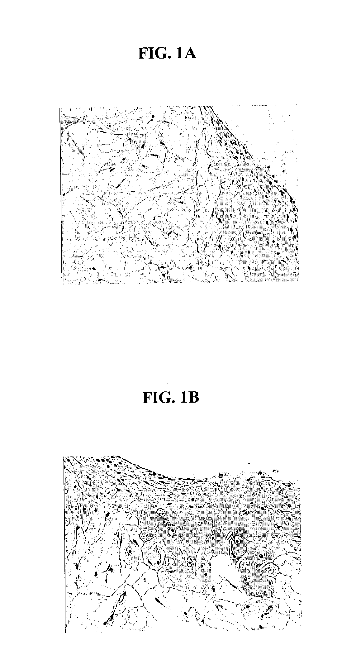
Healthy cartilage tissue from articulating joints was obtained from bovine shoulders. The cartilage tissue, which was substantially free of bone tissue, was minced using scalpel blades to obtain small tissue fragments in the presence of 0.2% collagenase. The size of the tissue fragments varied but on average should be approximately 1×1 mm in dimension. The minced tissue was then distributed uniformly on a 4×5 cm synthetic bioresorbable polycaprolactone / polyglycolic acid (PCL / PGA) scaffold. Ethylene oxide sterilized polymer scaffolds, were pre-soaked for 4 hours in Dulbecco's Modified Eagle's Medium prior to distribution of tissue fragments. The scaffold loaded with minced fragments was then placed in a 10 cm cell culture dish containing chondrocyte growth medium. The chondrocyte growth medium consisted of Dulbecco's modified eagles medium (DMEM-high glucose) supplemented with 20% fetal calf serum (FCS), 10 mM HEPES, 0.1 mM nonessential amino acids, 20 mg / ml of L-proline, 50 mg / ml as...

example 2

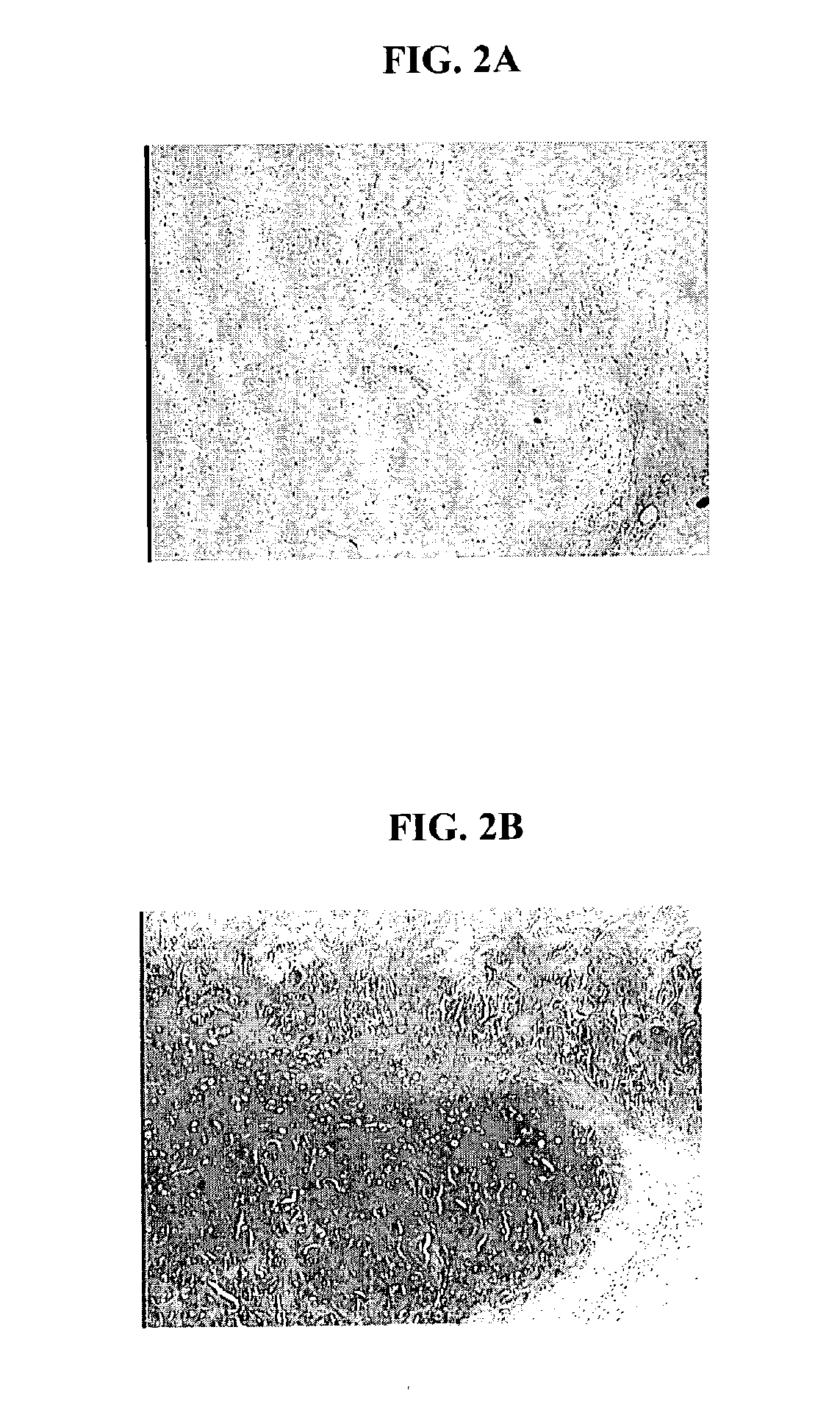
The bioresorbable scaffolds containing minced cartilage tissue and cells from Example 1 were also implanted into SCID mice. The objective was to evaluate the chondrocytic ingrowth of minced cartilaginous tissues into polymer scaffolds in vivo. Polymer scaffolds 5 mm in diameter, were subcutaneously implanted bilaterally in the lateral thoracic region of SCID mice. The implanted scaffold was permitted to support cell growth for four weeks. The subcutaneous implantation sites with their overlying skin were then excised and preserved in 10% buffered formalin fixative. Following fixation, each implantation site was processed for histology. Histological sections were stained with Hematoxylin and eosin, and Safranin-O. FIGS. 2 A and B show that abundant cells were distributed within the scaffold. The cells displayed chondrocyte-like morphology, as evidenced by the intense positive staining for Safranin O of the synthesized matrix.

example 3

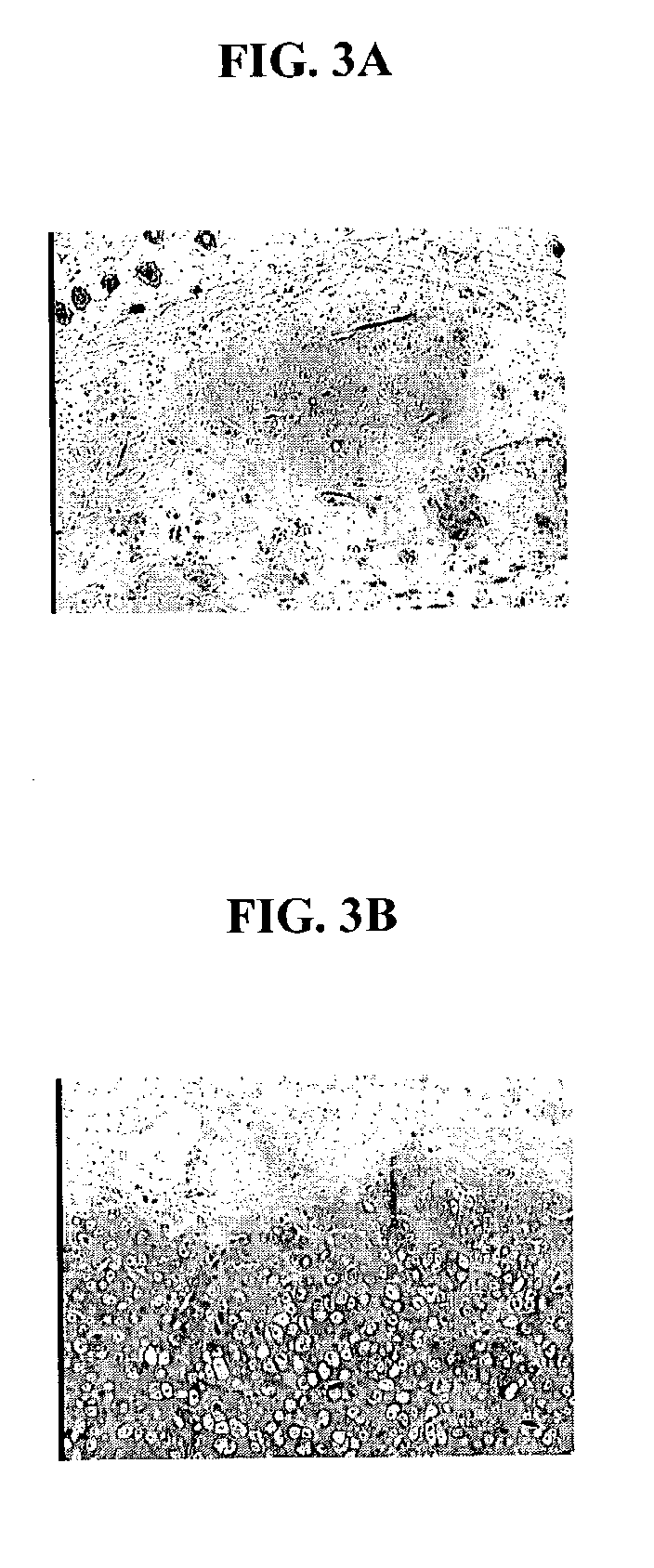
Minced cartilage tissue prepared according to the method described in Example 1 was distributed uniformly on a 4×5 cm synthetic bioresorbable polycaprolactone / polyglycolic acid (PCL / PGA) scaffold. Minced cartilage tissue fragments were adhered to the scaffolds with 1 mL of platelet rich plasma (PRP, Human). Sixty microliters (60 units) of thrombin were used to induce clot formation in the PRP. Control scaffolds loaded with minced cartilage fragments alone and scaffolds loaded with minced cartilage fragments adhered by PRP, were cultured in vitro for 1 week, and then implanted into SCID mice as described in the Example 2. FIG. 3A is a photomicrograph of a control scaffold loaded with minced tissue. FIG. 3B is a photomicrograph depicting a scaffold loaded with minced tissue and PRP. FIG. 3B demonstrates that PRP is beneficial in promoting the migration of the chondrocyte cells, and PRP is also beneficial in promoting the maintenance of the differentiated phenotype of the chondrocyte c...


PUM

PropertyMeasurementUnit
aspect ratioaaaaaaaaaa
diameteraaaaaaaaaa

Abstract

A biocompatible ligament repair implant or scaffold device is provided for use in repairing a variety of ligament tissue injuries. The repair procedures may be conducted with ligament repair implants that contain a biological component that assists in healing or tissue repair. The biocompatible ligament repair implants include a biocompatible scaffold and particles of viable tissue derived from ligament tissue or tendon tissue, such that the tissue and the scaffold become associated. The particles of living tissue contain one or more viable cells that can migrate from the tissue and populate the scaffold.

Description

FIELD OF THE INVENTIONThe present invention relates to biocompatible tissue implant devices for use in the repair of tissue injuries, as well as methods for making and using such biocompatible tissue implant devices.BACKGROUND OF THE INVENTIONInjuries to soft tissue, such as cartilage, skin, muscle, bone, tendon and ligament, where the tissue has been injured or traumatized frequently require surgical intervention to repair the damage and facilitate healing. Such surgical repairs can include suturing or otherwise repairing the damaged tissue with known medical devices, augmenting the damaged tissue with other tissue, using an implant, a graft or any combination of these techniques.One common tissue injury involves damage to cartilage, which is a non-vascular, resilient, flexible connective tissue. Cartilage typically acts as a “shock-absorber” at articulating joints, but some types of cartilage provide support to tubular structures, such as for example, the larynx, air passages, and...

Claims


Application Information

Patent Timeline
Patent Type & Authority Applications(United States)
IPC IPC(8): A61F2/08A61F2/00A61K35/12A61L27/00A61F2/28A61L27/36
CPCA61F2/08A61L27/3604A61L2430/10A61L27/3683A61L27/3662
Inventor BINNETTE, FRANCOISHWANG, JULIAZIMMERMAN, MARKMELICAN, MORA CAROLYNNE
Owner DEPUY MITEK INC
Why Eureka
Try Eureka