Drug-eluting catheter and method of manufacturing the same
a technology of dilatation catheter and nanoparticles, which is applied in the direction of catheters, dilators, surgery, etc., can solve the problems of difficult uniformaly adhesion of a sufficient number of nanoparticles, difficult control of the rate at which the drug is released, and inability to effectively inhibit an inflammatory process occurring during a period of 1 to 3 days after the catheter, etc., to achieve the effect of efficient introduction of bioactive substances into cells and easy control
- Summary
- Abstract
- Description
- Claims
- Application Information
AI Technical Summary
Benefits of technology
Problems solved by technology
Method used
Image

Examples
example 1
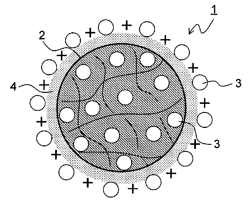
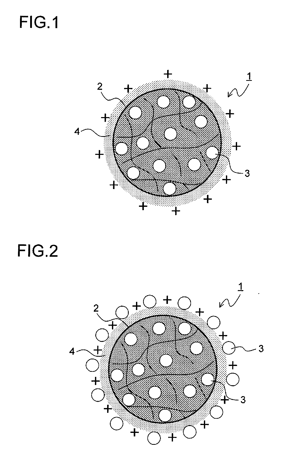
[0171]50 mg of the NFκB decoy indicated by sequence number 1 was dissolved in 6 ml of purified water. 1 g of lactic acid / glycolic acid copolymer (PLGA: PLGA7520 (product name) made by Wako Pure Chemical Industries, Ltd. with a molecular weight of 20000 and a lactic acid / glycolic acid molar ratio of 75 to 25), which was a biocompatible polymer, was dissolved in 43 ml of acetone, which was a good solvent for the acid copolymer, and thus a polymer solution was obtained. Then, the aqueous solution of the NFκB decoy was added to and mixed with the polymer solution, and thus a mixed solution was obtained. 5 g of a 2 weight percent cationic chitosan (Moiss Coat PX (product name) made by Katakura Chikkarin Co., Ltd.) aqueous solution was mixed with 25 ml of a 2 weight percent polyvinyl alcohol (PVA: Poval 403 (product name) made by Kuraray Co., Ltd. with a degree of polymerization of about 300 and a degree of saponification of about 80 mole percent) aqueous solution, and thus a poor solvent...
example 2
[0173]50 mg of the NFκB decoy indicated by sequence number 1 was dissolved in 6 ml of purified water. 1 g of lactic acid / glycolic acid copolymer (PLGA: PLGA7520 (product name) made by Wako Pure Chemical Industries, Ltd.), which was a biocompatible polymer, was dissolved in 43 ml of acetone, which was a good solvent for the acid copolymer, and thus a polymer solution was obtained. Then, the aqueous solution of the NFκB decoy was added to and mixed with the polymer solution, and thus a mixed solution was obtained. 5 g of a 2 weight percent cationic chitosan (Moiss Coat PX (product name) made by Katakura Chikkarin Co., Ltd.) aqueous solution was mixed with 25 ml of a 2 weight percent polyvinyl alcohol (PVA: Poval 403 (product name) made by Kuraray Co., Ltd.) aqueous solution, and thus a poor solvent was obtained. The mixed solution was dropped into this poor solvent at a constant rate of 4 ml / minute at a temperature of 40° C. while stirred at 400 rpm, and then the good solvent was diff...
example 3
Assembly of the Balloon Catheter Main Body
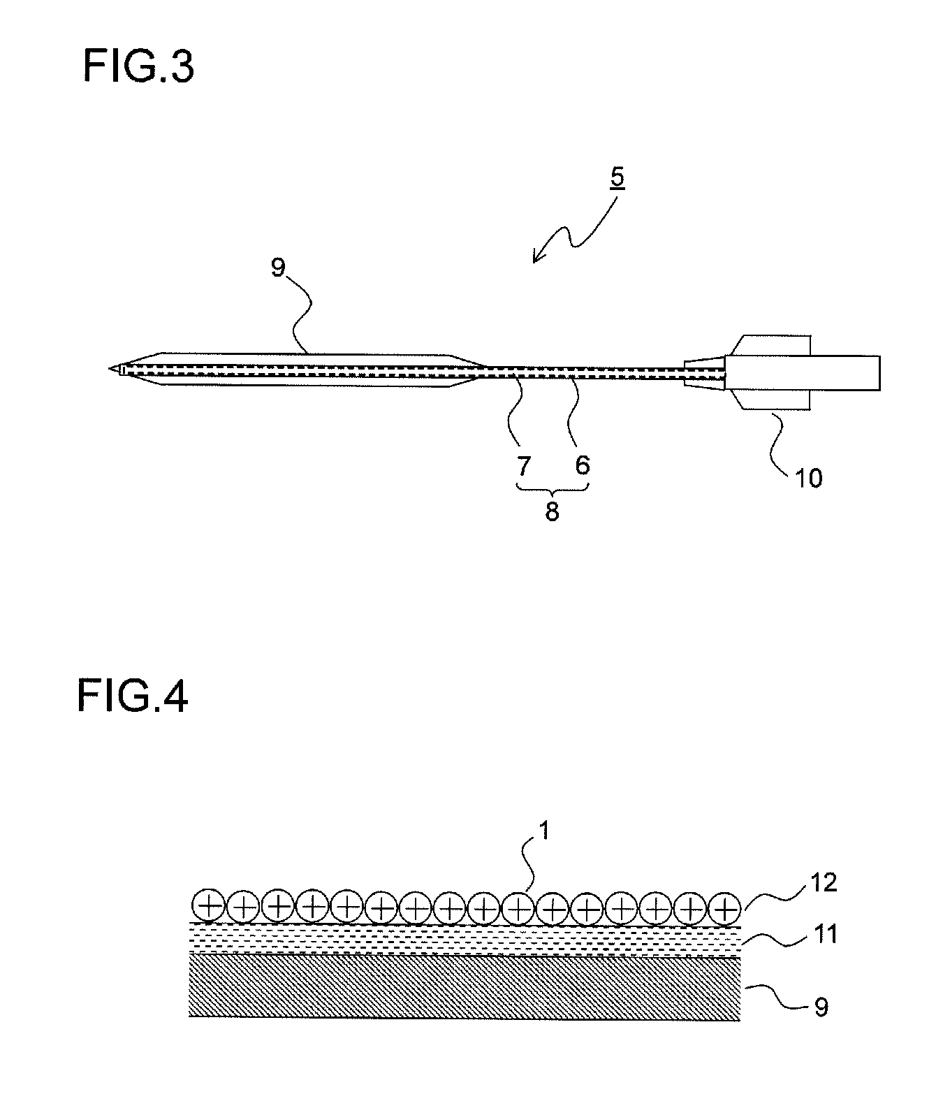
[0176]Hexafluoroisopropanol (HFIP) was applied to part of the polyamide balloon to be adhered and was melted, and the part was adhered to the end of the catheter shaft. A core wire was inserted through the end of the catheter to prevent the entrance of the nanoparticle dispersion solution, and the catheter main body was melted and sealed, with the result that the catheter main body as shown in FIG. 3 was produced.
(The Coating of the Balloon Portion with a Maleic Anhydride Polymer)
[0177]The balloon portion of the catheter main body produced was immersed in ethanol (99.5%) for five seconds, and then its surface was wiped with Kimwipes (product name) impregnated with ethanol and was vacuum-dried within a dryer (55° C., 2 hours). Thereafter, in pretreatment for facilitating the coating, the balloon portion was immersed for one hour in a 4 weight percent methyl ethyl ketone solution of hexamethylene-1,6-diisocyanate (HMDI), and was further vacuum...
PUM
| Property | Measurement | Unit |
|---|---|---|
| Mass | aaaaa | aaaaa |
| Mass | aaaaa | aaaaa |
| Mass | aaaaa | aaaaa |
Abstract
Description
Claims
Application Information