System and method for in vitro blood vessel modeling

a technology system, applied in the field of system and method of in vitro blood vessel modeling, to achieve the effect of reducing non-physiological shear stress on the porous membran

Active Publication Date: 2011-03-03
CHARLES STARK DRAPER LABORATORY +1
View PDF7 Cites 31 Cited by
  • Summary
  • Abstract
  • Description
  • Claims
  • Application Information

AI Technical Summary

Benefits of technology

[0009]In a certain embodiment of the present invention, the in vitro blood vessel model includes smooth muscle and luminal channels having offset inflow and outflow ponds that reduce non-physiological shear stress on the porous membrane.

Problems solved by technology

The cause of DIVI is poorly understood but its occurrence often halts in vivo testing of candidate drugs.

Method used


Image

  • System and method for in vitro blood vessel modeling
  • System and method for in vitro blood vessel modeling
  • System and method for in vitro blood vessel modeling

Examples


Embodiment Construction

[0020]The present invention provides an in vitro model of a blood vessel, through which blood or culture media flows at physiological rates, that allows the investigation of a variety of vascular diseases and physiological mechanisms.

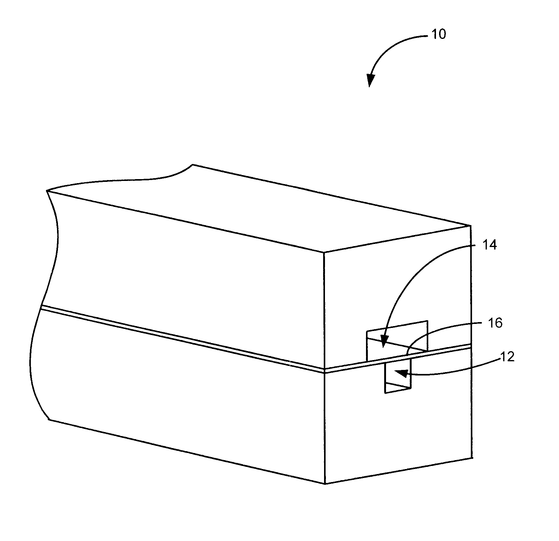
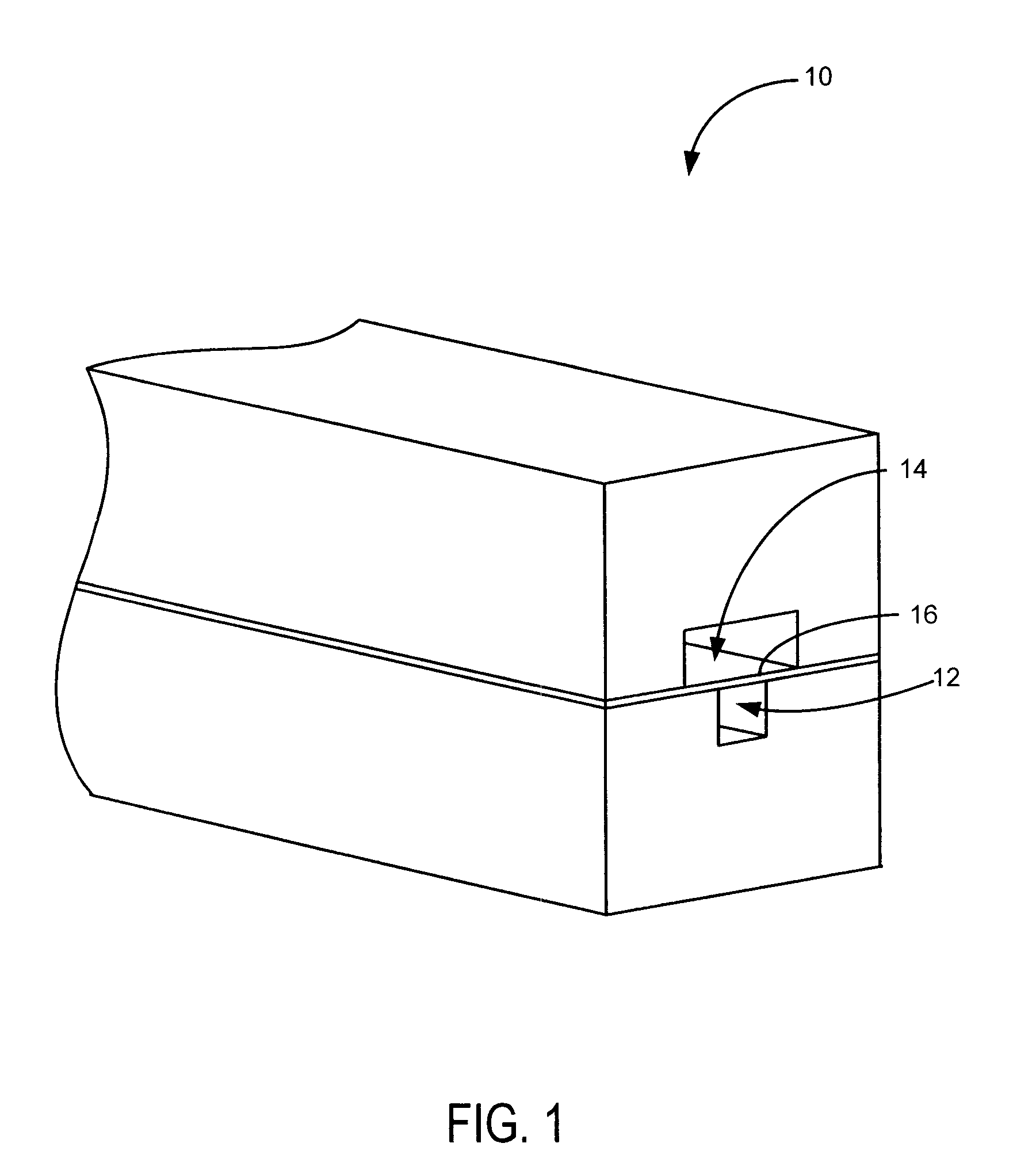
[0021]Referring to FIGS. 1-2, the present invention includes a microfluidic device 10 having a luminal channel 12 separated from a smooth muscle channel 14 by a porous membrane 16.

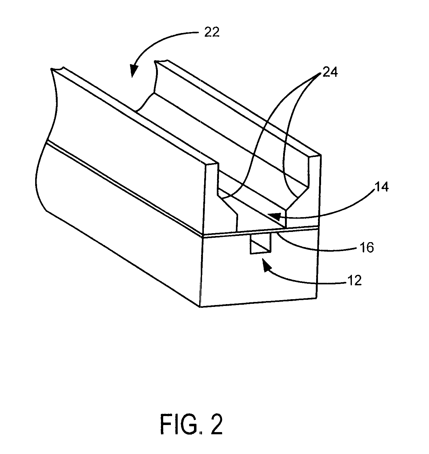
[0022]The luminal channel 12 models the lumen of a blood vessel and may also be referred to as the vascular channel or vascular lumen. The smooth muscle channel 14 is designed to allow SMC growth and models the medial layer of the blood vessel. It is contemplated that SMC growth may be better facilitated by employing an open smooth muscle channel 22, which provides access to increased volumes of growth media, and beveled sides 24, which allow cells to more readily come into contact with the membrane. This arrangement provides more uniform seeding of the device and minimizes th...


PUM

PropertyMeasurementUnit
porosityaaaaaaaaaa
porosityaaaaaaaaaa
pore sizesaaaaaaaaaa

Abstract

The present invention provides an in vitro blood vessel model for investigation of drug induced vascular injury and other vascular pathologies. The in vitro blood vessel model provides two channels separated by a porous membrane that is coated on one side by an endothelial cell layer and is coated on the other side by a smooth muscle cell layer, wherein said model is susceptible to the extravasation of red blood cells across said porous membrane due to drug induced vascular injury.

Description

CROSS-REFERENCE TO RELATED APPLICATIONS[0001]This application claims the benefit of U.S. Provisional Application 61 / 065,356, filed Feb. 11, 2008, and U.S. Provisional Application 61 / 103,117, filed Oct. 6, 2008, both of which are incorporated herein in their entirety for all purposes.FIELD OF THE INVENTION[0002]The present invention relates to a system and method for creating an in vitro blood vessel model to investigate drug induced vascular injury and other vascular pathology.BACKGROUND OF THE INVENTION[0003]Blood vessels are a tissue central to many disease states. One such disease state is drug induced vascular injury (DIVI), a pathological insult to blood vessels that occurs after the administration of a drug. DIVI causes smooth muscle cell (SMC) death and hemorrhage and is characterized by the extravasation of red blood cells (RBCs) from the vascular lumen into surrounding SMC layers. This phenomenon occurs without overt damage to the endothelial cells (ECs). The cause of DIVI ...

Claims


Application Information

Patent Timeline
Patent Type & Authority Applications(United States)
IPC IPC(8): C12Q1/02G09B23/30
CPCA61L27/18A61L27/38A61L27/507A61L27/56B01L3/5027C12M23/34C12M25/02C12M35/08G09B23/28C12M21/08C08L69/00B01L3/502707B01L2200/027B01L2300/0681G01N33/5088
Inventor HOGANSON, DAVID M.VACANTI, JOSEPH P.
Owner CHARLES STARK DRAPER LABORATORY
Why Eureka
Try Eureka