Medical image processing apparatus and method and computer-readable recording medium

a technology of medical image and recording medium, which is applied in the field of medical image processing apparatus and method and computer-readable recording medium, can solve the problems of imposing an extremely heavy burden on users, insufficient matching position accuracy, and troublesome operation for searching the image reading report for words, etc., and achieves the effect of accurately identifying a medical imag

Active Publication Date: 2011-03-31
FUJIFILM CORP
View PDF8 Cites 49 Cited by
  • Summary
  • Abstract
  • Description
  • Claims
  • Application Information

AI Technical Summary

Benefits of technology

[0012]In view of the foregoing circumstances, it is an object of the present invention to make it poss...

Problems solved by technology

However, since several hundred to several thousand medical images are obtained in a series of radiography, the operation for matching the slice positions of slice images to be compared with each other imposes an extremely heavy burden on users.
Therefore, the accuracy of matching the...

Method used


Image

  • Medical image processing apparatus and method and computer-readable recording medium
  • Medical image processing apparatus and method and computer-readable recording medium
  • Medical image processing apparatus and method and computer-readable recording medium

Examples


first embodiment

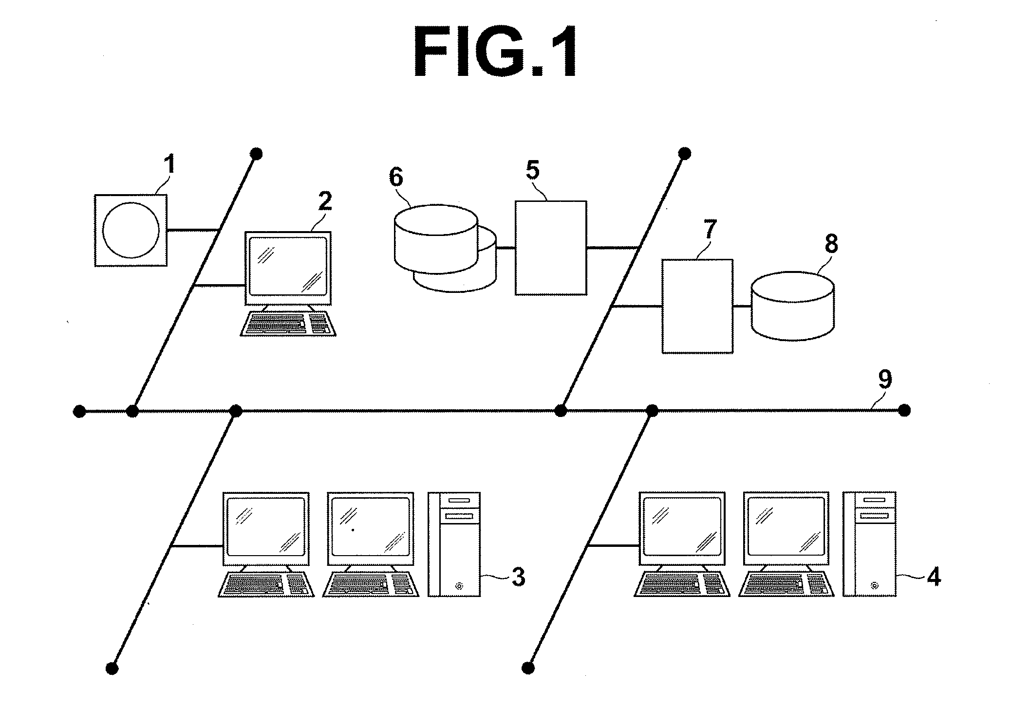
[0098]Hereinafter, embodiments of the present invention will be described with reference to drawings. FIG. 1 is a schematic diagram illustrating the configuration of a medical information system to which a medical image processing apparatus according to the present invention has been applied. The system is used to perform radiography on a region to be examined of a subject (patient) based on an examination order given by a doctor in a clinical department by using a known ordering system, and to store data or images obtained by radiography. Further, the system is used by a radiologist or doctor in a radiology department, who is specialized in image reading, to read images and to prepare a report on the result of image reading. Further, the system is used by the doctor in the clinical department who requested radiography. The doctor in the clinical department uses the system to view (retrieve) the report on the result of image reading and to observe, in detail, an image on which image...

second embodiment

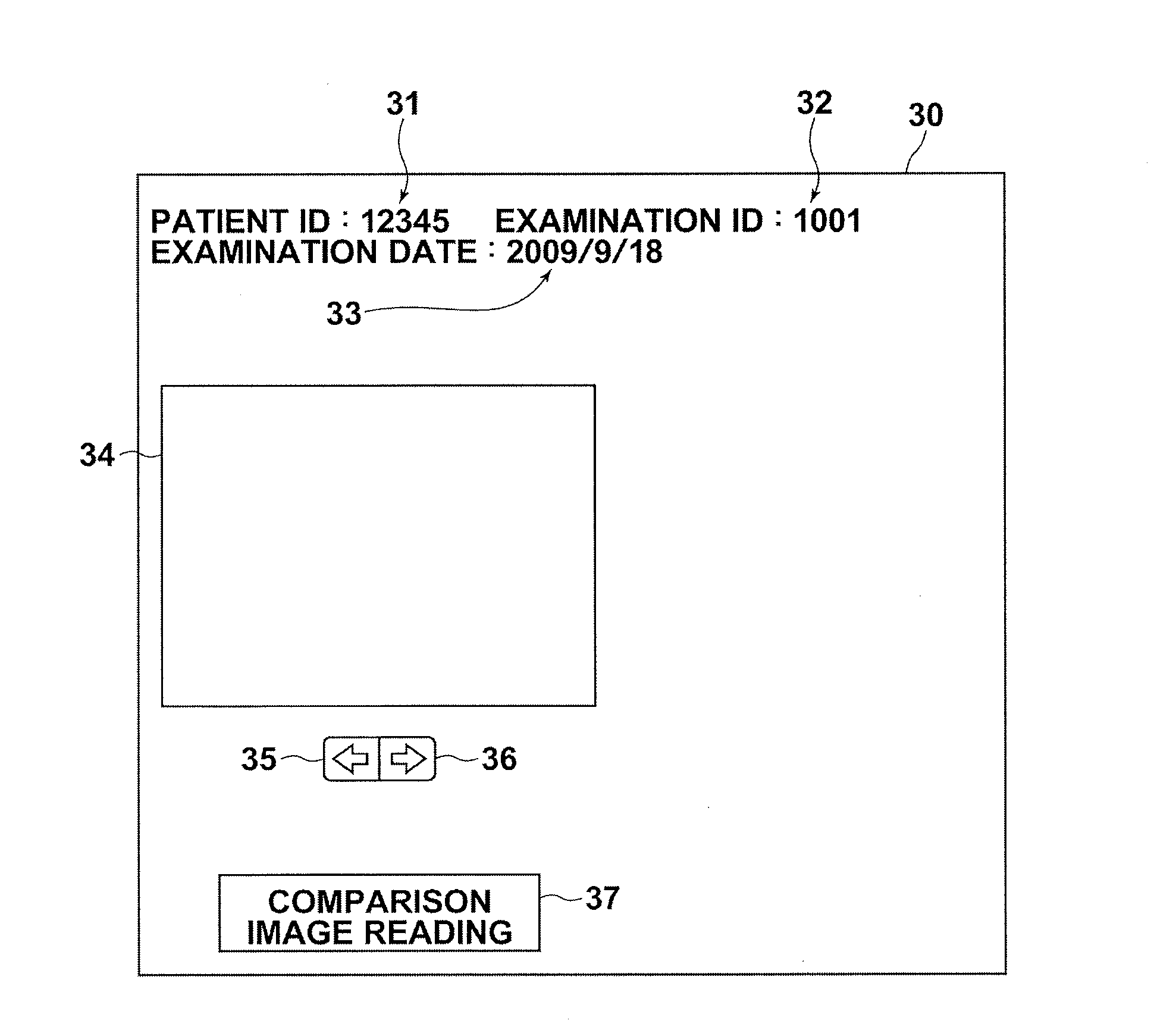
[0142]In the second embodiment, the image reading report RPT illustrated in FIG. 7 is displayed, and the keyword in the text of the finding is specified by clicking or the like, and an image linked to the keyword is displayed in the display screen 30 of the diagnosis target image. Alternatively, the display screen 30 may be displayed in advance. In that case, when an image at a slice position of an image reading target displayed in the area 34 is linked to the keyword in the text of the finding in the image reading report RPT, a frame 39 may be displayed around the image displayed in the area 34, as illustrated in FIG. 17, so that it is possible to distinguish that the image is linked to the image reading report RPT. Alternatively, a mark may be provided in a part of the image displayed in the area 34 instead of the frame 39. Accordingly, when the frame 39 or the image to which a mark is provided is displayed in the area 34, the user can recognize that the image is linked to the ima...


PUM


Abstract

A target-viewpoint position in a first-medical-image having multiple viewpoints is determined. Information about an anatomical structure and/or a lesion in the first-medical-image is obtained, as first information. Further, information about an anatomical structure and/or a lesion in a finding with respect to at least one second-medical-image of the same examined person as the first-medical-image, the information being related to the first information, is obtained, as second information, from image-reading-report information including the finding. Each of the at least one second-medical-image has multiple viewpoints, and was obtained at different time from the first-medical-image. The image-reading-report information is correlated with an image of a viewpoint position at which the finding was prepared. An image of a corresponding viewpoint position, which corresponds to the target-viewpoint position, is determined based on the viewpoint position of the image correlated with the image-reading-report information from which the second information has been obtained.

Description

BACKGROUND OF THE INVENTION[0001]1. Field of the Invention[0002]The present invention relates to a medical image processing apparatus and method appropriate for comparison reading of medical images of an examined subject (patient), and a computer-readable recording having stored therein a program for causing a computer to execute the medical image processing method.[0003]2. Description of the Related Art[0004]In recent years, modalities using various techniques, such as an X-ray CT (Computed Tomography) apparatus, an ultrasonic (US) diagnosis apparatus, an MRI (Magnetic Resonance Imaging) apparatus, a PET (Positron Emission Tomography) apparatus and a SPET (Single-Photon Emission Tomography) apparatus, were used in medical image fields in addition to an X-ray radiography apparatus. As the speed of the modalities became higher, and the performance of the modalities improved to cope with multi-slice tomography or the like, it became possible to perform radiography on a plurality of re...

Claims


Application Information

Patent Timeline
IPC IPC(8): G06K9/00G16H30/40
CPCG06F19/321G06F19/3487G06F19/322G16H10/60G16H15/00G16H30/40
Inventor NAKAMURA, KEIGO
Owner FUJIFILM CORP
Why Eureka
Try Eureka