Supercharge Your Innovation With Domain-Expert AI Agents!

Method and System for Shape-Constrained Aortic Valve Landmark Detection

Active Publication Date: 2011-04-28
SIEMENS HEALTHCARE GMBH
View PDF12 Cites 32 Cited by
  • Summary
  • Abstract
  • Description
  • Claims
  • Application Information

AI Technical Summary

Benefits of technology

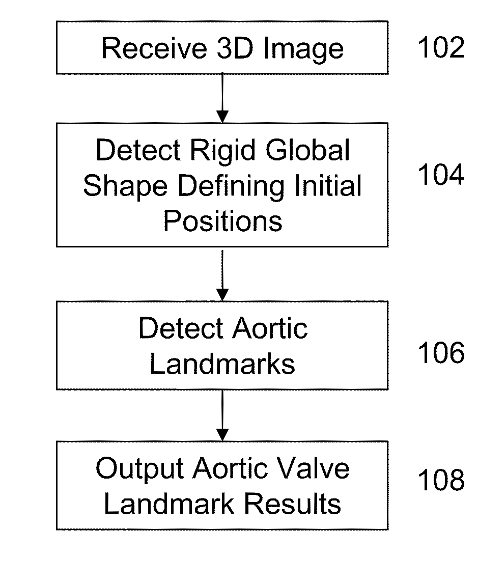
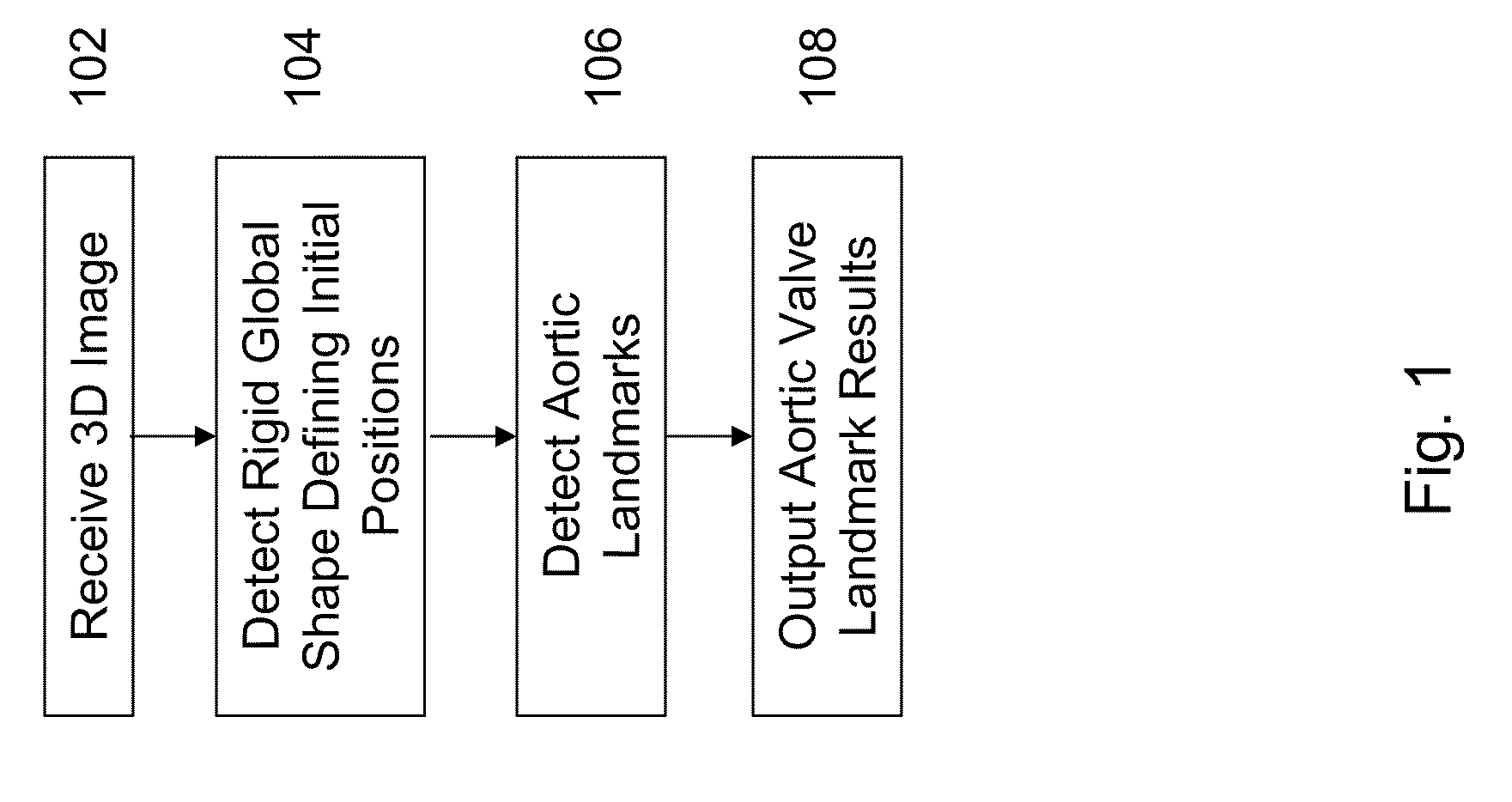
[0007]The present invention provides a method and system for automatic aortic valve landmark detection. Embodiments of the present invention automatically determine aortic valve landmarks in a robust and efficient fashion. Embodiments of the invention achieve robust detection of aortic valve landmarks by defining landmarks, assigning labels to each detected landmark, and refining the landmark detections based on initial landmark estimates.

Problems solved by technology

However, contrast agents are typically toxic and it is preferable that contrast agent usage be minimal.

Method used


Image

  • Method and System for Shape-Constrained Aortic Valve Landmark Detection
  • Method and System for Shape-Constrained Aortic Valve Landmark Detection
  • Method and System for Shape-Constrained Aortic Valve Landmark Detection

Examples


Embodiment Construction

[0016]The present invention is directed to a method and system for shape-constrained aortic valve landmark detection in 3D medical images, including, but not limited to, DynaCT images, cardiac CT images, and cardiac magnetic resonance imaging (MRI) images. Embodiments of the present invention are described herein to give a visual understanding of aortic valve landmark detection. A digital image is often composed of digital representations of one or more objects (or shapes). The digital representation of an object is often described herein in terms of identifying and manipulating the objects. Such manipulations are virtual manipulations accomplished in the memory or other circuitry / hardware of a computer system. Accordingly, it is to be understood that embodiments of the present invention may be performed within and by a computer system using data stored within the computer system.

[0017]DynaCT has recently emerged as an imaging modality offering several advantages over traditional co...


PUM


Abstract

A system and method for performing shape-constrained aortic valve landmark detection using 3D medical images is provided. A rigid global shape defining initial positions of a plurality of aortic valve landmarks is detected within a 3D image. Each of the plurality of aortic valve landmarks is detected based on the initial positions.

Description

[0001]This application claims the benefit of U.S. Provisional Application No. 61 / 253,882, filed Oct. 22, 2009, the disclosure of which is incorporated herein by reference.BACKGROUND OF THE INVENTION[0002]The present invention relates to medical imaging of the heart, and more particularly, to automatic detection of aortic valve landmarks in medical images of the heart.[0003]Aortic valve disease is the most common valvular disease in developed countries, affecting up to 1.8% of the global population. A damaged aortic valve may be remedied by implantation of an artificial aortic valve. During transcatheter valve implantation surgery, in which implantation of an artificial aortic valve occurs, 2D fluoroscopic images are captured in real time by a C-arm system to provide guidance to physicians performing the implantation. The aortic root structure is distinguishable from its background for only a short period of time after a contrast agent is applied. However, contrast agents are typical...

Claims


Application Information

Patent Timeline
IPC IPC(8): G06K9/00
CPCG06K9/6209G06K9/00214A61B6/032A61B6/4441A61B6/503A61B6/504A61B6/5211G16H50/20G06V20/653G06V10/7553
Inventor ZHENG, YEFENGJOHN, MATTHIASBOESE, JANCOMANICIU, DORIN
Owner SIEMENS HEALTHCARE GMBH
Features
  • R&D
  • Intellectual Property
  • Life Sciences
  • Materials
  • Tech Scout
  • Unparalleled Data Quality
  • Higher Quality Content
  • 60% Fewer Hallucinations
Learn More