
Methods and devices for determining lumen occlusion

a technology of hysterosalpingocontrast and lumen, applied in the field of enhanced emission hysterosalpingocontrast sonography, can solve the problems of increasing costs, putting a burden on patients, and difficulty for patients

Inactive Publication Date: 2011-06-09
BAYER HEALTHCARE LLC
View PDF6 Cites 62 Cited by
  • Summary
  • Abstract
  • Description
  • Claims
  • Application Information

AI Technical Summary

Benefits of technology

The patent describes a method for confirming the blockage of body lumens, specifically the fallopian tubes, using a contrast agent containing microbubbles. This technique is called contrast enhanced ultrasonography. The technical effect is the ability to accurately confirm the placement and effectiveness of an intrafallopian contraceptive device.

Problems solved by technology

The inability to confirm the placement of the device at the time of placement within the doctor's office creates difficulty for the patient and increases costs by requiring the patient to come in again for another placement procedure of the intrafallopian contraceptive device if the placement was not done properly in the first place.
This must also be performed in a hospital and may require a follow-up visit to a doctor, placing a burden on the patient and creating further expense.
Visualizing the intrafallopian contraceptive devices by ultrasound performed with saline may be difficult because saline looks the same as other fluids in the body.
It therefore introduces a greater amount of uncertainty in the determination of whether the devices have been properly positioned or whether the fallopian tubes or other type of body lumen is occluded.

Method used


Image

  • Methods and devices for determining lumen occlusion
  • Methods and devices for determining lumen occlusion
  • Methods and devices for determining lumen occlusion

Examples


Embodiment Construction

[0015]In the following description numerous specific details are set forth in order to provide a thorough understanding of the present invention. One of ordinary skill in the art will understand that these specific details are for illustrative purposes only and are not intended to limit the scope of the present invention. Additionally, in other instances, well-known processing techniques and equipment have not been set forth in particular detail in order to not unnecessarily obscure the present invention.

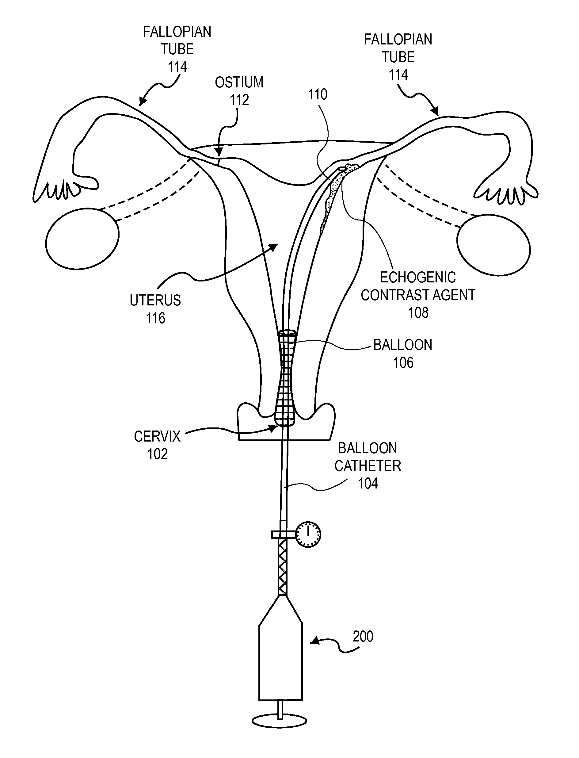
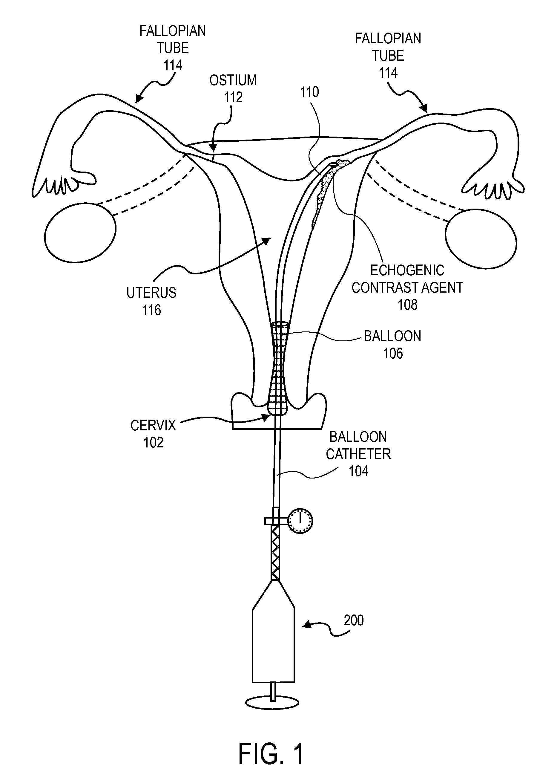
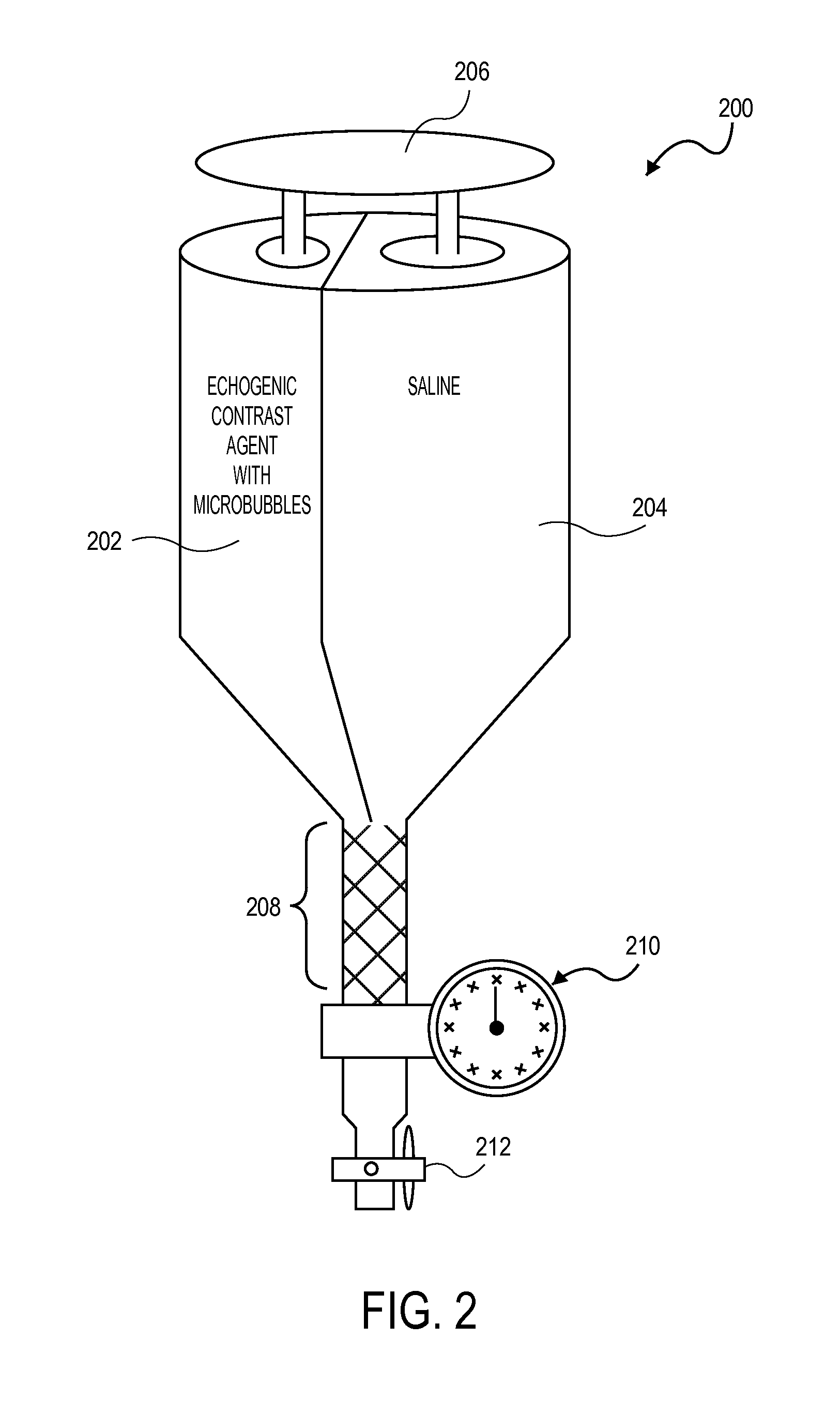
[0016]Embodiments of the present invention describe methods of determining the occlusion of body lumens. In one particular embodiment, the occlusion of the fallopian tubes by an intrafallopian contraceptive device may be confirmed by contrast enhanced ultrasonography (also known as stimulated acoustic emission hysterosalpingo-contrast sonography). In these embodiments a contrast agent containing microbubbles is used.

[0017]Contrast enhanced ultrasonography may be used to determine th...


PUM


Abstract

Embodiments of the present invention describe methods of determining the occlusion of body lumens and apparatuses for doing so. In one particular embodiment, the occlusion of the fallopian tubes by an intrafallopian contraceptive device may be confirmed by contrast enhanced ultrasonography (also known as stimulated acoustic emission hysterosalpingo-contrast sonography). In these embodiments a contrast agent containing microbubbles is used.

Description

[0001]This application claims the benefit of U.S. provisional patent application Ser. No. 60 / 692,497, filed Jun. 20, 2005.BACKGROUND[0002]1. Field[0003]The present invention relates to the field of hysterosalpingo-contrast sonography and in particular the field of enhanced emission hysterosalpingo-contrast sonography.[0004]2. Discussion of Related Art[0005]Female contraception and / or sterilization may be affected by transervically introducing an object (e.g. a coil) into a fallopian tube to inhibit conception. Devices, systems and methods for such a contraceptive approach have been described in various patents and patent applications assigned to the present assignee. For example, U.S. Pat. No. 6,526,979 and U.S. Pat. No. 6,634,361 describe devices that are transcervically inserted into an ostium of a fallopian tube and mechanically anchored within the fallopian tube, both of these patents are hereby incorporated by reference. The devices described in these patents and patent applica...

Claims


Application Information

Patent Timeline
Patent Type & Authority Applications(United States)
IPC IPC(8): A61B8/00A61B8/12
CPCA61B8/0833A61B8/0841A61B8/481A61M25/10A61M5/007A61M5/19A61F6/20
Inventor CONNOR, VIVIANESINCLAIR, EDWARDSWANN, BETSY
Owner BAYER HEALTHCARE LLC
Learn More