Method of ultrasound imaging

a technology of ultrasound imaging and ultrasound contrast, applied in the field of ultrasound imaging, can solve the problems of limiting the use of ultrasound contrast-enhancing agents, difficult to obtain useful images of organs or structures of interest, and poor quality of traditional ultrasound images, so as to improve the quality and usefulness of ultrasound imaging

Inactive Publication Date: 2005-04-05
AMERSHAM INT PLC
View PDF56 Cites 58 Cited by
  • Summary
  • Abstract
  • Description
  • Claims
  • Application Information

AI Technical Summary

Benefits of technology

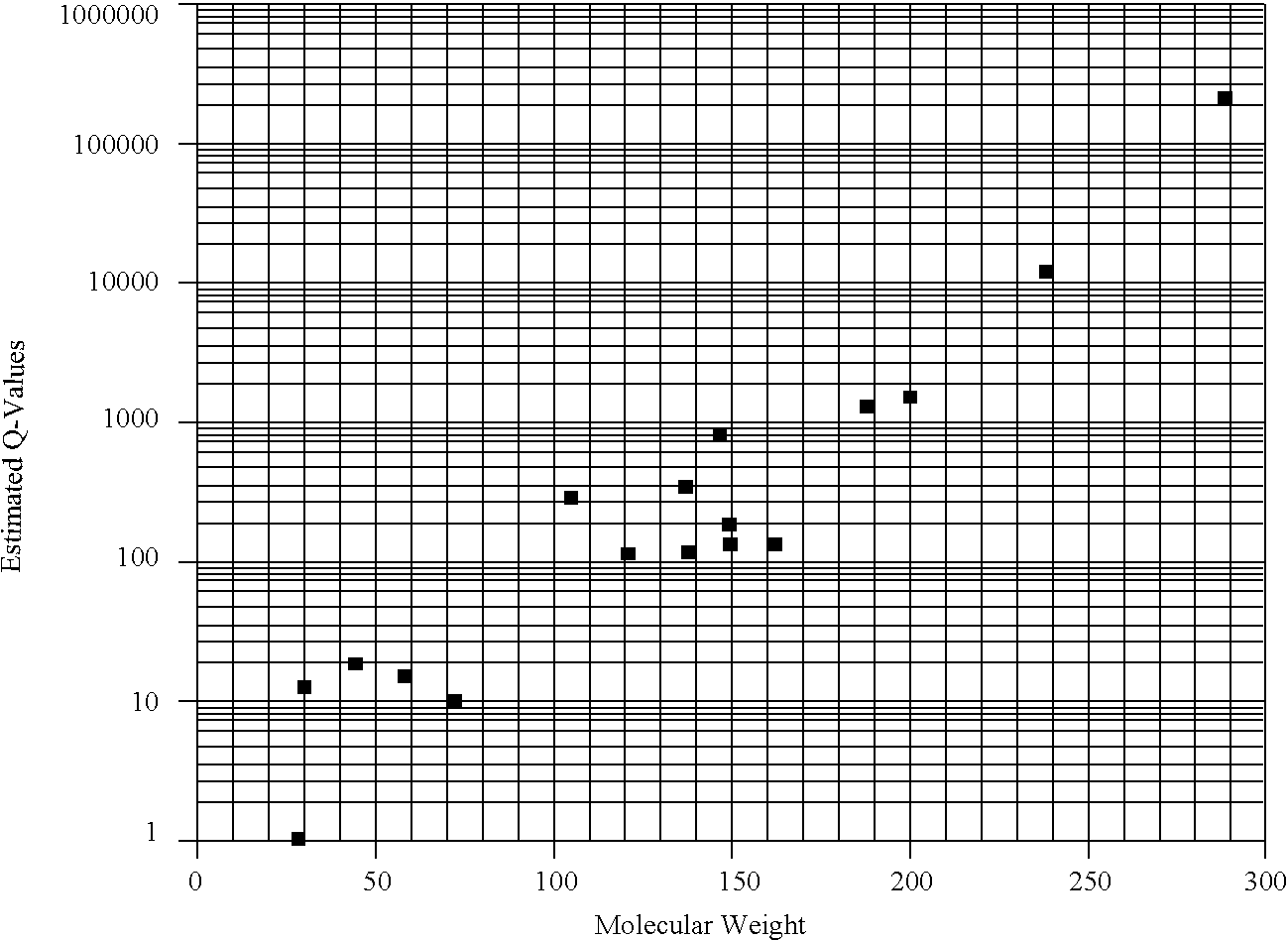
The method that is the subject matter of this invention requires that calculations be made, consistent with the equations provided herein, based on the intrinsic physical properties of a gas and a liquid. Particularly, the density of a gas, the solubility of a gas in solution, and the diffusivity of a gas in solution, which in turn is dependent on the molar volume of the gas and the viscosity of the solution, are used in the equations disclosed b

Problems solved by technology

For some medical conditions, obtaining a useful image of the organ or structure of interest is especially difficult because the details of the structure may not be adequately discernible from the surrounding tissue in an ultrasound image produced by the reflection of ultrasound waves absent a contrast-enhancing agent.
Additionally, traditional ultrasound images are notoriously poor in quality and resolution.
Despite the known advantages, however, the rapid. dissolution of free gas bubbles in solutions such as blood or many aqueous intravenous solutions, severely limits their use as an ultrasound contrast-enhancing agent.
The most important limitations are the size of the microbubble and the length of time that a microbubble will exist before dissolving into the solution.
An additional obstacle exists for ultrasound imaging of the cardiovascular system.
Primarily because of this tradeoff between bubble size and life span, many researchers concluded that free gas microbubbles were not useful as a contrast-enhancing agent for ultrasound diagnosis of certain parts of the cardiovascular system.
However, liquids may exhibit some backscatter if large volumes of a liquid agent are present presumably because the term a in the first bracketed quantity in the above equation may become sufficiently large.
Nevertheless, in light of the above equation and the following, it is appreciated by those skilled in the art that pure liquids are relatively inefficient scatterers compared to free gas microbubbles.
However, techniques based on attenuation contrast as a means to measur the contrast enhancement of a liquid agent are not well-developed and, even if fully developed, may suffer from limitations as to the internal organs or structures with which this technique can be used.
Presently, this technique is only experimental.
Thus, use of these suspensions has been limited to certain cell types in which the particles have the tendency to coagulate because. unless the suspension becomes highly concentrated in particular tissue, the contrast enha

Method used


Image

  • Method of ultrasound imaging
  • Method of ultrasound imaging
  • Method of ultrasound imaging

Examples


example 1

An ultrasound contrast agent was prepared using decafluorobutane as the microbubble-forming gas. A solution was prepared containing:

sorbitol 20.0 gNaCl 0.9 gsoy bean oil 6.0 mLTween 20 0.5 mLwater q.s.100.0 mL

A soapy, clear, yellow solution was afforded with stirring. A 10 mL aliquot of this solution was taken up in a 10 mL glass syringe. The syringe was then attached to a three-way stopcock. A second 10 mL syringe was attached to the stopcock and 1.0 cc of decafluorobutane (PCR, Inc., Gainesville, Fla.) was delivered to the empty syringe. The stopcock valve was opened to the solution-containing syringe and the liquid and gas phases mixed rapidly 20-30 times. A resulting milky-white, slightly viscous solution was obtained.

example 2

The gas emulsion obtained in Example 1 was diluted with water (1:10 to 1:1000), placed in a hemocytometer, and examined under the microscope using an oil immersion lens. The emulsion consisted of predominately 2-5 micron bubbles. The density was 50-100 million microbubbles per mL of original undiluted formulation.

example 3

The formulation of Example 1 was prepared and echocardiography performed in a canine model. A 17.5 kg mongrel dog was anesthetized with isoflurane and monitors established to measure ECG, blood pressure, heart rate, and arterial blood gases according to the methods described by Keller, M W, Feinstein, S B, Watson, D D: Successful left ventricular opacification following peripheral venous injection of sonicated contrast agent: An experimental evaluation. Am Heart J 114:570d (1987).

The results of the safety evaluation are as follows:

MAXIMUM PERCENTAGE CHANGE IN MEASUREDPARAMETER WITHIN 5 MIN POST INJECTIONAORTICPRESSURESYS-DIASTO-BLOODTOLICLICGASESHEARTDOSEmm HgMEANPaO2PaCO2pHRATE0.5 mL+6, −14+9, 0+8, −632958.17.26+10 −191.0 mL+9, −2+5, −1+4, −4+1, −42.0 mL+5, −3+5, −1+5, −10, −13.0 mL+6, −2+7, 0+4, −30, −34.0 mL+5, −1+3, −3+5, −30, −35.0 mL0, −10+1, −30, −4+1, −17.0 mL0, −130, −80, −931328.67.360, −1

All changes were transient and returned to baseline values typically within 3-6 minut...


PUM


Abstract

Disclosed herein are agents for enhancing the contrast in an ultrasound image. These agents are extremely small bubbles, or “microbubbles,” comprised of specially selected gases. The microbubbles described herein exhibit long life spans in solution and may be produced at a size small enough to traverse the lungs, thus enabling improved ultrasound imaging of the cardiovascular system and other vital organs. Also disclosed herein is a method for selecting gases from which contrast agents may be produced. The method is based on calculations using inherent physical properties of gases and describes a means to associate the properties of a gas with the time for dissolution of microbubbles comprised of the gas.

Description

DESCRIPTIONThis invention relates to agents that enhance the contrast in an ultrasound image generated for use in medical diagnosis. The contrast-enhancing media disclosed herein are comprised of extremely small gas bubbles which are present in a solution that is infused into the body during or just before an ultrasound image is generated. This invention is also directed to a method for enhancing such images by selecting gases from which a collection of free gas microbubbles can be prepared that have novel and superior properties. These microbubbles, composed of the gases whose selection is enabled by the process of this invention, may be extremely small in size and yet survive in the bloodstream long enough to allow contrast-enhanced imaging of parts of the cardiovascular system, peripheral vascular system, and vital organs previously believed to be inaccessible to free gas microbubbles.BACKGROUNDWhen using ultrasound to obtain an image of the internal organs and structures of a hu...

Claims


Application Information

Patent Timeline
IPC IPC(8): A61K49/22G06F17/30
CPCA61K49/223
Inventor QUAY, STEVEN C.
Owner AMERSHAM INT PLC
Why Eureka
Try Eureka