Device and method for inhibiting complement activation

a technology of complement activation and device, applied in the field of complement activation, can solve the problems of cellular activation and pathological consequences, and achieve the effects of preventing neutrophil activation, reducing the level of properdin in the blood, and reducing the level of properdin

Inactive Publication Date: 2011-06-30
NOVELMED THERAPEUTICS
View PDF18 Cites 15 Cited by
  • Summary
  • Abstract
  • Description
  • Claims
  • Application Information

AI Technical Summary

Benefits of technology

[0009]In another aspect of the invention, the blood contacted with the anti-complement antibody is incapable of activating the alternative complement pathway when returned to the subject. The removal of the complement protein in the blood prevents activation of neutrophils, monocytes, basophils, lymphocytes, and platelets via the alternative pathway.
[0010]In a further aspect of the invention, the anti-complement antibody can reduce the level of properdin in the blood. The reduced levels of properdin in blood can decrease levels of C3a, C5a, Bb, C5b-9 as a result of decreased alternative complement pathway activation during extracorporeal circulation. The reduced levels of properdin can also reduce cellular activation in blood from the subject following extracorporeal circulation.

Problems solved by technology

All these cell types are known to have receptors for anaphylatoxins, which cause cellular activation leading to pathological consequences.

Method used


Image

  • Device and method for inhibiting complement activation
  • Device and method for inhibiting complement activation
  • Device and method for inhibiting complement activation

Examples


example 1

Anti-Complement Monoclonal Antibody for the Device

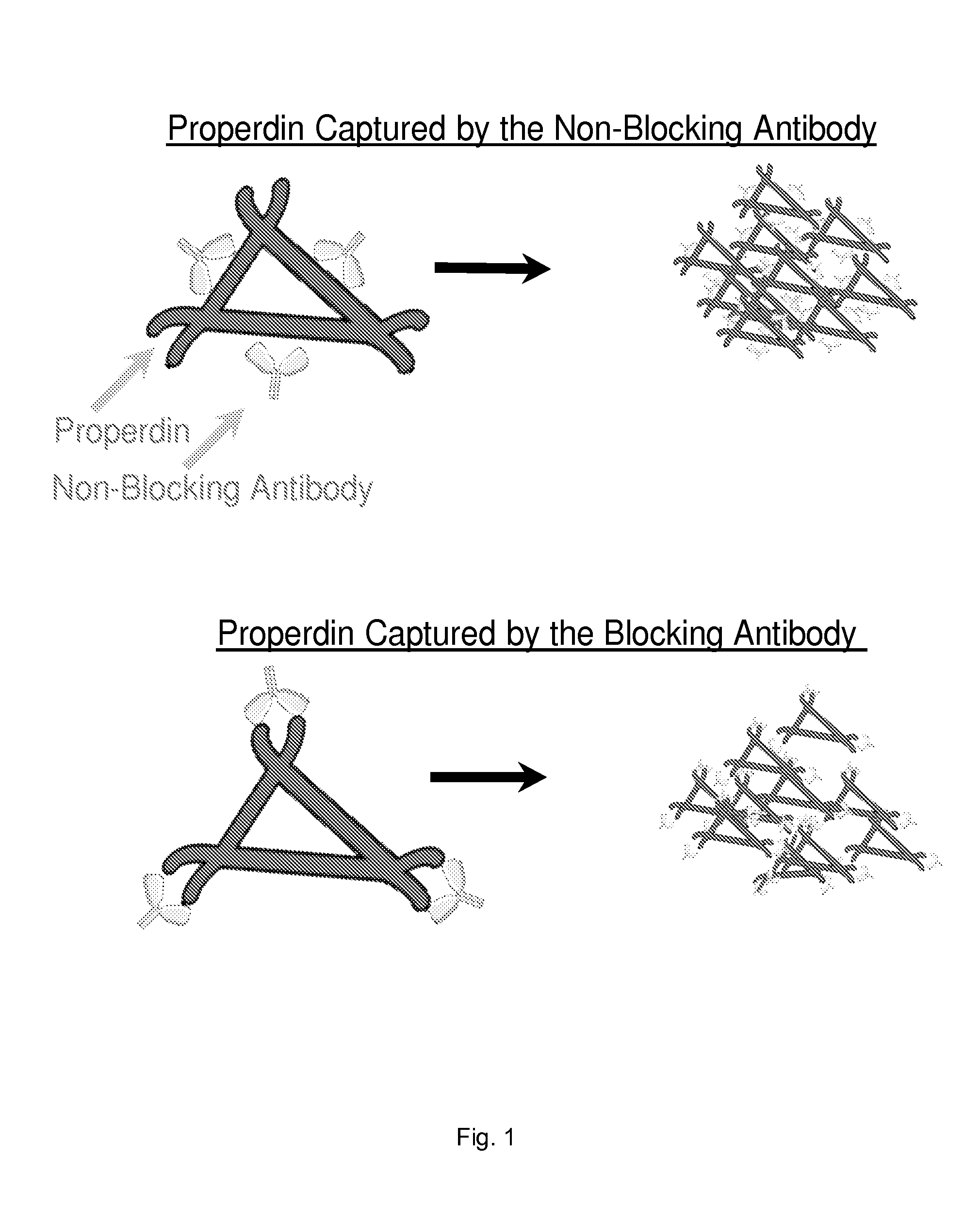
[0064]We selected an anti-P antibody that blocks the AP activation. The anti-P antibody is described in PCT / US08 / 68530. This antibody binds properdin and blocks properdin function. Properdin plays a role in AP activation and therefore, blockade of properdin function inhibits the AP. FIG. 1 shows a trimer of properdin monomer. Anti-P binds the TSR-1, which is represented in the Figure as corners of the trimer. Based on the molar ratio of anti-P to properdin, the model shown perfectly fits the anti-P used. This particular model also shows that if anti-P is immobilized onto the matrix and correctly oriented, it should bind the trimer and retain it onto itself. As a result, the blood / plasma samples passing through should become depleted of properdin. Since properdin plays a critical role AP, the blood and plasma should not activate the AP during blood transit through the extracorporeal circuit.

[0065]If non-blocking anti-P, which binds pr...

example 2

Schematics of how the Anti-P Coated Bead Looks Like when Trimers of Properdin are Extracted from the Blood

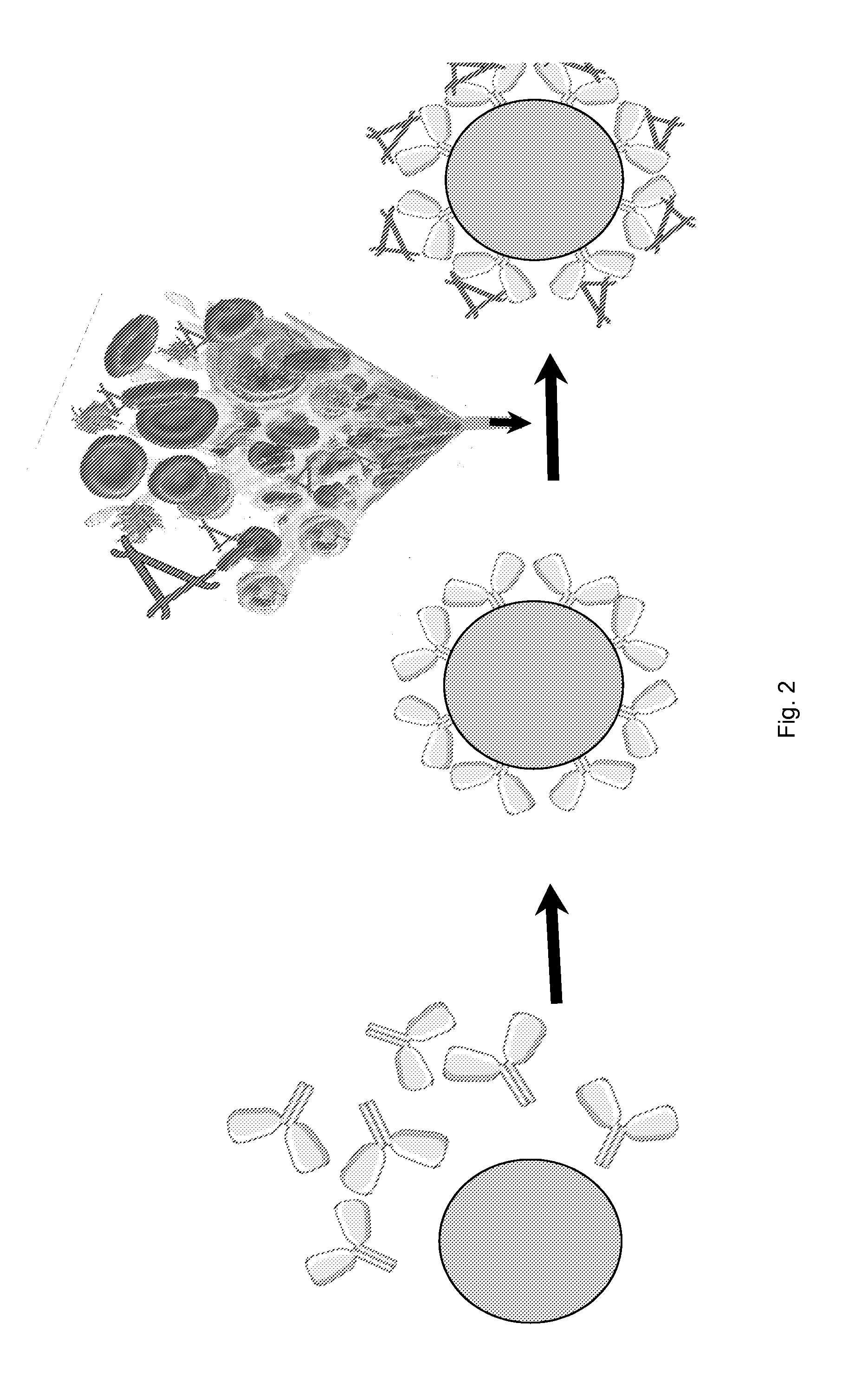
[0067]Bead matrix (CELLTHRUBIGBEADS (Sreogene Corporation) or any bead with large diameter of nearly 300 microns) uncoated or coated with protein G is incubated with the anti-P monoclonal antibody to generate anti-P coated beads. Whole heparinized blood containing functional AP complement proteins is passed through the device. The anti-P coated onto the beads bind properdin from plasma. The flow through should have no AP activity.

example 3

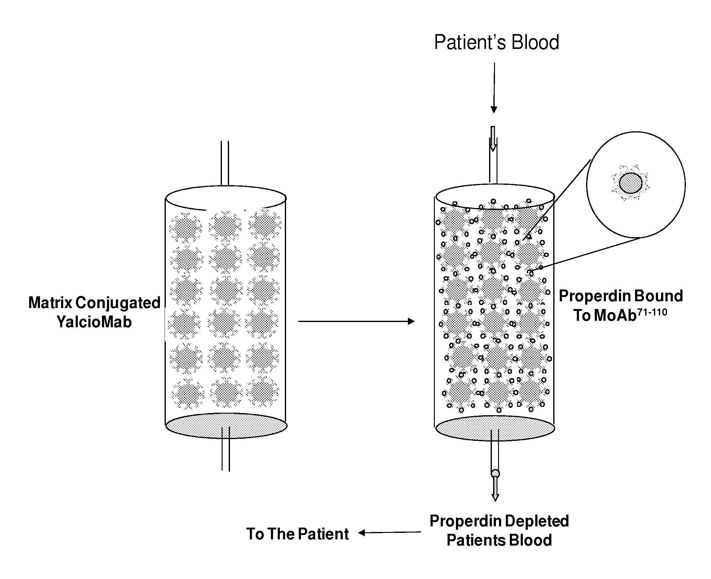
Schematics of Anti-P Coated Beads in a Column. View of Column Before and After Blood Passes Through. Absence of Properdin in Flow Through

[0068]FIG. 3 illustrates two columns. The first column only has anti-P conjugated to the beads. The second column illustrates zoomed-out version of anti-P coated neads with retained properdin. An inset shows the zoom-in portion of the single bead with retained properdin. FIG. 4 shows that the blood that has been through the device is depleted off properdin. The outlet from the device is being poured into the container. The trimer triangles are missing from the flow through.


PUM

PropertyMeasurementUnit
diameteraaaaaaaaaa
temperatureaaaaaaaaaa
volumeaaaaaaaaaa

Abstract

An extracorporeal device for inhibiting alternative complement pathway activation includes a support structure an anti-complement antibody disposed on or within the support structure and a first conduit for conducting blood of the subject to the anti-complement antibody. The anti-complement antibody can bind to the complement protein and remove the complement protein from the blood.

Description

RELATED APPLICATION[0001]This application claims priority from U.S. Provisional Application No. 61 / 095,822, filed Sep. 10, 2008, the subject matter, which is incorporated herein by reference.FIELD OF THE INVENTION[0002]The present invention relates to complement activation. Particularly, the present invention relates to a method for inhibiting complement activation via an extracorporeal device.BACKGROUND OF THE INVENTION[0003]It is estimated that approximately twenty million procedures involving extracorporeal blood circulation are performed annually. Approximately 10,000 renal transplantations are performed annually in the U.S. along with about 500,000 open-heart surgeries, both procedures requiring extracorporeal blood treatment. In addition, approximately ninety thousand plasmapheresis procedures are carried out annually in the U.S. Blood oxygenation has been used to treat patients suffering from acute respiratory failure and infants with diaphragmatic hernia. It is likely that n...

Claims


Application Information

Patent Timeline
Patent Type & Authority Applications(United States)
IPC IPC(8): A61M1/34
CPCA61K2039/505C07K2317/76C07K2317/54C07K16/18A61M1/3679A61M1/3486
Inventor BANSAL, REKHA
Owner NOVELMED THERAPEUTICS
Why Eureka
Try Eureka